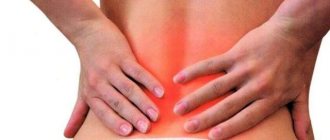
Back pain
- a common complaint. Usually we talk about back pain when pain is felt in the lumbar region. Sometimes in such cases they say that “the back is pinched.” According to recent studies, out of every five people, four have experienced such pain at least once in their lives. Read more about lower back pain>>>
Another typical location of back pain is under the right or left shoulder blade or between the shoulder blades. Read more about pain under the shoulder blade>>>
With age, back pain is more common - among older people, every second person experiences such pain from time to time.
Back pain can have a different character: it can be sharp, stabbing, burning, aching, pulling, and can radiate to other parts of the body (for example, to the chest, legs, abdomen, genitals). The pain may be occasional or constant.
When listening to pain, a person usually asks himself two questions: should he see a doctor, and which doctor should he go to for back pain? Let's try to answer these questions.
Symptoms of nonspecific pain in the spine
A sharp lumbago in the lumbar region, pain when turning the body, a slight aching pain in the back - these are what patients most often complain about.
Causes
- prolonged uncomfortable position at the desk;
- carrying heavy loads without taking into account the correct biomechanics of the body;
- overweight (body mass index greater than or equal to 25);
- weakened back muscles (lack of physical fitness);
- sudden movements leading to microtrauma of the spine;
- stress and depression.
Treatment
Help before diagnosis
In case of traumatic injuries, the victim should be placed on a backboard and given an anesthetic. For pain of non-traumatic origin, functional rest is required. Patients need to avoid sudden movements and take regular breaks when working in a static position. In the absence of signs of an acute condition or severe inflammatory phenomena, short-term use of NSAIDs and the use of local agents are acceptable. Sharp, increasing pain, disturbances in general condition, and neurological disorders are reasons to immediately consult a specialist.
Conservative therapy
Therapeutic tactics are determined by the nature and stage of the pathology. In case of injuries, a protective regime is prescribed, and various methods of traction are occasionally used. The basis of treatment for most traumatic and non-traumatic injuries are medications and physiotherapeutic techniques. The following medications are used:
- NSAIDs
. Effective for acute and chronic pain. Eliminate pain, reduce the severity of inflammation. Prescribed in tablets, injections, and local forms. - Local anesthetics
. Local anesthetics alone or in combination with other drugs (usually glucocorticosteroids) are injected into the affected area during a therapeutic blockade. - Antibiotics
. Indicated for infectious processes. As a rule, they are administered by injection. They are selected taking into account the sensitivity of the pathogen. - Neurotropic vitamins
. A good result is provided by B vitamins, which enhance the effect of other medications and increase the amount of endogenous compounds with an analgesic effect.
Physiotherapy is prescribed after acute symptoms have resolved. Ultrasound, magnetic therapy, electrical stimulation, acupuncture, and other methods are used. Patients are recommended massage, exercise therapy, and manual therapy if indicated.
Symptoms of specific pain in the spine
Before finding out the cause, more persistent symptoms should be a reason to see a doctor:
- pain in the spine that does not go away for several days;
- deterioration of health when moving;
- irradiation of pain to the limb;
- a feeling of numbness in the affected area.
Thus, lumbago in the legs is characteristic of compression of the spinal cord (this may be a symptom of a herniated disc). A dull local pain near the spine may indicate arthrosis of its joints. The sensation of pain intensity is individual for each patient. Therefore, it is important to consult a competent doctor in time.
Which specialists should I contact?
If you want to be sure that you will receive high-quality medical care, contact the CELT Pain Clinic. Describe the nature, accompanying symptoms and causes of lower back pain - and, if necessary, you will receive a referral for examination to a specialist to solve a specific problem, without endless walking in circles. These may be doctors of the following specialties:
- traumatologist-orthopedist;
- neurologist;
- neurosurgeon;
- urologist;
- phthisiatrician;
- oncologist;
- rheumatologist;
- nephrologist;
Diagnostics
Determination of pain characteristics and assumptions about their source are carried out by a doctor during a face-to-face consultation.
What the doctor will tell you:
- onset of dorsalgia and patient age;
- the nature of the pain (does not decrease in a lying position, with rest, or change of position);
- pain is related to previous injury;
- increases over time;
- history of oncology;
- pain due to fever and weight loss;
- “stiffness” in the morning;
- damage to the spinal cord (this is indicated by paralysis, dysfunction of organs, sensitivity).
Next, the necessary list of tests and examinations is compiled to clarify the diagnosis. Our multifunctional center has comprehensive programs for diagnosing back pain. It is recommended for patients:
- whose age is over 65 years;
- early menopause has occurred;
- frequent injuries and fractures occur;
- there is a vitamin D deficiency;
- there are chronic rheumatic diseases or endocrine pathologies, etc.;
- and there are also bad habits (smoking, alcoholism, excessive coffee drinking).
As part of the program, the patient is advised by an experienced rheumatologist. The level of vitamin D, total and ionized calcium and inorganic phosphorus is examined, and osteodensitometry is performed.
Spinal deformities
Spinal column deformity means any significant deviation from the normal curves of the spine. The most common are
- Scoliosis
- Hyperkyphosis
- Hyperlordosis
There are various causes of pathological curvature of the spine. Some children are born with congenital scoliosis or congenital hyperkyphosis.
Sometimes nerve and muscle diseases, injuries, or other conditions cause spinal deformities (such as cerebral palsy).
Most often (up to 80-85%) scoliosis is “idiopathic” (without an obvious cause). Idiopathic scoliosis develops gradually but can progress rapidly during the growing years of adolescence.
Scoliosis
The term scoliosis was first used to describe this spinal deformity by Hippocrates in 400 BC. It is a progressive disease with an unknown cause (idiopathic) in 80% of cases, although there is evidence that genetics and nutrition play a role. Women are 10 times more likely to develop scoliosis than men. Scoliosis is often accompanied by twisting of the spine, which leads to deformation of the costal arches and chest. Scoliosis usually begins to appear during adolescence. Conservative treatment is quite effective for grade 1-2 scoliosis. In cases of severe deformation (grade 3-4) and in cases of progressive scoliosis in adolescence, surgical treatment is recommended (the earlier surgical treatment is performed, the better the long-term results).
Hyperkyphosis
Mild kyphosis is the natural curvature of the thoracic spine, while hyperkyphosis is an excessive forward tilt of the thoracic spine (slouching). Hyperkyphosis is common in older people and is usually associated with the presence of osteoporosis and previous vertebral compression fractures. Causes of hyperkyphosis can also be injuries, diseases of the endocrine system and other diseases. In adolescence, hyperkyphosis may occur, such as Scheuermann-Mau disease, which is characterized by a wedge-shaped deformation of three or more vertebrae in the thoracic spine. As a rule, conservative treatment for Scheuermann-Mau disease is quite effective, but when the angle of deviation from the axis is more than 60 degrees, surgical treatment is recommended.
Hyperlordosis
Lordosis is a natural inward curve in the lumbar spine, and hyperlordosis is a pathological increased curve in the lumbar spine. Hyperlordosis is usually accompanied by an abnormal anterior tilt of the pelvis and is often accompanied by excessive protrusion of the buttocks. Symptoms may include pain and numbness if there is compression of nerve structures. As a rule, hyperlordosis is caused by weakness of the back muscles, hyperextension, for example, in pregnant women, in men with excessive visceral fat. Hyperlordosis is also associated with puberty.
Treatment for hyperlordosis is usually not required unless nerve structures are affected.
Treatment of spinal pain
Effective relief of the patient from dorsalgia is the task of experienced
doctors.
The Federal Scientific and Clinical Center of the Federal Medical and Biological Agency of Russia provides a full range of treatment for neurological diseases using a multidisciplinary approach, individual programs and complexity. Our advantages also include the use of the most advanced diagnostic methods and the ability to provide emergency care for spinal pain.
Conservative
treatment includes various techniques, including the use of non-steroidal anti-inflammatory drugs and muscle relaxants, as well as physiotherapeutic effects (amplipulse, darsonvalization, electrophoresis).
The period of subsequent rehabilitation requires mandatory personal participation from the patient. It is necessary to regularly perform a set of therapeutic exercises to strengthen the muscular frame of the spine and prevent relapse.
Surgical
treatment is carried out in extremely difficult cases, when conservative treatment options have not led to the desired results, and the pain continues.
BACK PAIN: painful muscle spasms and its treatment with muscle relaxants
Back pain is one of the most common complaints that patients present in general medical practice. They are often caused by spinal osteochondrosis - a degenerative lesion of the intervertebral disc cartilage and reactive changes in the adjacent vertebral bodies. Damage to the intervertebral disc develops as a result of repeated injuries (heavy lifting, excessive static and dynamic load, falls, etc.) and age-related degenerative changes. The nucleus pulposus, the central part of the disc, dries out and partially loses its shock-absorbing function. The fibrous ring, located along the periphery of the disc, becomes thinner, cracks form in it, towards which the nucleus pulposus moves, forming a protrusion (prolapse), and if the fibrous ring ruptures, a hernia. Currently, drugs have been created that have a structure-modifying effect on cartilage tissue (the old name is chondroprotectors). A typical representative of the group is the drug chondro, prescribed in a course of 4 months (the effect lasts 2 months after discontinuation). In the affected spinal segment, relative instability of the spine occurs, osteophytes of the vertebral bodies develop (spondylosis), and ligaments and intervertebral joints are damaged (spondyloarthrosis). Herniated intervertebral discs are most often observed in the lower lumbar discs, less often in the lower cervical and upper lumbar discs, and extremely rarely in the thoracic discs. Disc herniations into the vertebral body (Schmorl's hernias) are not clinically significant; disc herniations in the posterior and posterolateral direction can cause compression of the spinal root (radiculopathy), spinal cord (myelopathy at the cervical level) or their vessels.
In addition to compression syndromes, reflex (muscular-tonic) syndromes are possible, which are caused by impulses from receptors in response to changes in the discs, ligaments and joints of the spine - painful muscle spasm. Reflex muscle tension initially has a protective nature, since it leads to immobilization of the affected segment, but later this factor becomes the cause of pain. Unlike compression syndromes of spinal osteochondrosis, which are relatively rare, painful muscle spasms occur during the life of almost every second person.
A classic example of a painful muscle spasm is lumbago (lumbago), which is characterized by a sharp, shooting pain in the lower back, usually developing during physical activity (lifting heavy objects, etc.) or awkward movement. The patient often freezes in an uncomfortable position, and an attempt to move leads to increased pain. The examination reveals tension in the back muscles, usually scoliosis, flattening of the lumbar lordosis or kyphosis.
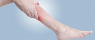
Lumbodynia - back pain - and lumboischialgia - pain in the back and along the back of the leg - develop more often after physical activity, awkward movement or hypothermia, less often - without any reason. The pain is aching in nature and intensifies with movements in the spine, certain postures, and walking. Lumboischialgia is characterized by pain in the buttock, in the posterior parts of the leg, not reaching the toes. The examination reveals pain, tension in the muscles of the back and posterior group of leg muscles, limited mobility of the spine, often scoliosis, and symptoms of tension (Lasegue, Wasserman, etc.).
At the cervical level, reflex muscular-tonic syndromes may occur: cervicalgia and cervicobrachialgia, which often develop after physical activity or awkward neck movement. Cervicalgia is pain in the cervical region, which often spreads to the back of the head (cervicocranialgia). Cervicobrachialgia is pain in the cervical region spreading to the arm. Characteristically, the pain intensifies with movements in the neck or, conversely, with a prolonged static position (at the movies, after sleeping on a thick, high pillow, etc.). During the examination, tension in the cervical muscles is revealed, restriction of movements in the cervical spine, and pain on palpation of the spinous processes and intervertebral joints on the side of pain are often observed.
When a nerve root is compressed (radiculopathy), in addition to painful muscle spasms and limitations in mobility in the spine and limbs, sensory, reflex and (or) motor disturbances are detected in the area of the affected root. At the lumbar level, the fifth lumbar (L5) and first sacral (S1) roots are most often affected, less often - the fourth lumbar root and very rarely - the upper lumbar roots. Radiculopathies of the lower cervical roots are much less common.
Painful muscle spasm also occurs with another fairly common cause of pain in the back and limbs - myofascial pain caused by the formation of so-called trigger zones in the muscles and (or) associated fascia. Myofascial pain is manifested by muscle tension and the presence of trigger points in them, which are identified through manual examination of the muscles. An active trigger point is a constant source of pain that increases with palpation in the muscle; a latent trigger point causes pain only when palpated. For each muscle there is an independent myofascial syndrome with a characteristic localization of pain when the trigger zone is irritated, spreading beyond the projection of the muscle to the skin surface. There are no focal neurological disorders, except in cases where tense muscles compress the nerve trunk.
It is important to remember that back pain may be the only symptom of a spinal cord tumor, syringomyelia and other spinal cord diseases. Pain occurs with destruction of the vertebrae and damage to the nerve roots due to infectious processes (tuberculous spondylitis, spinal epidural abscess), neoplasms (primary and metastatic tumors of the spine, myeloma), dysmetabolic disorders (osteoporosis, hyperparathyroidism, Paget's disease). Back pain can be a consequence of a spinal fracture, congenital or acquired deformities (scoliosis, etc.), spinal stenosis, spondylolisthesis, ankylosing spondylitis.
It is possible for various somatic diseases (heart, stomach, pancreas, kidneys, pelvic organs, etc.) through the mechanism of referred pain.
The examination of a patient with back pain requires careful consideration. Any back pain cannot be attributed to “osteochondrosis” - a condition that is detected by X-ray examination in most middle-aged and elderly people. Neurological manifestations of spinal osteochondrosis and myofascial pain are characterized by painful muscle spasms and limited mobility of the spine.
The diagnosis of reflex and compression complications of osteochondrosis is based on clinical data and requires the exclusion of other possible causes of back pain. X-rays of the spine are used mainly to exclude congenital anomalies and deformities, inflammatory diseases (spondylitis), primary and metastatic tumors. X-ray CT or MRI can identify a disc herniation, determine its size and location, and also detect spinal stenosis and a spinal cord tumor.
The diagnosis of myofascial pain is based on clinical findings (identification of painful muscle tension in one or more muscles) and requires the exclusion of other possible causes of pain; differential diagnosis with reflex syndromes (muscular-tonic syndromes) due to spinal osteochondrosis often causes difficulties; a combination of these diseases is possible.
Treatment of reflex syndromes and radiculopathy due to osteochondrosis is based in the acute period on ensuring rest - the patient is advised to avoid sharp bends and painful positions. Prescribed bed rest for several days until the sharp pain subsides, a hard bed (a shield under the mattress), taking centrally acting muscle relaxants, and, if necessary, additional analgesics and non-steroidal anti-inflammatory drugs. To facilitate movement during this period, you should wear a neck or lumbar corset (fixing belt). You can use physiotherapeutic analgesic procedures, rubbing in pain-relieving ointments, compresses with a 30-50% solution of dimexide and novocaine, novocaine and hydrocortisone blockades. As pain subsides, a gradual increase in physical activity and muscle strengthening exercises are recommended.
In the chronic course of reflex syndromes and radiculopathies, manual therapy, reflexology, physiotherapeutic treatment, and sanatorium-resort treatment can be effective. Surgical treatment (removal of a herniated disc) is necessary in those rare cases when compression of the spinal cord or cauda equina roots occurs. Surgical treatment is also indicated for discogenic radiculopathy, accompanied by severe paresis, and in case of long-term (more than three to four months) lack of effect from conservative treatment and the presence of a large disc herniation. To prevent exacerbations of osteochondrosis, it is recommended to avoid provoking factors (lifting large loads, carrying a heavy bag in one hand, hypothermia, etc.), and regularly engage in therapeutic exercises.
For myofascial pain, it is necessary to keep the muscle at rest for several days. As treatment, you can prescribe muscle stretching exercises (post-isometric relaxation), physiotherapy, reflexology or local injection of anesthetics into trigger zones, compresses with dimexide and anesthetics.
As already noted, for both acute pain and chronic pain syndromes, the treatment of painful muscle spasms is of great importance. Tonic muscle tension can not only cause pain in itself, but can also cause deformation and limit the mobility of the spine, as well as cause compression of the nerve trunks and vessels passing nearby. For its treatment, in addition to non-steroidal anti-inflammatory drugs, analgesics (for example, nimulide in the form of a transdermal gel for local therapy or in the form of lingual tablets for acute pain syndrome), physiotherapy and therapeutic exercises, muscle relaxants are used as first-line drugs - drugs that can break the “vicious circle" of pain syndrome [2].
To treat painful muscle spasms, muscle relaxants are used orally or parenterally. By reducing reflex muscle tension, muscle relaxants reduce pain, improve motor functions and facilitate physical therapy. Treatment with muscle relaxants begins with the usual therapeutic dose and continues as long as the pain syndrome persists; As a rule, the course of treatment lasts several weeks. A number of studies have proven that in the case of painful muscle spasms, adding muscle relaxants to standard therapy (non-steroidal anti-inflammatory drugs, analgesics, physiotherapy, therapeutic exercises) leads to a more rapid regression of pain, muscle tension and improved spinal mobility.
Mydocalm, baclofen and sirdalud are used as muscle relaxants. Muscle relaxants are usually not combined with each other. To relieve painful muscle spasms, you can also use diazepam (Seduxen, Relanium) in an individually selected dose.
Baclofen has a muscle relaxant effect mainly at the spinal level. The drug is close in structure to γ-aminobutyric acid (GABA); it binds to presynaptic GABA receptors, leading to a decrease in the release of excitatory amino acids (glutamate, aspratate) and suppression of mono- and polysynaptic activity at the spinal level, which causes a decrease in muscle tone; baclofen also has a moderate central analgesic effect. It is well absorbed from the gastrointestinal tract, the maximum concentration in the blood is reached 2-3 hours after administration. The initial dose is 15 mg per day (in three doses), then the dose is increased by 5 mg every day until the desired effect is obtained, the drug is taken with meals. Usual doses for the treatment of painful muscle spasms are 20-30 mg. The maximum dose of baclofen for adults is 60-75 mg per day. Side effects often include drowsiness and dizziness. Sometimes nausea, constipation, diarrhea, and arterial hypotension occur; Caution is required when treating elderly patients.
Sirdalud (tizanidine) is an α-2 adrenergic receptor agonist. The drug reduces muscle tone due to suppression of polysynaptic reflexes at the level of the spinal cord, which can be caused by inhibition of the release of excitatory amino acids and activation of glycine, which reduces the excitability of spinal cord interneurons; Sirdalud also has a moderate central analgesic effect. When taken orally, the maximum concentration of sirdalud in the blood is reached within an hour; food intake does not affect its pharmacokinetics. The initial dose of the drug is 6 mg per day in three doses, the average therapeutic dose is 12-24 mg per day, the maximum dose is 36 mg per day. Side effects include drowsiness, dizziness, and a slight decrease in blood pressure; Caution is required when taking the drug in elderly patients.
Mydocalm (tolperisone) has been widely used for a long time in the treatment of reflex and compression complications of degenerative changes in the spine (osteochondrosis, spondylosis, spondyloarthrosis) and myofascial pain [3]. Mydocalm has a predominantly central muscle relaxant effect. A decrease in muscle tone when taking the drug is associated with a depressive effect on the caudal part of the reticular pharmacy and suppression of spinal reflex activity. The drug has a moderate central analgesic effect and a slight vasodilator effect. Mydocalm intake begins with 150 mg per day three times a day, gradually increasing the dose until the effect is obtained, in adults usually up to 300-450 mg per day. For a quick effect, the drug is administered intramuscularly at 1 ml (100 mg) twice a day or intravenously at 1 ml once a day.
The effectiveness and safety of the use of mydocalm for painful muscle spasms was proven in a double-blind, placebo-controlled study [4]. In eight study centers, 110 patients aged 20 to 75 years were randomized to receive mydocalm 300 mg per day or placebo in combination with physical therapy and rehabilitation for 21 days. The pain threshold of pressure, measured using a special device (Pressure Tolerance Meter) at 16 symmetrical points of the torso and limbs, is considered as an objective criterion for the effectiveness of treatment. In addition, patients subjectively assessed their condition based on the intensity of pain, the feeling of muscle tension and spinal mobility; the doctor also assessed muscle tension and spinal mobility. Before the start of treatment and after its completion, a detailed clinical and laboratory examination was carried out, including an ECG, blood pressure measurement, and a biochemical blood test for 16 indicators.
According to research results, the use of mydocalm significantly reduces painful muscle spasms, measured objectively by instrumental methods. The difference between the treatment and placebo groups, which was observed as early as the fourth day, gradually increased and became statistically significant on the 10th and 21st days of treatment, which were chosen as endpoints for evidence-based comparison. An analysis of the subjective assessment of treatment results given by doctors and patients after its completion (21 days) showed that in the group of patients receiving Mydocalm, the treatment results were significantly more often assessed as very good, while in the placebo group there was no effect significantly more often. According to the subjective assessment of the results of treatment given by patients after its completion (after 21 days), no significant differences were identified regarding the tolerability of mydocalm and placebo. The vast majority of patients had good tolerability of mydocalm. ECG results, biochemical and hematological parameters in the group of patients taking both mydocalm and placebo also did not differ.
It is important to note that more than half (62%) of the patients included in the study were receiving other types of therapy before the study began, and most of them (68%) did not experience improvement. This demonstrates the effectiveness of Mydocalm in the treatment of painful muscle spasms that are resistant to other types of therapy.
Parenteral administration of mydocalm can quickly relieve pain and reduce muscle tension. In vertebrogenic muscular-tonic syndrome, intramuscular administration of 100 mg mydocalm relieves pain after 1.5 hours, and treatment with mydocalm for a week at 200 mg/day intramuscularly, and then for two weeks at 450 mg/day orally has a significant advantage over standard therapy; At the same time, therapy with mydocalm not only reduces pain, but also relieves anxiety and increases mental performance [1].
For painful muscle spasms, the advantages of mydocalm, in addition to its effective muscle relaxant and analgesic effect, are the absence of side effects and good interaction with non-steroidal anti-inflammatory drugs, which in many cases makes it possible to reduce the dose of the latter and, as a result, weaken or even completely eliminate their side effects without reducing the effectiveness of treatment.
An important advantage of mydocalm over other muscle relaxants is the absence of sedation and muscle weakness when taking it. This benefit was proven in a double-blind, placebo-controlled study [5]. The study included 72 healthy volunteers aged 19 to 27 years (mean age 21.7 years). The study was carried out over eight days, during which time volunteers randomized to receive 150 or 450 mg of mydocalm per day in three divided doses or placebo, also in three divided doses. Neuropsychological studies are carried out in the morning on the first and last (eighth) days of the study before and after taking mydocalm after 1.5, 4 and 6 hours or placebo. The results of the study did not show any significant differences in the speed of sensorimotor reactions and the speed of performing various psychological tests 1.5, 4 and 6 hours after taking mydocalm at a dose of 50 or 150 mg or placebo. Similar studies conducted on the eighth day from the start of taking mydocalm also did not show significant differences compared to the placebo group. This indicates the good tolerability of mydocalm and the possibility of prescribing it in cases where the patient’s occupation requires maintaining quick reactions and the ability to concentrate, including when driving a car.
Thus, painful muscle spasm is one of the most common causes of back pain (due to reflex syndromes of osteochondrosis or myofascial pain). In such cases, it is recommended to use muscle relaxants in combination with various medications, physiotherapy and therapeutic exercises. In recent years, the effectiveness and safety of the muscle relaxant mydocalm has been proven, which does not cause a sedative effect and is available in a form for parenteral administration to quickly relieve pain.
Literature.
- Avakyan G.N., Chukanova E.I., Nikonov A.A. The use of mydocalm in the relief of vertebrogenic pain syndromes // Journal. neurol. and psychiatrist. 2000. No. 5. P. 26-31.
- Parfenov V. A., Yakhno N. N. Neurology in general medical practice. - M., 2001.
- Parfenov V. A. Mydocalm in neurological practice // Treatment of nervous diseases. 2002. No. 2. P. 10-12.
- Pratzel HG, Alken RG, Ramm S. Efficacy and tolerance of repeated doses of tolperisone hydrochloride in the treatment of painful reflex muscle spasm: results of a prospective placebo-controlled double-blind trial // Pain. 1996. Vol. 67.- P. 417-425.
- Dulin J., Kovacs L., Ramm S. et al. Evaluation of sedative effects of single and repeated doses of 50 mg and 150 mg tolperisone hydrochloride. Results of a prospective, randomized, double-blind, placebo-controlled trial // Pharmacopsychiat. 1998. Vol. 31. P. 137-142.
V. A. Parfenov, Doctor of Medical Sciences, Professor of MMA named after. I. M. Sechenova T. T. Batysheva, Candidate of Medical Sciences Polyclinic for Rehabilitation No. 7 of Moscow
Reasons for appearance
Almost all degenerative-dystrophic changes in the musculoskeletal system, in general, and the spine in particular, are a consequence of overload. As a result of overload, in addition to gradual mechanical destruction, which with age begins to prevail over recovery processes, a disruption in the nutrition of anatomical structures (intervertebral discs, joint cartilage) occurs.
Excess weight, heavy physical labor, regular sports with predominant axial loads (weightlifting, running, jumping), prolonged static loads (“sedentary” work, especially at the computer), poor sleep conditions - all these are factors that overload the musculoskeletal system. .
Overload of the supporting structures of the spine and joints is aggravated by: impaired spring function of the feet (longitudinal, transverse flatfoot), smoothness or absence of shock-absorbing physiological curves of the spine, the presence of pathological ones (scoliosis, hyperkyphosis), weakness of the paravertebral muscles. A predisposing factor is also burdened heredity - the presence of relatives suffering from “osteochondrosis”, “radiculitis”, “arthrosis”, etc.
Prevention of pain
Preventing pain is much more effective than treating various diseases. What rules should be followed in order to reduce the likelihood of spinal pain in the middle of the back?
- Avoid excessive stress on the spine . The efforts should correspond as much as possible to the state of the back muscles: if they are weakened for various reasons, then the maximum load should decrease proportionally. If there is a need to be in one position for a long period of time, then it should be physiological, the back should be straight without unnatural bends and torsions.
- Lead a healthy lifestyle, do physical exercises . It is not necessary to try to greatly increase the load to achieve records. The main thing is to exercise regularly and enjoy the process.
- Periodically undergo medical examinations, especially for elderly people of both sexes . Never stop treatment after the first signs of improvement; the course prescribed by the doctor must be completed completely.
Back exercises
Official medicine advises paying close attention to the spine from infancy. A child’s lifelong health is largely established in the first three years. During this period, with modernly discovered deviations, the treatment results are the most positive. Children should be taught to exercise and pay attention to correct posture.
Important. The attitude of medicine towards support corsets for children is currently negative. They can be used only for a short time to eliminate crisis phenomena.
Children's corsets are recommended for short-term use
Why is it not recommended to wear corsets for a long time? The fact is that corsets take the load off the back muscles; they atrophy over time and cannot perform their natural physiological functions. It takes a lot of time and effort to restore muscle tone.
How does the disease manifest itself?
The disease can begin with acute pain in any part of the back, which intensifies with active movements and bending. But more often it all starts with a “tolerable” feeling of discomfort in the interscapular, lumbar, and shoulder girdle areas. Discomfort intensifies in the evening, with weakness of the paravertebral muscles, or in the morning, as a result of poor sleep conditions.
The spinal column bears a colossal mechanical load, but at the same time it is a complex anatomical formation in which circulatory, supporting, and spinal structures innervating the entire human body are closely intertwined. That is why changes in the spine, which lead to compression or irritation of the spinal elements, can manifest as pathology of any internal organs (headache, vascular dystonia, hypertension, arrhythmia, sexual dysfunction, etc.). The ancients argued: “if there are many diseases and they cannot be cured traditionally, then there is only one disease and the spine needs to be treated.”
Causes of spinal pain in the middle of the back
As already mentioned, such pain can be due to various reasons, in some cases requiring consultation with a doctor.
- Mechanical reasons . Most often, complex mechanical injuries resulting from various emergency events.
- Consequences of diseases . They can either be caused by problems with the spinal column or nearby muscles, or be one of the clinical manifestations of a disease of the internal organs.
Any damage to the spine of a mechanical nature causes pain and interferes with a full life
In order to choose the right path to localize and eliminate pain, you should familiarize yourself with the causes in more detail.
The causes of pain can be very different
Pain arising after mechanical injuries
Most often, excessive physical activity leads to such negative consequences. If a person leads a sedentary lifestyle and does not exercise, then the muscular system degrades over time. The body is designed in such a way that it supports only those organs and systems that are necessary for its functioning at a given stage; everything unnecessary gradually degrades. In this way, nature seeks to minimize energy losses for the maintenance of vestigial organs.
Consequences of a sedentary lifestyle
A sedentary lifestyle signals to the central nervous system that there is no need to supply nutrients to a large volume of muscle mass, including the muscles of the spine. They decrease in volume, oxidative and reduction processes slow down, and blood flow and natural chemical reactions deteriorate. In this condition, the muscular system of the spine has difficulty coping even with normal loads, and any excess causes various injuries.
- Rupture of muscle tissue . May be extensive or minor. Pain appears, which disappears after tissue restoration.
- Damage to the vertebrae or intervertebral discs . Muscles in normal condition take most of the load on themselves; if they are weakened, then all efforts are transferred to the spine. As a result, mechanical damage to the spine appears, nerve bundles and endings are irritated, and pain signals are transmitted to the brain.
Intervertebral disc
Another group of mechanical damage is injury resulting from falls, car accidents or other emergencies. There are often cases when the spine in the middle of the back hurts after a sharp turn of the body, deep bending or other too amplitude movement. These problems affect not only ordinary patients, but also professional athletes who do not follow the recommended training regimens.
Signs of a spinal injury
Back muscles along the spine hurt
Well-developed back muscles are a sign of a healthy spine, since the muscle corset relieves some of the stress and further stabilizes the spine. With chronic diseases of the spine, excessive loads are placed on the muscles, which weak and atrophied muscles are not able to withstand. As a result, muscle spasm occurs, and due to metabolic disorders in the muscles, pain occurs. This condition is called myofascial syndrome, which is characteristic of spinal osteochondrosis, hernias and protrusions of intervertebral discs.
Postoperative pain
Spinal surgeries are usually accompanied by extensive trauma - to stabilize and restore the integrity of the vertebrae, doctors may resort to installing metal structures, screwing screws into the vertebrae and removing non-viable tissue. The pain in this case is caused not only by the disease itself, but also by the surgery undergone. With proper rehabilitation and postoperative care, the pain will gradually go away. However, if in the postoperative period you neglect the instructions of doctors and ignore mandatory exercises, the pain can become chronic.
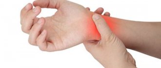
Pain and arthritis
Arthritis, like osteochondrosis, is a very complex disease that requires constant attention from the patient and the doctor. There are mild and severe forms of the disease; depending on the patient’s condition, a treatment plan is developed. Patients should know that in most cases the doctor must apply existing protocols; deviations are possible only in exceptional cases and after consultation of several specialists or a medical council. Diseases of bone tissue gradually progress, which causes increased pain. The treatment is long-term and eliminates only the pain, and not the cause of its occurrence.
Diagnosis of rheumatoid arthritis