Read about the diagnosis and treatment of cholelithiasis at the link.
Get help right away if you develop signs and symptoms of a serious complication related to gallstones, such as:
- Abdominal pain is so severe that you cannot sit still or find a comfortable position
- Yellowing of the skin and whites of the eyes (jaundice)
- High temperature with chills
Number to call an ambulance in Moscow – 103
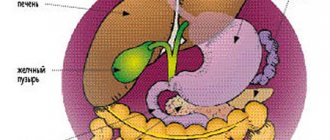
How does the liver work?
The liver is the largest gland in humans. It is located in the upper floor of the abdominal cavity on the right. It consists of two main lobes - right and left, which in turn are divided into 8 segments.
In an adult, it weighs about 1/40 of the total body weight (approximately 1.6 kg in men and 1.2 kg in women). The liver has a dual blood supply: approximately 80% of the blood entering the liver comes from the portal vein (venous blood collected from all unpaired abdominal organs), the rest (oxygenated arterial blood) comes from the hepatic artery. Having entered the portal of the liver, both vessels give rise to multiple branching smaller vessels. At the exit from the liver, 3-4 hepatic veins are formed, which flow into the inferior vena cava.
The main structural unit of the liver is the hepatic lobule, which consists of liver cells - hepatocytes. This cell is one of the most important biochemical laboratories of the body. The hepatocyte takes part in the metabolism of proteins, carbohydrates, fats, bile acids, and vitamins. Neutralizes toxic substances coming from the intestines, followed by the release of associated products into the intestinal lumen. An important function of the hepatocyte is the synthesis of bile, which is involved in digestion.
Consequences of choledocholithiasis
After treatment and following the doctor’s recommendations, the patient quickly returns to his normal life. However, if you do not seek medical help promptly, complications may arise.
- The presence of stones provokes inflammatory processes that are severe and painful.
- Since the bile and pancreatic ducts connect before entering the duodenum, if the mouth is blocked by a stone, the ducts become swollen and damage to the pancreas is possible. Acute pancreatitis can result in the death of the patient.
- Communicating with the liver, the inflamed biliary system disrupts the functional activity of this organ (and the liver participates in digestion, hematopoiesis, deposits necessary substances, utilizes and deactivates unnecessary ones, and so on). As a result, there is an imbalance in the functioning of almost all body systems.
What is bile and why is it needed?
Bile is a secretion produced by hepatocytes. The color of bile ranges from light yellow to dark green. Bile contains bile pigments - bilirubin and biliverdin, cholesterol, fatty acids, lecithin, mucin.
Bile is the main participant in digestion. It neutralizes hydrochloric acid that enters the duodenum from the stomach, thereby creating favorable conditions for the action of pancreatic juice and parietal digestion in the jejunum.
Bile acids emulsify fats, allowing them to be absorbed in the intestines.
Bile has a bactericidal effect that prevents the proliferation of pathogenic microflora.
How are the bile ducts arranged and how do they work?
The bile ducts are a system of ducts that originate from hepatocytes and gradually gather into interlobular, segmental and right and left lobar ducts, which unite into the common hepatic duct.
One of the stages of bile removal is its accumulation in the reservoir - the gallbladder. The main function of the gallbladder is to concentrate bile and release it into the lumen of the duodenum during digestion. The volume of this organ is about 80 ml.
The gallbladder is located on the lower surface of the liver. It is a hollow muscular organ, externally covered with a serous membrane and internally lined with mucous membrane. The structure of the gallbladder is divided into the bottom, body and neck of the bladder, which passes into the cystic duct.
Main types of stones
Gallstones vary in size, shape, color and chemical composition.
Size
. There are no large single stones in the common bile duct (common bile duct), which is due to the diameter of the cystic duct. Smaller ones are lighter and leave the bladder with a flow of bile. Large ones remain at the bottom under their own weight.
Form.
If stones with “edges” are found in the ducts, it can be argued that they came from a bladder: this shape appears from the close adhesion of the stones to each other. Basically, the shape of bile formations is round, oval.
Color
depends on the chemical composition:
- cholesterol - predominantly yellow, greenish, golden in color;
- bilirubin (pigment) - soft and black in color;
- calcium (calcareous) formations are whitish, sometimes their surface is like an egg shell;
- with parasitic or bacterial infection - brown.
The classification is based on the predominance of one of the chemical elements. At the same time, the other two are also present. In addition to the main ones, dead cellular elements, protein parts and other components are mixed into the structure.
Why do gallstones form?
The exact cause of cholelithiasis has not yet been fully elucidated. Only a few factors are known that increase the likelihood of its occurrence.
- Genetic: family predisposition increases the risk of cholelithiasis by 4-5 times;
- Congenital anomalies of the biliary tract;
- Gender - the risk of developing gallstones in women is approximately 2-3 times higher, which is associated with the influence of estrogens on the ability to form gallstones;
- Age - the maximum frequency of clinical manifestations of cholelithiasis is recorded at the age of 40-69 years;
- Dietary (high-calorie diet, poor in plant fiber, as well as with an excess of simple carbohydrates, animal proteins, foods containing cholesterol);
- Use of medications (oral contraceptives, fibrates, ceftriaxone, somatostatin, nicotinic acid);
- Concomitant diseases and conditions (obesity - a tendency to form gallstones in persons with a body mass index of more than 30 is 3 times higher, type 2 diabetes mellitus, hypothyroidism, cirrhosis of the liver, etc.);
- Pregnancy - the risk of developing cholelithiasis increases during pregnancy, especially with repeated pregnancies (the likelihood of stone formation increases by 10-11 times). During pregnancy, bile heterogeneity, or the so-called biliary sludge, develops in 20-30% of patients, stones - in 5-12% of cases.
There is a so-called 5F rule that describes patients at greatest risk of developing stones:
1. Female (female);
2. Fat (overweight/obesity);
3. Forty (age 40 years or more);
4. Fertile (fertile women - that is, pregnant and giving birth);
5. Fair (light-skinned blondes).
Dietary recommendations
- Sugar is a source of endogenous cholesterol and should be avoided.
- Increase your intake of proteins balanced in amino acid composition.
- Increase your intake of plant proteins: oatmeal and buckwheat, seaweed.
- Accustom yourself to regularly eating plant fibers.
- Avoid eating legumes, animal fats, and coffee.
- Skipping breakfast increases the risk of developing gallstones.
- Vitamins E and C reduce the likelihood of gallstone formation.
Unfortunately, in order to forget about gallstone disease in later stages, it is not enough just to follow the correct regimen and diet. Any doctor will tell you that “this is only an additional therapy, not the main treatment.”
How are gallstones formed?
The process of stone formation is quite well studied. The key links are:
- An increase in the lithogenicity of bile (the ability to form stones) means an increase in the content of cholesterol and bilirubin in it.
- Impaired contractile function of the gallbladder.
- Increased pressure in the bile duct system.
Mucin is a natural gel, constantly secreted by the wall of the gallbladder; when lithogenicity increases, it begins to “absorb” cholesterol crystals, thereby forming a “fossilization nucleus.” The formed suspension, with impaired contractile function, continues to attach salt crystals encrusted with calcium salts. Thus, gallstones can grow up to 5-7 mm per year.
Causes of stones
When a lot of cholesterol begins to accumulate in the bile, the disease gradually begins to develop. A large accumulation of cholesterol can result from:
- obesity;
- frequent consumption of fatty foods;
- stagnation of bile.
Bile stagnation can be functional or mechanical. In the latter case, the pathology can be caused by:
- tumor;
- an increase in the size of neighboring organs;
- the appearance of scars;
- inflammatory reactions leading to swelling of the walls of the organ.
How does gallstone disease occur?
The main factors influencing the course of the disease are the nature of nutrition and the size of the stones. Gallstone disease can occur both in the form of chronic stone carriage and in acute, and sometimes in complicated, form.
Where does pain come from with gallstone disease?
By themselves, gallstones do not cause any pain and can be an accidental finding during medical examination. When food enters the duodenum, the mechanoreceptors are irritated and cause contraction of the gallbladder. The stones wedge into the cervix, causing a prolonged spasm, which manifests itself as pain. When taking antispasmodic drugs, the calculus can “move” back from the cervix and the attack of hepatic colic is stopped. If the stone remains in the cervix, then inflammation begins to develop against the background of obstruction.
Chronic calculous cholecystitis is an asymptomatic stone carrier. The size of the stones can vary from 2 – 3 mm to 5 cm. This stage is characterized by an almost complete absence of clinical manifestations of the disease. Stones can remain in the lumen of the gallbladder for years without causing any symptoms. However, if there is an error, discomfort may occur in the right hypochondrium, which can be relieved independently or while taking antispasmodic drugs.
Hydrocele of the gallbladder - occurs against the background of obstruction of the neck of the gallbladder by a calculus, cutting off the latter from the entry of bile. The bladder is filled with mucus secreted by the mucous membrane and is not involved in digestion.
Acute calculous cholecystitis - occurs with prolonged obstruction of the neck of the gallbladder by a calculus. The pain syndrome persists against the background of ineffective conservative treatment. The patient is bothered by vomiting, which does not bring relief; pain can be felt behind the sternum - cholecystocardial syndrome, in the right shoulder blade. Body temperature rises and chills appear. Inflammatory changes begin to develop in the wall of the gallbladder, which progress from catarrhal, phlegmonous and gangrenous forms. Against the background of the latter, perforation of the gallbladder or abscess may develop.
Choledocholithiasis and acute biliary pancreatitis. Choledocholithiasis develops as a result of migration of biliary sludge or small stones from the gallbladder into the common bile duct. Once in the common bile duct, small stones freely descend to the sphincter of Oddi, which is a release mechanism - a circular muscle cuff. Sludge and stones “wedge” into the sphincter, causing its long-term spasm, causing a violation of the outflow of bile - obstructive jaundice. The patient's skin and sclera become a rich yellow color. This condition requires emergency decompression of the bile ducts - restoration of bile outflow.
An increase in pressure in the common bile duct promotes the reflux of bile into the main pancreatic duct and intraductal activation of pancreatic juice enzymes - a trigger for the development of acute biliary pancreatitis.
Mirizzi syndrome . A fairly rare complication of gallstone disease. Develops as a result of prolonged stone bearing. First, a stricture forms, then a bedsore between the gallbladder and the common bile duct.
Mirizzi syndrome presents significant difficulties for surgical treatment. This is due to the fact that in the human body there is no plastic material to close the defect of the common bile duct. Surgical treatment of Mirizzi syndrome is often accompanied by the formation of an anastomosis between the small intestine and the common bile duct. When a stone migrates into the small intestine, acute intestinal obstruction may develop, requiring emergency surgical treatment.
In the photographs, in the lumen of the resected section of the small intestine there is a calculus that caused intestinal obstruction.
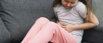
Clinical picture and symptoms of the disease
Most often, the first sign that forces the patient to see a doctor is pain in the right hypochondrium, of varying intensity. Cutting or stabbing in nature, the pain is most often constant, often radiating to the right shoulder blade, lower back, and forearm. In some cases (with cholecystocoronary Botkin's symptom), it can radiate beyond the sternum, resembling an attack of angina. It should be borne in mind that the intensity of pain is in no way an indicator of the severity of the process. For example, in some cases, severe pain may disappear, but mild pain does not mean a mild form of the disease.
Often the patient’s condition worsens after eating spicy or fatty foods; their consumption increases the need for bile to process food, which leads to contraction of the gallbladder. In any form of the disease, there is an increase in body temperature. In the form of short rises, the temperature rises to 37-38°C, and the patient often experiences pain. However, during an acute attack accompanied by chills, the temperature can rise to 38-40°C.
How to diagnose gallstone disease?
The gold standard for diagnosing gallstone disease is ultrasound examination of the abdominal cavity. This method allows you to identify stones in the gallbladder. Assess their size, shape and location. An important indicator is the thickness of the gallbladder wall. Normally, it does not exceed 2-3 mm. Thickening of the wall is an absolute sign of inflammatory changes - acute cholecystitis. An important criterion is the diameter of the common bile duct. If there are stones, its diameter increases from 3 – 6 to 20 mm.
Magnetic resonance imaging allows you to see the intra- and extrahepatic bile ducts, as well as the main pancreatic duct. This diagnostic method is used when there is a suspicion of the presence of stones in the common bile duct. This is necessary for treatment planning.
Multislice computed tomography is indicated for the development of acute biliary pancreatitis. This diagnostic method allows you to identify inflammatory changes in the pancreas and surrounding tissues.
Gastroduodenoscopy - this diagnostic method is especially valuable for complicated cholelithiasis, as it allows not only to evaluate the stomach, duodenum, but the flow of bile through the major duodenal papilla. If necessary, gastroduodenoscopy can easily be transformed from a diagnostic procedure into a therapeutic one. Using special instruments, you can “paint” the bile ducts and remove stones.
In the figure, the arrow indicates a stone in the common bile duct. Using a duodenoscope, a dissection of the sphincter of Oddi was performed in the duodenum. A radiopaque substance was introduced to allow visualization of the calculus. Using a special basket, the stone is captured and removed.
Diagnostics
The diagnosis is based on the results of instrumental studies and anamnesis data.
- Ultrasound examination (ultrasound) of the upper abdominal cavity is performed to diagnose cholelithiasis and calculous cholecystitis. During the study, in addition to stones in the gall bladder or ducts, the size of the gallbladder, the condition of its walls, and pathology of the liver or pancreas are determined.
- gastroduodenoscopy - allows you to determine diseases of the stomach, esophagus and duodenum.
- retrograde cholangiography (x-ray examination using a contrast agent) is performed in the presence of complications.
- transgastric ultrasound of the ducts is necessary when diagnosing choledocholithiasis.
Treatment of gallstone disease
Treatment of gallstone disease is only surgical. Conservative treatment, taking choleretic drugs promotes the migration of stones from the gallbladder into the common bile duct and can cause acute biliary pancreatitis and obstructive jaundice.
Depending on the access and equipment used, cholecystectomy can be performed openly, laparoscopically through punctures in the anterior abdominal wall, and robotically using the DaVinci apparatus. Each method has its own indications and limitations.
Open cholecystectomy, this type of surgical intervention is indicated if there are contraindications for laparoscopic cholecystectomy. Such as severe cardiovascular or respiratory failure, abdominal adhesive disease, the presence of internal biliodigestive fistulas, Mirizzi syndrome.
Laparoscopic cholecystectomy is the gold standard in the treatment of gallstone disease. This method is low-traumatic and provides excellent visualization and functionality. Modern high-tech equipment reduces the risk of intraoperative complications.
One of the options for laparoscopic surgery is robotic cholecystectomy using the DaVinci device. Allows you to isolate extrahepatic bile ducts with pinpoint accuracy, thereby protecting the patient from a dangerous complication - intersection of the common bile duct.
Special categories of patients
Elderly and senile patients require special attention. Many concomitant diseases can cause refusal of planned surgical treatment in favor of surgical treatment “for health reasons” in other clinics. Our clinic has accumulated extensive experience in providing care to patients of the older age group. After preoperative evaluation and preparation, older patients can expect to undergo surgical intervention.
Patients suffering from diabetes with a high body mass index require increased attention. Against the background of diabetes mellitus, the body's reparative capabilities are significantly reduced, which, along with severe pain and prolonged bed rest, can lead to a number of undesirable complications.
In both groups of patients, laparoscopic cholecystectomy is the operation of choice, because minimally invasive access can significantly reduce surgical trauma and significantly speed up the process of patient activation. As a rule, you can get out of bed 3-4 hours after surgery. Thanks to early activation, the risk of developing cardiovascular complications and abdominal adhesive disease is significantly reduced. The use of special extended instruments allows laparoscopic surgery to be performed on patients with a body mass index of more than 40 kg/m2.
How is laparoscopic cholecystectomy performed?
The photographs show in detail the stages of laparoscopic cholecystectomy.
The picture shows the stage of mobilization of the neck of the gallbladder, its isolation from the adhesions with the duodenum.
In this image, using a laparoscopic instrument, the peritoneum in the area of the neck of the gallbladder is opened and the cystic duct is mobilized.
In this image, the cystic duct is highlighted and a clip is applied to it.
After applying two clips to the remaining part of the cystic duct and one to the outgoing part, it can be crossed.
After crossing the cystic duct, the cystic artery is isolated and clipped.
Now that the cystic artery has been crossed, all that remains is to separate the gallbladder from the bed and remove it from the abdominal cavity.
This image shows the separation of the gallbladder from its bed in the cervical region. Seemingly simple to perform, it requires very careful work to avoid liver damage.
The final stage of the operation. The gallbladder is separated from the bed and removed from the abdominal cavity.
What do gallbladder stones look like?
The photographs show removed gallbladders with extracted stones. The appearance of stones is varied. They differ in shape, size, appearance and structure.
Transvaginal laparoscopic cholecystectomy using the NOTES method
Rice. 6. Scheme of laparoscopic transvaginal cholecystectomy using NOTES technology
Rice. 7. View of the anterior abdominal wall during laparoscopic cholecystectomy using SILS technology.
Since 2007 in France, and since 2008 in the Russian Federation, a unique method of removing the gallbladder has been practiced - transvaginal cholecystectomy using NOTES technology. The operation is performed without any punctures of the abdominal wall, therefore, there are no postoperative scars. The essence of the surgical intervention is access to the affected organ through a small puncture (1 cm) of the posterior vaginal fornix. The intervention is performed using laparoscopic instruments and optics introduced into the peritoneal cavity through this access. After removing the gallbladder through the same access, one suture is placed on the puncture. The synthetic suture material used in this case dissolves within 3-4 weeks.
The advantages of this method are:
- in the absence of pain after surgery;
- the patient's motor activity is not impaired;
- short rehabilitation period, patient hospitalization lasts only one day;
- excellent cosmetic effect;
After 7-10 days after surgery, a person can begin to work, and they can play sports on the 14th day. Among the restrictions in the postoperative period should be the need to exclude intimate relationships for a month. At the same time, the genital organs (uterus, appendages, etc.) are not affected during transvaginal cholecystectomy, so their functionality remains unchanged.
Complications after laparoscopic cholecystectomy
The most common intraoperative complications are the intersection of the cystic artery and the common bile duct.
Bleeding from the transected cystic artery is stopped by clipping and does not cause technical difficulties for an experienced surgeon.
Damage to the common bile duct requires reconstructive intervention, which is associated with significant difficulties. In the long term, such a surgical injury can lead to a significant decrease in quality of life and disability.
Performing cholecystectomy using minimally invasive and high-tech equipment can significantly reduce the risks of intraoperative complications.
Postoperative period
In case of an uncomplicated course of the postoperative period, early activation of the patient is practiced, 3-4 hours after the operation. Eating is allowed on the second day. Discharge from the hospital occurs within 3-4 days, and depends on the individual threshold of pain sensitivity. Removal of sutures is not required, as skin wounds are sutured with cosmetic sutures and absorbable sutures.
A return to normal lifestyle and physical activity is possible within 10–14 days, after complete healing of skin wounds.
Appearance after laparoscopic cholecystectomy:
Single-port laparoscopic cholecystectomy SILS
The use of the NOSE technique is technically impossible in cases where patients have undergone many surgical interventions on the pelvic organs in the past. Therefore, an equally effective method of minimally invasive cholecystectomy was developed, which has been used in America since 2008, and since 2009 such operations have been performed by domestic surgeons. We are talking about removing the gallbladder through a puncture in the navel area (SILS technology).
Single-port laparoscopic cholecystectomy
consists of performing an operation using a special device (port), which is a soft plastic apparatus that is inserted into the abdominal cavity through a puncture. Port diameter 23-24 mm. It is through this that laparoscopic instruments are inserted; the diameter of the laparoscope does not exceed 5 mm. Upon completion of the operation, a cosmetic suture is applied to the small wound. Surgical intervention using SILS technology (single-port surgery), in contrast to conventional laparoscopic access (multi-puncture), has a number of advantages:
- fewer punctures on the abdominal wall;
- less pain after surgery;
- shorter rehabilitation period;
- better cosmetic effect;
The advantages of this method of surgery are especially noticeable when there are multiple stones in the gallbladder - with the usual method, the surgeon must increase the puncture to remove stones and the diseased organ.
The choice of the appropriate surgical treatment method depends on the individual characteristics and health status of the patient. Visiting a clinic with qualified and experienced specialists guarantees the highest possible treatment results.
What questions can you ask the doctor at your appointment?
- Do I need surgery?
- Is it possible to dissolve stones?
- Why is it better not to dissolve stones?
- Is it possible to simply remove the stones and leave the bubble?
- How will my life change after cholecystectomy?
- Will there be dietary restrictions?
- Will it hurt after the surgery?
- What kind of anesthesia will it be?
- When can I eat fatty heavy foods and alcohol?
- Are dressings necessary after discharge from the hospital?
You can contact the clinic of coloproctology and minimally invasive surgery for diagnosis and treatment of gallstone disease. Thanks to modern operating rooms and highly qualified staff, the clinic provides all types of treatment for gallstone disease, including robotic surgeries.
Treatment under the compulsory medical insurance policy is possible.
Sign up for a consultation and receive quality treatment as soon as possible.
If you have:
Nothing hurts. A deformation in the gallbladder was accidentally discovered
Deformation of the gallbladder is often related to anatomical features and can be asymptomatic for a long time. At the same time, there may be a violation of the outflow of bile from the gallbladder, oversaturation of bile with cholesterol, the formation of sediment, putty-like bile, and the formation of biliary sludge, which is the most important condition for the formation of gallstones. Over time, complaints may arise:
- for a periodic feeling of discomfort or dull pain in the right hypochondrium
- bitterness in the mouth
- flatulence
- unstable stool with a tendency to diarrhea.
In this case, an active lifestyle, physical exercise, and normalization of body weight are recommended. A consultation with a gastroenterologist is necessary in order to select drug therapy (if necessary), recommendations for proper nutrition, and further observation.
Nothing hurts. We accidentally discovered flakes, thick bile, and biliary sludge in the gallbladder
In approximately half of patients, biliary sludge does not cause any symptoms and is detected only by ultrasound of the gallbladder. Many patients do not attach importance to this pathology and do not consult a doctor. Meanwhile, the long-term existence of biliary sludge in more than half of patients can be complicated by biliary pancreatitis, dysfunction or stenosis of the sphincter of Oddi, acute cholecystitis, cholangitis, and gallbladder shutdown. 20% develop gallstones. To prevent the development of cholelithiasis and complications, timely consultation with a gastroenterologist-hepatologist is recommended. As a result, the causes that contribute to the formation of biliary sludge and the development of complications will be identified and eliminated.
There are complaints. Deformation of the gallbladder, flakes, thick bile, biliary sludge in the gallbladder were detected
If complaints arise, it is often not enough to make do with recommendations for lifestyle changes, moderate physical activity, and normalization of body weight. A consultation with a gastroenterologist is necessary in order to select drug therapy that will improve the excretion of bile from the gallbladder, relieve pain, prevent the formation of gallstones and the development of complications in the future, and, as a result, avoid surgical treatment in advanced cases.
The stones were identified a long time ago, but nothing bothers me
Latent (asymptomatic) stone carriage requires long-term observation by a gastroenterologist-hepatologist:
- to determine indications for gene therapy - medicinal dissolution of stones using bile acids (in the presence of cholesterol stones)
- for an ultrasound scan, which will determine the size and shape of the gallbladder, the thickness of its wall, the number of stones and their size
- to determine the dynamics of these indicators over time
If necessary, a joint consultation with a surgeon is held and indications for surgical treatment are determined.
There are complaints. Gallstones detected
Immediately consult a gastroenterologist, where a specialist will determine:
- indications for drug dissolution of stones
- will select therapy to relieve the patient’s complaints
- will reveal metabolic disorders underlying stone formation.
The success of conservative therapy for cholelithiasis is determined by strict adherence to recommendations and the correct selection of litholytic therapy. The effectiveness of treatment is monitored by a gastroenterologist-hepatologist using ultrasound examination, which must be carried out throughout the entire course of treatment. After completing the drug course of dissolving stones, in rare cases, relapse of stone formation is possible. Therefore, to prevent relapse, the gastroenterologist creates supportive and preventive therapy.
Heaviness, discomfort, pain under the “spoon” and in the right hypochondrium
These complaints are quite nonspecific and may be present in diseases of the liver, pancreas, stomach, duodenum, gall bladder (including cholelithiasis).
If you are concerned about heaviness, discomfort, pain in the pit of the stomach and in the right hypochondrium, you need to seek help from a gastroenterologist-hepatologist who:
- will find out the history of the disease
- will conduct objective research
- will determine the scope of necessary additional examination
There are stones, there was 1 colic
Mandatory observation by a gastroenterologist-hepatologist:
- to prescribe the necessary treatment
- to control the situation and constant monitoring
This simple measure:
- reduces the risk of recurrent colic
- prevents the development of disease and complications
There are stones, there were 2 colics
Repeated biliary colic, a relapsing course of the disease, increases the risk of complications and the risk of death by almost 4 times.
In this case, mandatory observation by a gastroenterologist-hepatologist together with a surgeon is indicated:
- to determine treatment tactics
- if necessary – to resolve the issue of surgical treatment
It is necessary to remove the gallbladder - what to do?
When preparing for a planned cholecystectomy, a joint consultation with a gastroenterologist-hepatologist and a surgeon is necessary:
- to determine the presence of indications and contraindications for surgical intervention
- if necessary - to draw up a plan for additional examination
In case of a decision on surgical treatment, it is important to perform a number of instrumental and laboratory tests, which are included in the standard examination before surgery.
The gallbladder has already been removed. Do I need to see a doctor, and which specialist should I see?
The gallbladder is an important organ of the gastrointestinal tract. In its absence, the following are often observed:
- change in hormonal function
- change in concentration function
- disorders associated with changed conditions of food absorption in the intestines.
This leads to disturbances in motility and secretory function of the stomach, ulcerative lesions of the stomach or duodenum, duodenitis, pancreatitis, intestinal lesions, disturbances in the metabolism of fat-soluble vitamins, protein and carbohydrate metabolism, fat metabolism and calcium metabolism.
Patients who have undergone cholecystectomy (that is, surgical removal of the gallbladder) require constant monitoring by a gastroenterologist-hepatologist.
In most cases, removal of the gallbladder does not relieve the patient of metabolic disorders underlying stone formation. The separated bile contains many cholesterol crystals and remains thick and viscous. After surgery, the pathological processes underlying cholelithiasis occur in new conditions: due to the absence of the gallbladder, the physiological function it performs is no longer possible, the motility of the biliary tract is impaired, and there is no regulation of the processes of bile formation and bile excretion.
Loss of the physiological role of the gallbladder, namely the lack of concentration of bile during the interdigestive period and its release into the duodenum during meals, is accompanied by impaired excretion of bile and indigestion. Changes in the chemical composition of bile and its chaotic entry into the duodenum disrupt the digestion and absorption of lipids, reduce the bactericidal properties of intestinal contents, which leads to microbial contamination of the duodenum and weakened growth of normal intestinal microflora.
In this regard, the patient may be concerned about:
- nausea
- vomit
- heartburn
- bitter feeling in the mouth
- flatulence
- unstable chair
- constipation
- frequent loose stools
- stomach ache
Excessive bacterial growth in the intestines leads to disturbances in protein, carbohydrate and fat metabolism, calcium metabolism and fat-soluble vitamins. This results:
- to frequent heavy diarrhea
- to weight loss
- to osteoporosis (disorders of bone mineralization)
- to vitamin deficiencies, which are manifested by loss of skin elasticity, fine wrinkles, peeling, depigmentation, dry skin, damage to the lips (hyperemia, swelling, peeling, formation of cracks and crusts, weeping in the corners of the mouth), damage to the tongue (ulceration of the papillae, the appearance of cracks), damage to gums (looseness and bleeding, weakening of teeth and their loss)
At least once every 4 months - with this frequency, regular observation by a gastroenterologist-hepatologist and ultrasound of the bile ducts (dynamic echo-choledochography) after cholecystectomy are recommended for early diagnosis of possible complications.
A year after removal of the gallbladder, the same symptoms appeared: pain in the right side, nausea, loose stools
In patients after removal of the gallbladder, the existing clinical manifestations may be associated with a number of factors:
- changes in the chemical composition of bile
- impaired excretion of bile into the duodenum
- impaired motility of the biliary tract
- excessive growth of pathogenic microflora in the intestines
- disruption of food digestion and nutrient absorption
In this case it is necessary:
- consultation with a gastroenterologist-hepatologist
- specialized ultrasound examination (dynamic echo-choledochography)
- subject examination
- complex treatment
- further long-term follow-up