Optic neuritis is an inflammatory process that damages, often suddenly, nerve fibers.
Two forms of the disease
- Intrabulbar, when the localization of inflammation occurs in the initial part of the nerve and does not extend beyond the tissues of the eyeball.
- Retrobulbar, in which the visual canals located outside the eye are acutely affected.
Classification of the disease and its symptoms
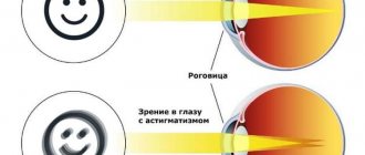
The optic nerve is located in such a way that one part of it is inside the orbit, connecting to the eyeball, and the other goes into the cranial cavity, from where it continues further into the brain.
With neuritis,
either the intracranial part of the nerve or the part that runs in the orbit can be affected .
In the second case, nephritis is retrobulbar , and in turn this disease is divided into several types:
- Interstitial - with this neurosis, in addition to the nerve itself, inflammatory processes also affect adjacent connective tissues , as well as glial cells;
- Retrobulbar orbital is a pathological inflammatory process that affects exclusively the intraorbital part of the optic nerve;
- Transverse retrobulbar neuritis - inflammation affects all fibers of the optic nerve ;
- Retrobulbar axial - partial damage to the optic nerve of only the part that begins outside the eyeball .
Attention! When the part of the nerve that is located outside the orbit and goes into the skull is inflamed, they speak of intracranial neuritis.
Regardless of the type of pathology, its symptoms mostly the same and begin to appear already in the first hours of the inflammatory process (extremely rarely the symptoms are long in coming and can develop within 24 hours).
Here are the signs that indicate the obvious presence of this disease:
- The affected eye cannot perceive colors normally : they appear unnatural.
- In the first few hours after the onset of the inflammatory process, a so-called “mesh” may appear in front of the eyes .
- In the dark the affected eye begins to see significantly worse.
- A sharp decrease in visual acuity in either one affected eye or two.
- The level of photophobia increases , as well as intolerance to white color, when looking at which the patient’s eyes begin to hurt.
- When you rotate or move your eyes, pain appears in the area of the eyeball.
- The field of view narrows significantly , and the central areas may completely disappear from the field of view.
In addition to these signs, there is a general deterioration in the condition, including weakness, headache and increased body temperature.
Optic neuritis
The clinical picture depends on the severity of the inflammatory process. With mild inflammation, the optic disc is moderately hyperemic, its boundaries are unclear, the arteries and veins are somewhat dilated. A more pronounced inflammatory process is accompanied by a sharp hyperemia of the disc, its borders merge with the surrounding retina. White spots appear in the peripapillary zone of the retina and multiple hemorrhages. Usually the disc does not proliferate with neuritis. The exception is cases of neuritis with edema. Neuritis is characterized by early impairment of visual functions, expressed in decreased visual acuity and changes in the visual field. The degree of decrease in visual acuity depends on inflammatory changes in the papillomacular bundle. There is usually a narrowing of the visual field, which may be concentric or more significant in one area. Central and paracentral scotomas appear. Narrowing of the peripheral boundaries of the visual field can be combined with scotomas. Also characteristic is a sharp narrowing of the field of vision to red, and sometimes a complete lack of color perception. When neuritis passes into atrophy, the disc turns pale, the arteries narrow, exudate and hemorrhages resolve.
Neuritis of various etiologies can occur with characteristic clinical symptoms. An edematous form of inflammation of the optic nerve is characteristic of relapses of neurosyphilis . In the early period of secondary syphilis, neuritis occurs either with mild changes in the disc in the form of hyperemia and blurred boundaries, or in the form of severe papillitis with a sharp decrease in visual functions. A very rare form is papular neuritis, in which the disc is covered with a massive grayish-white exudate protruding into the vitreous body.
Tuberculous neuritis manifests itself in the form of solitary optic disc tubercle or ordinary neuritis. The solitary tubercle is a grayish-white tumor-like formation located on the surface of the disc and spreading to the surrounding retina.
Optic neuritis in acute infectious diseases has approximately the same clinical picture.
With retrobulbar neuritis at the onset of the disease, the fundus of the eye can sometimes be normal. More often there is slight hyperemia of the optic nerve head, its boundaries are unclear. These changes can be more pronounced, as with neuritis. In rare cases, the picture resembles a congestive optic disc. In this case, the disc is enlarged in diameter, its boundaries are not defined, the veins are dilated and tortuous. Retrobulbar neuritis most often develops in one eye. The second eye may become sore some time after the first. Simultaneous disease of both eyes is rare.
According to the clinical course, acute and chronic retrobulbar neuritis . In the first case, a sharp decrease in visual acuity occurs quickly (within 2-3 days); in the chronic course of the process, visual acuity decreases gradually. Acute retrobulbar neuritis is characterized by pain behind the eyeball and when the eye is pressed into the orbit. Visual acuity after its initial decrease begins to recover after a few days. Only in rare cases does this not happen and the eye remains practically blind.
Usually, with retrobulbar neuritis, a central absolute scotoma is determined in the field of vision in white and other colors. At the beginning of the disease, the scotoma is large in size; later, if visual acuity increases, it decreases, becomes relative and, with a favorable course of the disease, may disappear. In some cases, the central scotoma turns into a paracentral annular scotoma. The disease leads to descending atrophy of the optic disc, most often in the form of temporal blanching of half of the disc due to damage to the papillomacular bundle. If there are changes in the disc, atrophy may be secondary.
Some features during the course have retrobulbar neuritis of toxic origin. One of the most common causes of these neuritis is poisoning with methyl alcohol or liquids containing methyl alcohol. Against the background of general symptoms of poisoning (unconsciousness or coma in severe cases of poisoning, nausea, vomiting in milder cases), after 1-2 days a sharp decrease in visual acuity in both eyes develops, sometimes to complete blindness; At the same time, dilation of the pupils is observed, their reaction to light is weakened or absent. The fundus is normal or there is slight hyperemia of the optic nerve head.
In rare cases, a picture of ischemic neuritis is observed - the disc is pale, its borders are blurred, the arteries are sharply narrowed. The further course of the process may be different. During the first month after poisoning, vision may improve. Following this, significant deterioration of vision occurs again, up to blindness. Decreased visual acuity is caused by the development of optic nerve atrophy.
Alcohol and tobacco intoxication causes damage to the papillomacular bundle. Occurs with chronic alcoholism or when smoking strong varieties of tobacco containing large amounts of nicotine. It is observed more often in men over 30 years of age. The disease occurs as a chronic retrobulbar neuritis, the fundus of the eye is often normal. Mild hyperemia of the optic disc is much less common. A relative central scotoma appears in the field of view with normal peripheral boundaries. It often has the shape of a horizontal oval running from the point of fixation to the blind spot. It is characteristic that with a complete cessation of drinking alcohol or smoking, there is an increase in visual acuity and a decrease in scotoma. However, pallor of the temporal half of the optic disc persists.
Retrobulbar neuritis in diabetes mellitus has a chronic course and usually occurs in men. The defeat is almost always bilateral. Visual acuity decreases slowly. Central absolute or relative scotomas appear with normal peripheral boundaries of the visual field. The optic discs are initially normal, but subsequently develop temporal pallor.
Symptoms
One of the main symptoms of ocular neuritis is a sharp deterioration in vision - it is observed in more than 90% of patients. Most often, only one eye is affected.
The second obligatory symptom is considered to be a violation of color vision - that is, the ability to perceive all the colors of the spectrum, interpret them correctly and distinguish between tones and shades. In the case of inflammation of the optic nerve, the picture acquires problems similar to color blindness; symptoms can manifest themselves in the form of complete color blindness or be characterized by a partial impairment of color perception.
A manifestation such as the positive Uthoff phenomenon is also a symptom of ocular neuritis. This is a clinical syndrome characteristic of patients with multiple sclerosis, which consists of a worsening of the mental and neurological symptoms of the disease - patients feel increased fatigue, weakness, tiredness, impaired attention, memory and other cognitive skills. The relationship of the phenomenon to optic neuritis can be explained by the fact that there is a certain connection between this disease and multiple sclerosis. Thus, it is believed that in 50% of patients, neuritis is the first sign of multiple sclerosis, and in patients with this form of sclerosis, neuritis manifests itself in approximately 40-50% of cases.
A third of patients complain of the appearance of optical phenomena - a kind of visual hallucinations, similar to the effect of the use of hallucinogenic or psychotropic substances, but caused by inadequate stimulation in the parts of the visual analyzer. The most common manifestation of neuritis is photopsia, characterized by the appearance of false moving images before the eyes, such as sparks, flashes, luminous circles, and so on. When the eye is inflamed, patients feel pain in the orbit, nausea occurs, darkening of the eyes, and with neuritis the eye swells.
Treatment of optic neuritis
Patients with optic neuritis must undergo treatment in a hospital setting. Before choosing a treatment method, it is necessary to establish the cause of the disease. After it is clarified, treatment for inflammation of the nerve is prescribed and the general disease that caused this condition is additionally treated. Unfortunately, it is not always possible to find out the cause of neuritis.
Most often, anti-inflammatory drugs, antibiotics, detoxification therapy and vitamin therapy are used for this disease. Typically, the following medications are prescribed in the first week - streptomycin, neomycin, gentamicin, kanamycin, gentamicin. Corticosteroids can also be used additionally - prednisolone and dexamethasone solution 0.4% (retrobulbar injections). Additionally, the patient is prescribed B vitamins, solcoseryl intramuscularly, piracetam and dibazol.
The prognosis for treatment is quite favorable, so in most cases vision and other ocular functions are restored. But, if treatment is not started in a timely manner, then vision can be restored with great difficulty. In some cases, especially in the absence of the necessary therapy, the disease can lead to partial or complete loss of vision. This occurs due to inflammation of the optic nerve and its further atrophy. Therefore, you should not delay treatment; at the first symptoms of the disease, you should consult a specialist.
MagazinLinz.ru team
Diagnosis of the disease
As a rule, patients who have the first symptoms of optic neuritis turn to an ophthalmologist. The disease is considered an interdisciplinary pathology; an ophthalmologist or neurologist must take part in its treatment. If neuritis develops against the background of other pathologies, it is necessary to clarify the diagnosis and carry out specific therapy for the primary diseases. Source: Visualization of the optic nerve in the diagnosis and monitoring of retrobulbar neuritis. Yuryeva T.N., Burlakova E.V., Khudonogov A.A., Ayueva E.K., Sukharchuk O.V. Acta Biomedica Scientifica, 2011. p. 133-136. Then the appropriate specialists are involved in the treatment - an immunologist, an otolaryngologist, an infectious disease specialist, a phthisiatrician.
The first step in diagnosing optic neuritis is collecting anamnesis, external examination of the patient, and palpation. During the medical history, the doctor will clarify the presence of concomitant pathologies, the time of onset of the disease, what complaints the patient has (pain, decreased visual acuity, changes in color perception, the appearance of “blind” spots), how quickly the symptoms developed and how severe they are, whether one eye or both is affected.
External examination and palpation may often not provide additional data. Pain, forward displacement of the eyeball, and limitation of its movements may occur with retrobulbar neuritis, but are not obligatory.
Next, the doctor proceeds to an ophthalmological examination. It includes:
- determination of visual acuity;
- the study of color perception is carried out using Rabkin’s polychromatic tables;
- study of pupil reaction to light;
- measurement of intraocular pressure, which can be a symptom of glaucoma and other diseases that provoke the development of neuritis;
- biomicroscopy – examination of the anterior segment of the eye to exclude its pathology;
- ophthalmoscopy (examination of the fundus of the eye) after instillation of drops that dilate the pupil;
- computer examination of visual fields at 120 points;
- study of visual fields using kinetic perimetry.
To clarify the diagnosis, the following methods are used:
- electrophysiological diagnostics - study of the threshold of electrical sensitivity of the retina and visual evoked potentials;
- ultrasound examination of the eyes, MRI of the orbit of the eye and brain;
- coherence tomography of the optic nerve;
- fluorescein angiography of the retina.
Laboratory diagnostics:
- general blood analysis;
- blood for HIV, syphilis, rheumatoid factor;
- blood culture for sterility;
- PCR studies;
- histological, immunochemical analysis.
If the patient has concomitant diseases, he is prescribed consultations with specialists.
Helpful information
To understand the essence of the problem of people diagnosed with optic neuritis, it is worth taking a closer look at the optic nerve. It is known that anatomically it has a process structure. It represents the neurons of the retina. The latter can absorb visualization and transmit information about it in the form of nervous vibrations. Their transportation is transmitted along axons to the visual part of the cerebral zone. All parts of a nerve are made up of millions of branches. The origin comes from the disk, which is located on the retina. It can be seen during a routine examination by a doctor. That part located in the inner region of the orbit is called intrabulbar. When the optic nerve leaves the orbit and makes its way to the cavity of the cranium, this part of it is called retrobulbar. The ending takes place in the visual centers of the midbrain and diencephalon. Along its entire length there are dense membranes, closely connected with nearby parts of the orbit, brain and cerebral surfaces. Therefore, quite often neuritis manifests itself in the formation of inflammatory anomalies.
Forecasts
The outcome of the pathology largely depends on the conditions when exactly the disease was detected and how quickly treatment was started. Relapse may occur in 25% of patients.
In 3% of patients, vision does not return. In other cases, the disease is completely curable. There are only 0.1% of hopeless cases in practice.
Optic neuritis is a serious and very dangerous disease that requires long-term treatment. The more timely the patient receives treatment, the greater the chance of a full recovery.
Common eye infections, causes and treatment
Among the most common infectious ophthalmic diseases are conjunctivitis, blepharitis, dacryocystitis, inflammation of the optic nerve, keratitis, stye, and several different types of purulent eye infections. Their development is caused by the penetration of viruses, bacterial and fungal agents into the visual organ. This is precisely the main provoking factor.
The natural barrier that protects the visual organs from infections is the eyelids. When a person blinks, the conjunctiva is moistened and cleansed - the tear fluid contains substances that neutralize many pathogens. Despite protection, eye infections sometimes occur. What causes them?
Easy penetration of infection into the visual organs occurs due to the following phenomena:
- insufficient hygiene procedures (hands, face, contact lenses);
- pathologies that disrupt the composition and integrity of the tear film;
- various injuries to the visual organs;
- decreased immunity and diseases that weaken the immune system.
Reasons for development
- Inflammatory processes of the visual organs: uveitis, iridocyclitis, choriodiditis, etc.
- Traumatic effects on the orbital bone, infectious effects;
- Sinus disease: frontal sinusitis, sinusitis;
- Specific infectious diseases: neurosyphilis, gonorrhea, diphtheria;
- Brain diseases of the inflammatory process: meningitis, encephalitis;
- Multiple sclerosis;
- Oral diseases: periodontitis, caries;
- Aggravated by alcohol.
Causes
The main causes of neuritis:
- infectious (viruses, bacteria, their toxins);
- exposure to toxic factors
- metabolic disorders (diabetes mellitus, deficiency of vitamins B1 and B6);
- allergic reactions;
- injuries;
- oncological diseases;
- hereditary diseases;
- diseases of the spine leading to mechanical compression of the nerve trunk (intervertebral hernia, tunnel syndromes, etc.).
The mechanism of nerve damage when exposed to various unfavorable factors is associated with the launch of a cascade of inflammatory reactions. Swelling of the nerve trunk occurs, damage to the myelin sheath and subsequently to the axial cylinder, which in turn leads to the death of the nerve cell.
Make an appointment
Treatment
The treatment process is prescribed by an ophthalmologist and a neurologist.
First of all, the root cause is affected. And, for alcohol lovers, this is bad news. So, in case of bacterial inflammation, a course of antibiotics is prescribed, and in case of viral infection, antiviral drugs are prescribed.
This is followed by additional therapy in the form of a course of various supportive medications, the action of which is designed to slow down the course of the disease, relieve symptoms and facilitate the recovery process.
Signs of infectious eye diseases
There are many types of infectious diseases of the visual organs, all of them are accompanied by specific symptoms, which can be used to make an accurate diagnosis. But there are some common clinical manifestations that in most cases indicate an infection. Among them:
- redness of the visual organ;
- discharge of pus;
- the formation of dried crusts in the corners of the eyes after sleep;
- feeling of a foreign body in the visual organ;
- swollen eyelids and peeling of the skin around the eyes;
- discomfort and pain syndrome;
- severe sensitivity to light, photophobia;
- tearfulness;
- decreased visual acuity.
It should be noted that all this may be signs of other pathologies not caused by infection. Therefore, it is not recommended to engage in self-diagnosis and choose treatment without a doctor’s prescription. If you experience symptoms of infectious eye diseases, you should immediately visit an ophthalmologist, who will prescribe therapy.
Symptoms and signs
This disease occurs unexpectedly and most often unilaterally (in one eye.
Intrabulbar form
After the process is started, the patient feels the first symptom after 1-2 days. Further, although it progresses, visual field defects and the appearance of “blind spots” in the central area of the image are noted. Visual acuity decreases.
Symptoms of the retrobulbar form
Less common. It is expressed in a sharp drop in visual functions, up to loss of vision. Characterized by accompanying headaches, fever, and weakness. Blind spots in the center of vision and narrowing of the visual periphery may also occur. Pain may occur in the eyebrow area.