Many people who care about their health try to lose weight. A correct lifestyle, which is directly related to health, can help them with this. It's no secret that in order to eat less you need to try to normalize the size of your stomach: a person's appetite and the amount of food he needs depends on the volume, condition and shape of the stomach.
In order to reduce the size of the stomach, obese people even undergo surgery. The size of the stomach is different for every adult and is constantly changing. The shape of this organ is also constantly subject to change. Why is this happening? This depends on many factors: on the amount of food consumed daily, on physiological characteristics, a history of stomach diseases, and even on the person’s physique.
How does the stomach work?
The stomach is the main one in the digestive chain.
The stomach is part of the digestive tract and is an organ made up of muscles. It is located in the upper third of the abdominal cavity and connects the esophagus and duodenum. An interesting fact is that the stomach can move when breathing. Its location directly depends on the fullness of neighboring organs. In a person who eats a small amount of food at one time, the stomach is located under the ribs. Main functions of the stomach:
- Chemical processing of food, the reagent of which is gastric juice. Gastric juice contains hydrochloric acid, chymosin, lipase and pepsin, which are digestive enzymes.
- Absorption of certain substances: water, sugar, salt.
- Synthesis of vitamin B12 to ensure iron absorption.
- Protecting the body from bacteria and microorganisms with the help of hydrochloric acid contained in gastric juice.
- The production of certain hormones and biologically active substances, including serotonin and histamine.
The main function of the stomach is to mix food with gastric juice and break down protein. All this is the beginning of a chain of a long process - digestion. In crushed and processed form, food is sent from the stomach to the duodenum. Gastric juice is a kind of reagent for food. It has neither color nor odor; up to two liters are produced per day. The amount of hormones produced by the stomach directly depends on the state of the chopped food. For example, more liquid food, bypassing processing, is immediately sent to the intestines, while coarse and solid food remains inside the stomach until it is completely processed.
The stomach has three sections: body, fundus and mucous membrane. The mucous membrane is a tissue consisting of three layers: its own, epithelial and muscle. The shell has folds, dimples and fields. The lining of the stomach is completely renewed every three days. This happens so that hydrochloric acid does not damage it. Its walls are protected by constantly produced mucus.
Lots of food - big belly!
The size of the stomach changes when food enters it.
When food enters the stomach, its size increases and the walls stretch. After the stomach has completed its function of processing food, it returns to its form. An empty stomach constantly moves, and at this time a person experiences a feeling of hunger. The shape of the stomach depends on the height and size of the person.
For example, in short and overweight people, the shape of the stomach resembles a horn. In tall and slender people, the shape of the stomach can be compared to a stocking. In people of standard build, the stomach is shaped like a hook. The size of the stomach can be easily adjusted. It depends on the amount of food a person consumes at one time. The walls of the stomach stretch and more and more food will be needed each time to become full (feeling of a full stomach). This can lead to an increase in the size of the stomach.
Healthy stomach size
The size of an adult's stomach when empty is no more than 500 ml; when full it can reach four liters. The average length of a healthy stomach is 25 cm, height is about 13 cm. The distance between the surfaces of the stomach (anterior and posterior) is about 8.5 cm. The distance between the lesser and greater curvature is 11 cm, in an empty stomach - 7.5 cm. In a hungry person The length of the stomach is 19 cm, while its walls touch each other.
Diagnostic methods
It is possible to understand the cause of stomach pain and determine the correct treatment tactics based on instrumental and laboratory studies. At the initial examination, the localization and nature of pain is determined by palpation, as well as the degree of its irradiation.
Next, a set of studies may be prescribed:
- Ultrasound - determines the condition of the organ wall;
- MRI, CT are more informative methods that are necessary if tumors are suspected;
- clinical and biochemical blood tests;
- urine and stool tests;
- probing of the stomach and duodenum with further examination of the contents (acidity is determined, microscopy and bacterial culture are performed).
Diagnosis begins with simpler methods that allow you to identify the most common stomach diseases. If necessary, additional studies are prescribed. The Clinical Institute of the Brain has all the conditions for conducting a comprehensive diagnosis of stomach pain.
What disease is an enlarged stomach a symptom of?
An enlarged stomach is not only a harbinger of obesity, this indicator can be a symptom of other diseases. Such diseases include:
- Stenosis of the pylorus of the stomach. In which the size of the stomach increases, a sour taste is felt in the mouth, a constant feeling of a full stomach appears, and frequent vomiting.
- Malignant neoplasms in the stomach (cancer). Increased stomach volume, heaviness, blood in the stool, weakness, loss of appetite.
- Hernia in the area of the stomach and esophagus. Pain while eating and while moving.
- Gastric obstruction. Obstruction of food, retention in the stomach, formation of polyps or tumors.
- Hyperplastic gastropathy. During the disease, there is an increase in the folds of the gastric mucosa in size, due to which the stomach becomes larger. Patients experience delayed digestion, a feeling of bloating, pain and heaviness.
- Menetrier's disease. Multiple polyps in the stomach. The stomach is enlarged, frequent pain, weight loss, nausea, diarrhea, bleeding.
If any of these symptoms occur, you should undergo a full examination.
Possible causes of gastric displacement
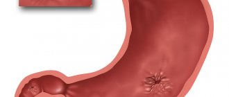
Structure of the stomach: schematically
In some cases, the stomach may change its location; even a slight displacement may indicate a serious pathology. Most often it occurs against the background of diseases of other internal organs; let’s look at the most common causes:
- Benign and malignant tumor formations. Stomach or duodenal cancer remains one of the most common and very dangerous forms, so at the slightest suspicion it is necessary to undergo a full examination.
- Perigastritis is an inflammatory process affecting the serous lining of the stomach. Its causes may be stomach ulcers, colitis, pancreatitis, and other inflammatory diseases of the digestive tract.
- Enlarged liver and spleen, affecting neighboring organs. The causes of liver enlargement can be hepatitis, impaired bile flow, cirrhosis, exposure to alcohol or drugs.
In all cases, treatment should be aimed at the underlying cause of the displacement of the stomach, it will allow the organs to return to their normal position. However, the stomach can change its location during pregnancy. During this period, due to the enlargement of the uterus, the stomach moves closer to the edge of the ribs, and because of this, its capacity is reduced.
A woman may feel an increased appetite, but if her stomach is full, she will feel serious pain. Because of this, during pregnancy it is especially important to eat small portions and avoid overeating. In addition, it is necessary to follow a daily routine to avoid unpleasant symptoms. After childbirth and the uterus shrinks in size, the organs will take their original position, and all negative symptoms will disappear without additional treatment.
How is the size of the stomach determined?
Fluoroscopy can be used to determine the size of the stomach.
To diagnose an enlarged stomach in modern medicine, the following diagnostic methods are used:
- X-ray
- Gastroscopy
- Computer diagnostics
To determine the size of the stomach using fluoroscopy, the patient is given a special liquid to drink. Then the specialist uses X-rays to obtain an image of the organ on a fluorescent screen. Today, X-ray television examination is used, during which the resulting image can be further processed in order to better diagnose the disease. The image remains in the device's memory and on the film.
Gastroscopy is performed to examine the inner surface of the stomach and its mucous membrane. A tube with a camera is inserted through the throat and esophagus into the patient's stomach. The image from the camera appears on the monitor. If necessary, the image is recorded. During the procedure, it is possible to conduct additional diagnostics: taking tissue for a biopsy. Computed tomography of the stomach has a number of advantages compared to other diagnostic methods:
- No special liquid is used during the study.
- Provides a more complete image in several projections compared to radiography.
- Does not cause pain during the procedure, compared to gastroscopy.
Pathology of the stomach and esophagus on ultrasound
Types of pathological changes in the stomach on ultrasound
Despite the difficulties of visualizing the stomach on ultrasound, due to the presence of a cavity in the organ, the accumulation of gases and the constantly changing shape due to peristalsis, the sonography method allows one to obtain fairly extensive and high-quality information about the condition of the stomach, on the basis of which various pathologies are identified.
Ultrasound signs of gastric pathology are the following:
- change in organ shape;
- change in the diameter of the gastric lumen;
- change in wall thickness;
- the presence of irregularities on the inner or outer surface of the stomach wall;
- the presence of bulges on the wall of the stomach;
- lack of peristalsis;
- presence of antiperistalsis.
The main sign of stomach pathology is thickening of its walls and expansion/narrowing of the lumen of the organ. Thickening of the walls of the stomach with a characteristic uneven echogenicity, when the outer part of the wall is hypoechoic compared to the hyperechoic inner part, is called a symptom of damage to a hollow organ. Currently, in the world medical literature, the terms “target”, “lesion halo”, “pseudo-kidney”, “bull’s eye”, “pathological type of cockade” are also used as synonyms for the term “symptom of lesion of a hollow organ”. All of the above terms refer to the same pathological condition of the stomach, and therefore can be used as equivalent. However, in the countries of the former USSR, the most commonly used term is “symptom of an affected hollow organ,” which we will also use.
Thus, thickening of the stomach wall, as a symptom of an affected hollow organ, can be diffuse and limited. With diffuse thickening, ultrasound shows an increase in wall thickness along its entire length. And with limited thickening, the wall of the stomach turns out to be thicker than normal only in a certain limited area. Unfortunately, the two main pathological signs of stomach diseases detected by ultrasound, namely the symptom of the affected hollow organ and the expansion/narrowing of the organ lumen, are nonspecific. This means that these pathological signs are inherent in many diseases, as a result of which it is impossible to accurately diagnose using ultrasound data alone.
In order to correctly diagnose a person’s existing stomach disease and choose one of the many pathologies that are characterized by a symptom of an affected hollow organ or a change in the lumen of the organ, it is necessary, in addition to the ultrasound results, to also evaluate the clinical symptoms that worry the patient. Only a combination of pathological changes on ultrasound with clinical symptoms will help make the correct diagnosis. Below, for general orientation in the problem, we will provide a table in which we will indicate which diseases of the stomach are characterized by the presence of symptoms on ultrasound of the affected hollow organ and changes in the lumen of the organ.
| Diseases characterized by an ultrasound symptom of an affected hollow organ with diffuse wall thickening | Diseases characterized by an ultrasound symptom of an affected hollow organ with limited wall thickening | Diseases that are characterized by narrowing or widening of the lumen of the stomach cavity on ultrasound |
| Swelling of the stomach wall | Pyloric stenosis | Scar stricture |
| Chronic gastritis type B | Ulceration of carcinoma | Inflammatory stenosis |
| Ménétrier's disease | Stomach ulcer | Tumor-induced stenosis |
| Infiltrative carcinoma | Varicose veins | Impaired evacuation of gastric contents |
| Malignant lymphoma | Gastric carcinoma |
Swelling of the stomach wall can be observed in acute pancreatitis, nephrotic syndrome, congestive heart failure, as well as protein deficiency in the body. Pyloric stenosis can be congenital or acquired due to inflammation, ulcers or tumors.
Ultrasound picture for various diseases of the stomach
Currently, there are six main pathological processes of the stomach that can be accurately diagnosed using ultrasound - these are erosive and ulcerative lesions, polyps, pyloric stenosis, duodenogastric reflux, malignant tumors and malformations. Below we present the characteristic features of the ultrasound picture for the six different above-mentioned pathologies of the stomach, as well as damage to the organ.
Ultrasound picture of the developmental defect “pyloric stenosis”. With congenital pyloric stenosis, a baby develops persistent vomiting of gastric contents at 2–4 weeks of life. Ultrasound shows a pronounced symptom of the affected hollow organ, namely: sharp thickening of the walls in the antrum, narrowing of the walls in the central part and the general appearance of the stomach as an “hourglass”, as well as the presence of contents in the stomach more than 24 hours after eating. Ultrasound picture with the developmental defect “duplication of the stomach” Using an ultrasound, it is possible to establish a doubling of the stomach only by performing a water-siphon test so that both cavities are filled with water and the doctor can clearly see them. A diverticulum is an area of protrusion in the wall of an organ.
Such diverticula can be single or multiple; on ultrasound they are visible only during a water-siphon test, when their lumen is filled with water and makes them visible. A diverticulum filled with water is an anechoic formation of any shape, bulging away from the wall of the stomach and connected to its cavity by a thin stalk. Damage to the stomach usually occurs due to mechanical impacts on the area where the organ is located, for example, from blows to the stomach, a fall from a height onto the stomach, etc. Such damage to the stomach can manifest itself as bruises, tears and ruptures of the walls of the organ. Less commonly, there are injuries caused by chemical substances, the picture of which on ultrasound is different from mechanical injuries.
Gastric contusion is characterized by limited, weakly echogenic thickening or bulging of the wall in the area of injury. The bruise is better visible when performing a water-siphon test than on an empty stomach. A rupture of the stomach wall is characterized by an uneven contour of the walls and their interruption in the area of \u200b\u200bthe rupture. When performing a water-siphon test, a rupture is easily diagnosed, since an ultrasound immediately shows the place where water leaks into the abdominal cavity. Chemical damage to the stomach wall is characterized by its thickening and doubling of the contour (an echo-negative stripe on the outside and an echo-positive stripe on the inside).
If, as a result of the damage, a cicatricial narrowing of the stomach cavity has developed, then an ultrasound will reveal fluid and food debris taken more than 14 hours ago. A gastric ulcer on an ultrasound looks like a hyperechoic thickening of the wall with its deformation and narrowing of the lumen. In addition, an excess amount of fluid on an empty stomach is recorded, a symptom of damage to a hollow organ. The following sizes of various parts of the stomach according to ultrasound are characteristic:
- outer diameter 16 – 22 mm;
- thickening of the wall of the gastric outlet from 6 to 10 mm;
- the distance between the walls in the thickening area is 7 – 12 mm;
- image factor 0.5 – 3.3 mm.
When performing a water-siphon test for a stomach ulcer, the following symptoms are characteristic:
- a clear, uneven contour of the wall in the area of the ulcer with hyperechoic inclusions;
- uneven thickening of the internal hyperechoic layer of the stomach up to 2 – 4 mm;
- the wall thickness in the area of ulcer or erosion is always more than 5 mm;
- indistinct layering of the wall in the area of the ulcer;
- inactive peristalsis.
Ultrasound picture for gastritis The walls of the stomach are evenly thickened, their echogenicity is uneven, with alternating hypoechoic and hyperechoic areas, more than 40 ml of fluid is detected in the cavity on an empty stomach, the diameter of the pylorus is more than 10 mm, signs of duodenogastric reflux. Stomach peristalsis is sluggish. Menetrier's disease is a giant hypertrophic gastritis, in which the gastric mucosa forms huge folds with polyp-like growths. Ultrasound shows a highly thickened wall with uneven echogenicity. Peristalsis is sluggish. A large amount of liquid and food taken more than 14 hours ago is found in the stomach.
A typical symptom is a lesion of a hollow organ with a thickening of the wall in the area where the polyp is located up to 20–28 mm, and a gap between the walls in the area of the thickening of 3–12 mm. There is also an uneven structure of the stomach wall in the area of thickening, the image coefficient in the area of the polyp is 0.9 - 9. The amount of fluid in the stomach on an empty stomach is normal. When performing a water-siphon test on ultrasound, a polypoid bulging of the wall into the lumen of the stomach is clearly visible. The structure of the polyps is heterogeneous, there is no layering.
Ultrasound picture for malignant tumors of the stomach
There is always a symptom of damage to a hollow organ above the area where the tumor is localized. The wall is thickened unevenly (from 10 to 30 mm), the thickening area is hypoechoic, heterogeneous, with impaired layering. The outer contour of the stomach wall in the area where the tumor is located is uneven, but clear. The lumen of the stomach in the tumor area is 2 – 16 mm. The lower border of the greater curvature of the stomach is lower than normal. The amount of fluid in the stomach on an empty stomach is either within normal limits or slightly increased. When performing a water-siphon test, uneven filling of the cavity with water and its insufficient expansion are noted. Rigidity of the thickened wall and sluggish peristalsis are also characteristic. The internal contour of the stomach is hyperechoic, fragmented and thickened more than 3 mm. In the area of the tumor, the outer and inner contours of the stomach are uneven, but clear. Pyloric stenosis is a narrowing of the outlet of the stomach, due to which the evacuation of food masses into the duodenum is impaired.
The causes of pyloric stenosis are usually tumors or a long course of a peptic ulcer, as a result of which scars are formed that tighten the walls of the stomach and reduce the lumen of its pyloric part. Pyloric stenosis is characterized by a shapeless hyperechoic formation with a sharp narrowing of the lumen of the affected area. At the initial stage, the walls of the stomach are thickened, but then they become thinner and the cavity expands. An ultrasound shows that the stomach is dilated and filled with contents of different ages. The amount of fluid on an empty stomach is more than normal, and there is sediment in it. The lower limit of the greater curvature of the stomach is significantly lower than normal. Peristalsis is either absent or weak. In the area of stenosis there is a symptom of damage to a hollow organ, and the wall is thickened to 7 - 22 mm, it has no layering, and the image coefficient is 0.6 - 8.5. When performing a water-siphon test for pyloric stenosis, the patient can only drink a small amount of liquid – no more than 300 ml, due to a feeling of fullness in the abdomen, nausea and sometimes vomiting. When liquid enters the stomach, its contents are mixed and sediment floats to the surface.
The lower border of the greater curvature drops even lower than when examined on an empty stomach. Peristalsis is sluggish or absent. It is impossible to visualize the normal layering of the stomach wall in the area of stenosis. Duodenogastric reflux is a pathological condition in which the contents of the duodenum are refluxed into the stomach. Reflux is detected during a water-siphon test, and on an empty stomach it can be seen on an ultrasound only if there is an accumulation of fluid in the patient’s stomach in an amount greater than normal. On ultrasound, reflux is visible by the retrograde movement of hyperechoic particles from the duodenum up the stomach after filling it with water. The presence of fluid in the stomach on an empty stomach in an amount of more than 40 ml may indicate a peptic ulcer, gastritis, pyloric stenosis or a malignant tumor. In order to understand exactly what disease the fluid in the stomach indicates in a particular case, you need to take into account other signs on ultrasound, as well as the clinical symptoms that the person has.
Pathological changes in the esophagus and ultrasound images in various diseases
Pathological changes in the esophagus on ultrasound are changes in the diameter, wall thickness, length of the abdominal organ, as well as the angle of His. Let's consider what ultrasound signs are characteristic of various pathologies of the esophagus. GER is a pathological reflux of stomach contents into the esophagus. And GERD is damage to the lining of the esophagus due to reflux. GER and GERD are characterized by expansion of the lumen of the esophagus on ultrasound and thickening of its wall. Chalazia is weak peristalsis, as a result of which food stagnates in the esophagus, moving only under the influence of gravity.
On ultrasound, chalazia of the esophagus is characterized by expansion of the lumen of the organ and thickening of its wall. Chalazia cardia is characterized by a gaping of the distal esophagus up to 6–14 mm, as well as visible flow of stomach contents into the esophagus. Cardiospasm is a pathological increase in the tone of the esophagus, as a result of which food does not move well to the stomach. Cardiospasm on ultrasound is characterized by expansion of the thoracic esophagus and at the same time a sharp narrowing of the esophageal-gastric junction to 3 - 5 mm. Stagnant contents are visible in the lumen of the esophagus, the wall of the organ is slightly thickened.