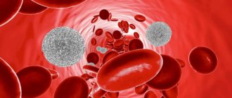
Monocytes are one of the types of white blood cells. Their functionality is of great importance for ensuring and maintaining the health of the human body.
They perform such important functions as antitumor, antiviral and others. However, often after taking blood tests you can hear from the doctor that the level of monocytes is increased. Unfortunately, few people know what this statement means and how to correct the situation.
And, most importantly, does such a phenomenon pose any danger in the body.
general information
Monocytes are cells belonging to the group of white blood cells. In healthy people, they make up about 3–8% of all white blood cells and are the largest blood cells found in the human bloodstream. Monocytes circulating in the blood have the ability to move from blood vessels to tissues, where they turn into macrophages, that is, phagocytic cells. Their main task is to fight bacteria and other microorganisms present in the body and produce substances that regulate the immune system.
A test that measures the number of monocytes in the blood is called a complete blood count with smear. If the result of this test shows an increased number of monocytes in your blood, you should consult your doctor as soon as possible, because such abnormalities can be caused not only by infections, but also by inflammatory, autoimmune or cancerous diseases.
blood test for monocytes in a child
Monocytosis in children
An increase in monocytes in childhood is also often associated with infectious pathologies, mainly of a viral nature. If monocytosis is diagnosed based on a blood test, this means that the body is actively fighting disease-causing processes.
Also, a similar condition is observed with helminthic lesions - enterobiasis, ascariasis and others. After the destruction of helminths, the content of monocytes returns to normal. The cause may also be associated with tuberculosis, but such pathologies are rare. Monocytosis can be observed due to oncological processes. The most common are leukemia and lymphogranulomatosis.
Other common reasons:
- poisoning;
- allergic reactions;
- inflammation in the gastrointestinal tract;
- autoimmune pathologies;
- rheumatoid type arthritis;
- Crohn's disease;
- infections due to surgical interventions.
Monocytes - what are they and what are their functions?
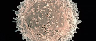
Monocytes, also referred to as MONO in blood counting, are one type of non-granular leukocytes, that is, agranulocytes. Monocytes are produced in the bone marrow and are the largest of all white blood cells. Monocytes present in the blood have the ability to move from blood vessels to surrounding tissues and organs, where they transform into macrophages.
Monocytes are highly migratory and can reach very distant areas of the body. Their surface is covered with receptors to which substances produced by the immune system - chemokines - can bind. As a result, these cells are activated in areas where inflammation occurs and they can cross cell membranes and reach different areas of the body to fight pathogens. Monocytes also have the ability to produce substances that regulate the immune system, such as tumor necrosis factor (TNF), interferon, and interleukins. Moreover, they skillfully carry out the process of phagocytosis of pathogenic microorganisms and dead cells.
Monocytosis often occurs against the background of infectious diseases
Changes in blood monocyte levels are most often the result of infection or bone marrow disorders. Elevated monocytes can be the result of bacterial, viral or fungal diseases, as well as malignant hematopoietic diseases such as leukemia and lymphoma. In turn, a decrease in the number of these cells can be the result of HIV infection, which leads to the destruction of monocytes and a sharp decrease in the body's immunity.
Determination of the level of monocytes in an adult , which are part of a blood test, should be carried out prophylactically, preferably once a year. It is especially recommended when the patient develops frequent, recurring infections and problems with the immune system, or when hematological diseases are suspected.
If the result of a basic blood test shows a change in the level of monocytes, a manual blood smear should be added to it, that is, an assessment of the structure of white blood cells under a microscope. This will allow you to judge whether these cells are normal or not.
Biological role in the body
The biological role of monocytes is very diverse. The largest phagocytes are necessary for many functions:
- Phagocytosis.
During this process, monocytes and macrophages identify proteins, bacteria, and viruses that are dangerous to the body, capture them and absorb them (phagocytose).
- Participation in the development of specific immunity.
Monocytes produce special cytotoxins, interferon, to counteract pathogenic viruses, bacteria and fungi.
- Assistance in the formation of allergic reactions.
Thanks to monocytes, specific elements of the compliment system are produced. Due to these processes, foreign proteins - allergens - are identified.
- Protection against tumors.
It is carried out through the production of tumor necrosis factor.
- Regulation of hematopoiesis and blood clotting processes
- carried out through the synthesis of specific elements.
Monocytes have special characteristics that distinguish them from neutrophils and other phagocytes:
- After absorbing a foreign element, monocytes do not die, but continue to function actively. In rare cases, the pathogen can defeat them.
- The life cycle of monocytes is longer than that of neutrophils.
- Monocytes have a pronounced antiviral effect, neutrophils have an antibacterial effect.
- When many monocytes are activated against a foreign agent, pus does not form.
- Chronic inflammatory process is a favorite place for concentration of macrophages and monocytes
Preparing for analysis
Because the monocyte count is part of the blood test, the preparation for it is the same as for other basic laboratory tests.
The patient should present to the laboratory on an empty stomach with a minimum 12-hour break from eating. 2-3 days before the examination, it is worth limiting the amount of intense exercise and the use of stimulants. You must inform the referring physician about all medications and dietary supplements you are taking, as they may affect the results obtained.
Symptoms of monocytosis
An increased content of monocytes can be reliably determined only by the results of a blood test. In this case, the disorder manifests itself with certain symptoms:
- apathy, decreased or lack of appetite;
- weight loss for no specific reason;
- high fatigue, general weakness of the body without an objective reason;
- depression;
- high level of anxiety, even panic states;
- emotional overexcitation for no specific reason;
- aversion to meat, aversion to it (occurs suddenly);
- bloating;
- constipation;
- diarrhea;
- discharge of large volumes of mucus with feces;
- dry cough that lasts for a long time (sometimes with blood discharge);
- muscle pain in the back area;
- pain in the legs;
- skin rash;
- rash in the genital area.
Norm for children and adults
Reference ranges, which should include both the total number of monocytes in the blood and their percentage in the fraction of all leukocytes, are determined individually for each diagnostic laboratory. However, they most often fall into the following ranges:
- for children under 4 years of age: 0.05-1.1 thousand/µl (2-7% leukocytes),
- for children from 4 to 18 years: 0.1–0.8 thousand/µl (2–7%),
- for adults: 0.1–0.8 thousand/µl (1–8%).
Elevated monocytes in the blood of a child - what to do
Table 1
| Index | Designation | In men | Among women |
| Red blood cells (x 1012/l) | R.B.C. | 4-5,1 | 3,7-4,7 |
| Average erythrocyte volume (fl or µm3) | MCV | 80-94 | 81-99 |
| Erythrocyte sedimentation rate (mm/h) | ESR | 2-15 | 2-10 |
| Anisocytosis of erythrocytes (%) | RDW | 11,5-14,5 | 11,5-14,5 |
| Hemoglobin (g/l) | HGB | 130-160 | 120-140 |
| Average hemoglobin level in erythrocyte (pg) | MCH | 27-31 | 27-31 |
| Average erythrocyte hemoglobin concentration (%) | MCHC | 33-37 | 33-37 |
| Color index | CPU | 0,9-1,1 | 0,9-1,1 |
| Hematocrit (%) | HCT | 40-48 | 36-42 |
| Platelets (x 109/l) | PLT | 180-320 | 180-320 |
| Average platelet volume (fl or µm3) | MPV | 7-11 | 7-11 |
| Reticulocytes (%) | RET | 0,5-1,2 | 0,5-1,2 |
| Leukocytes (x 109/l) | WBC | 4-9 | 4-9 |
Reasons for increased monocyte count
An elevated monocyte count in a child or adult can have various causes, and the interpretation of this result should always be made by a doctor, taking into account other blood parameters and blood smears.
The most common causes of an increase in the number of monocytes include:
- bacterial infections (for example, angina, tuberculosis, syphilis, Lyme disease, etc.),
- parasitic infections,
- viral (for example, mononucleosis) and protozoal infections,
- recovery from various types of infections,
- leukemia and lymphoma and other hematological diseases, such as multiple myeloma or myelodysplastic syndromes,
- cancer of the breast, ovaries, stomach and other organs, as well as tumor metastases,
- rheumatoid arthritis (RA), visceral systemic lupus and other collagen diseases,
- sarcoidosis,
- inflammatory bowel diseases and Crohn's disease,
- metabolic diseases,
- condition after removal of the spleen or other surgical procedures,
- alcoholic liver damage and other liver diseases,
- smoking,
- chronic stress,
- in infants - teething,
- taking certain medications, such as glucocorticosteroids.
Clinical significance of a blood test (excerpt from a lecture by Prof. E.B. Vladimirskaya)
Hematology:
12.03.2009
Doctor of Medical Sciences, Prof. E.B. Vladimirskaya Research Institute of Pediatric Hematology, Ministry of Health of Russia
Blood, being the internal environment of the body, carries within itself the stigmata of the vital functions of various organs and systems, the study of which is of undoubted clinical significance and is necessary for the diagnosis, prognosis of the course and monitoring of therapy of almost all internal human diseases.
The most accessible is the study of the morphological composition of blood; its results are included in the diagnostic algorithm for most pathological processes.
Since the first studies of blood under a microscope without the use of staining (the middle of the last century), blood cells have been divided into red - erythrocytes (based on the color of hemoglobin) and white - leukocytes. Leukocytes, in turn, are divided into cells containing specific granularity in the cytoplasm, and according to the ratio of this granularity to color, they are divided into (neutrophils, eosinophils and basophils) and those without - lymphocytes and monocytes. Based on the shape of the nucleus, the former are often called polymorphonuclear cells, and the latter are called mononuclear cells.
In recent years, clinical blood tests have increasingly been performed on automatic counters, which significantly increases the accuracy of calculations, but, however, does not negate the value of data obtained “manually” using light-optical microscopy. A comparison of the results of these two methods, together with the reference values of the indicators used, is presented in Table 1.
Table 1
| Automatic counting | Units _ | Normal limits | Manual counting |
| hgb -hemoglobin | g/liter | M: 132 - 173 F: 117 - 155 | Hb |
| rbc- red blood cells | 1012 /liter | M: 4.3 – 5.7 F: 3.8 – 5.1 | er. |
| hct-hematocrit | % | M: 39 – 49 F: 35 – 45 | ht |
| mcv - average volume red blood cell | 1 µm3 = 1 femtoliter | 80,0 – 95,0 | Spherical index (3,2-3,4) |
| mch - average hemoglobin content in an erythrocyte | picograms 1 g = 1012 picograms | 27,0 – 31,0 | Color index (0,85 – 1,0) Color.= Nb (g/l) x 3_____ Er (first three digits) |
| mchc – average concentration of HB in 1 erythrocyte | g/dl | 32,0 — 36,0 | |
| rdwwidth of distribution of red blood cells by volume | width histograms | 11,5 – 14,5 | No analogue |
| plt – platelets | *109 /l | 150 — 400 | Platelets |
| wbc- leukocytes | *109 /l | 4,5 –11,0 | Leukocytes |
| neu - neutrophils | *109 /l % | 1,8 – 5,5 47,0 – 72,0 | Neutrophils |
| lym – lymphocytes | *109 /l % | 1,2 – 3,0 19,0 – 37,0 | Lymphocytes |
| mon – monocytes | *109 /l % | 0,1 – 0,9 3,0 – 11,0 | Monocytes |
| eos – eosinophils | *109 /l % | 0,02 – 0,3 0,5 – 5,0 | Eosinophils |
| bas – basophils | *109 /l % | 0,0 – 0,07 0,0 –1,0 | Basophils |
When commenting on the data presented in the table, it is necessary to note:
- the vast majority of automatic counters do not detect young forms of leukocytes, normoblasts and reticulocytes - this data can only be obtained “manually”;
- normative values are never expressed in one figure; there is a limit of permissible fluctuations (it is presented in the table for all indicators), which fits 99.9% of the norm.
Let's analyze the clinical significance of individual blood test indicators.
Red blood counts.
Anemia is a decrease in hemoglobin below 120 g/l: mild - 110-120 g/l; average degree – 90-110 g/l; severe degree – below 90 g/l.
Based on the ratio of red blood indicators, 3 types of anemia are distinguished, which is the starting point for further diagnosis.
Microcytic-hypochromic anemia:
MCV < 80 Color.p. <0.85
MCH < 27; MCHC < 32
- Iron-deficiency anemia
- Anemia due to chronic inflammation
- Congenital spherocytic hemolytic anemia.
- Thalassemia.
Normocytic-normochromic anemias:
MCV 80 – 95
MCH = 27-31; MCHC = 32-36 Color.p. = 0.85-1.0
Acute blood loss.
- Anemia in chronic renal failure
- Anemia due to endocrine pathology.
- Anemia in cancer.
- Hemolytic anemias, immune and non-immune.
- Aplastic anemia.
- Myelodysplastic syndrome.
Hyperchromic macrocytic anemia.
MCV > 95
MCH>31; MCHC = 32-36 Color points > 1.0
- Megaloblastic B-12 is a deficiency (pernicious) anemia.
- Megaloblastic folate deficiency anemia.
- Autoimmune hemolytic anemia.
Thus, when detecting a decrease in hemoglobin, one should first determine the nature of anemia: normo-, hypo- or hyperchromic.
Let us dwell in more detail on iron deficiency anemia and anemia due to chronic inflammation, the diagnosis and treatment of which are the prerogative of general practitioners and do not require special hematological studies.
Iron deficiency anemia (IDA) is detected in 50% of women and 40% of men, representing one of the most common human diseases. The most common cause of IDA is a latent form of bleeding: in men – from the gastrointestinal tract and bronchopulmonary system, in women – menopause and metrorrhagia. Pregnancy is also one of the factors in the development of iron deficiency in women. Insufficient dietary intake of meat products is another significant and at the same time socially determined cause of the development of iron deficiency. In children born with a reserve of iron received from the mother in the last month of intrauterine development, further intake of iron occurs only from complementary foods. Thus, the reasons for the development of iron deficiency in young children may be prematurity, multiple pregnancies, late introduction of complementary foods, infections (increased iron consumption by microbial flora and malabsorption).
The clinical picture of IDA consists of the following clinical syndromes:
1. Anemia: weakness, drowsiness, fatigue, shortness of breath, palpitations, functional systolic murmur, floaters floating before the eyes.
2. Sideropenic syndrome:
· dryness, fragility, hair loss, early gray hair;
· flattening and brittleness of nails;
· dry skin, hyperkeratosis;
· painful, non-healing cracks in the corners of the mouth, tongue, fingers, toes, heels;
· perversion of taste and olfactory addictions;
· frequent infections;
· impaired swallowing, muscle weakness, sphincter weakness (urinary incontinence when laughing, coughing)
The diagnostic algorithm for IDA, in addition to a detailed clinical study to identify hidden bleeding, includes the mandatory determination of basic indicators of iron metabolism. The diagnosis of IDA is confirmed by the following values of these indicators:
- serum iron below 12.5 µmol/l
- total iron binding capacity (TIBC) above 64.4 µmol/l
- serum ferritin below 12 µg/l.
Correction of IDA is carried out by long-term oral treatment with iron preparations (6-8 weeks after hemoglobin restoration).
It is necessary to carry out a differential diagnosis between IDA and anemia in chronic inflammation.
Anemia during chronic inflammation in terms of the morphological composition of the blood is no different from IDA and is also accompanied by a decrease in serum iron content. However, its development is not based on exogenous iron deficiency, but on the impossibility of its utilization. Iron treatment for such anemia is contraindicated. Differential diagnosis is based on the study of iron metabolism indicators and is presented in Table 2.
Table 2. Differential diagnosis between IDA and anemia in chronic inflammation.
| Indicators | ZhDA | Anemia due to chronic inflammation |
| Serum iron | Reduced | Reduced |
| OZhSS | Increased | Normal or reduced |
| Serum ferritin | Reduced | Normal or increased |
To summarize the brief analysis of the clinical significance of the morphological parameters of red blood, we should dwell separately on reticulocytes and normoblasts.
A morphological sign indicating the hemolytic nature of the decrease in hemoglobin is an increase in the number of reticulocytes. With normal hemoglobin, the number of reticulocytes does not exceed 0.5-1.5%. The expected reticulocyte response to hemolytic anemia with intact hematopoiesis is presented in Table 3.
Table 3. Expected reticulocyte response to hemolytic anemia.
| Hematocrit, % | 45 | 40 | 35 | 30 | 25 | 20 | 15 |
| Hemoglobin, g/l | 130 | 120 | 115 | 100 | 83 | 66 | 50 |
| Reticulocytes, % | 0,5 | 1,5 | 5 | 10 | 15 | 20 | 30 |
Dynamic monitoring of reticulocyte levels is also necessary to assess the expected effectiveness of treatment for IDA and pernicious anemia. An increase in reticulocytes is naturally observed on days 5-8 of treatment with iron for IDA and is especially pronounced (up to 60%) on days 5-8 of treatment with vitamin B-12 for pernicious anemia. Such a hematopoietic response to the therapy of these diseases can also be considered as confirmation of the corresponding diagnosis of exjuvantibus.
Normoblastosis in peripheral blood is rare and always indicates a serious pathology. Its appearance is naturally observed in severe forms of hemolytic anemia and in patients who have undergone splenectomy. The detection of normoblasts in the blood of patients who do not suffer from this pathology should be a reason to search for oncological pathology.
about erythrocytosis when the following blood parameters are present: red blood cells above 5.7x10*12/l in men and 5.2x10*12/l in women, hemoglobin above 177 g/l and 172 g/l, respectively, hematocrit above 52% and 48 % respectively.
Primary erythrocytosis is considered to be rare genetically determined familial erythrocytosis and erythremia.
Secondary erythrocytosis, caused by increased formation of erythropoietin in response to arterial hypoxia or in certain tumors, is much more common.
Secondary erythrocytoses can be divided into the following groups:
1. Arterial hypoxia
- Altitude sickness
- Chronic pulmonary failure
- "Blue" heart defects
2. Tumors that produce erythropoietin
- Kidney tumors, hypernephroma
- Adrenal tumor
- Cerebellar hemangioma
- Ovarian cancer
3. Local renal ischemia
- Cyst
- Hydronephrosis
- Renal artery stenosis
4. Harmful production
- Cobalt poisoning
Treatment of secondary erythrocytosis requires elimination of their cause, but can also be symptomatic due to the threat of thrombosis. Symptomatic treatment of erythrocytosis is bloodletting.
White blood counts.
An increase or decrease in the number of different types of peripheral blood leukocytes can be judged only by changes in the absolute number of these formed elements.
Neutrophilia is an increase in the number of neutrophils more than 6x10*9/l.
Less commonly, neutrophilia is a manifestation of chronic myeloid leukemia, accompanied by clinical and hematological features specific to it (enlarged spleen, lymph nodes, blood rejuvenation, anemia, hyperthrombocytosis, bone marrow hyperplasia, the presence of a Ph chromosome and the chimeric c-abl-bcr gene).
Much more often, neutrophilia is a blood reaction to inflammation, the result of exposure to bacterial endotoxin and the release of inflammatory cytokines and chemokines by tissues. Neutrophilic leukocytosis can accompany any inflammation, bacterial, fungal and parasitic infections, necrotic tissue changes, hypoxemia, intoxication and tumors of various locations. With prolonged exposure to factors that induce neutrophilia, the bone marrow granulocyte reserve is depleted and young cells of the neutrophil series (band cells, metamyelocytes and myelocytes) begin to enter the blood. This blood condition is called leukemoid reaction of the neutrophil series. Sometimes it becomes necessary to make a differential diagnosis between such a reaction and the initial form of chronic myeloid leukemia. The absence of anemia, hyperthrombocytosis and a high level of alkaline phosphatase in neutrophils is characteristic of the leukemoid reaction.
Neutropenia is a decrease in the number of neutrophils less than 1.8 x 10*9/l.
Agranulocytosis - a decrease in the number of neutrophils less than 0.5 x 10*9/l.
Neutropenia can be primary (congenital and acquired), associated with blood diseases (acute leukemia, aplasia of hematopoiesis, cyclic neutropenia), and secondary, accompanying diseases, during which destruction and increased consumption of neutrophils occurs.
Secondary, reactive neutropenia includes immune and neutropenia in severe infections. Neutropenia in sepsis is usually accompanied by a rejuvenation of the leukocyte count and is a poor prognostic symptom, indicating depletion of hematopoiesis.
It is necessary to dwell on constitutional, so-called harmless neutropenias. About 4% of people have normal blood composition with a low content of neutrophils. This feature is associated with the genetically determined rapid movement of neutrophils into tissues, where they carry out their inherent protective functions. People with this blood composition are usually less susceptible to intercurrent infections and recover from them faster. However, often such patients, unfortunately, are the subject of close attention of doctors, are subjected to many unnecessary invasive studies, and they develop iatrogenic pathology. Thus, neutropenia, not accompanied by other blood changes and any clinical symptoms, does not require immediate intervention. Such patients require dynamic monitoring.
Separately, I would like to touch upon redistributive neutrophilia and neutropenia. The circulation of neutrophils has its own characteristics: half of the cells circulate with the blood (these cells are to be counted), while the other half is in the “marginal position” at the walls of the vessels. Irritation of the sympathetic system and vasospasm increase the number of circulating cells, while irritation of the parasympathetic system, on the contrary, reduces their number. Hence, stressful conditions contribute to transient neutrophilia (for example, neutrophilia in small children when screaming), and vagotonia - neutropenia.
Eosinophilia – an increase in the number of eosinophils above 0.4 x 10*9/l.
An increased release of eosinophils into the blood occurs under the influence of IL-4 and IL-5, which are formed in increased quantities during immunological tissue damage. Recently, the killer effect of eosinophils has been proven in some helminthiases and parasitic infections. Eosinophilia is a characteristic feature of collagenosis, allergies, many helminthic and parasitic infestations, immunodeficiency, especially hyper-IG-E syndrome, and some tumors.
Monocytosis – the number of monocytes is higher than 0.8 x 10*9/l.
Conditions often, but not always, associated with monocytosis include:
- Infections (especially tuberculosis, endocarditis, syphilis).
- Fever of unknown origin
- Various forms of neoplasia and myeloproliferative diseases.
- Chronic inflammation (especially cholecystitis and rheumatoid polyarthritis)
- Condition after splenectomy.
Lymphocytosis – an increase in the number of lymphocytes more than 4.0 x 10*9/l
Among malignant lymphoproliferative diseases with high lymphocytosis, the most common is chronic lymphocytic leukemia, a disease of people over 45 years of age. The distinctive feature of this lymphocytosis is its monoclonal nature and B-cell origin.
Secondary, reactive lymphocytosis , which is polyclonal in nature, accompanies many viral infections and some inflammatory and immune complex diseases. These include:
1. Lymphotropic viral diseases:
— infectious mononucleosis (atypical mononuclear cells, characteristic clinical picture);
- infectious lymphocytosis (asymptomatic epidemic form in young children - up to 20-30 thousand)
2. Cytomegalovirus infection (atypical mononuclear cells, characteristic clinical picture).
3. Childhood infections: whooping cough, chickenpox, scarlet fever prodrome.
4. Other viral infections: rubella, hepatitis, some respiratory adenoviral infections in the convalescent stage.
5. Inflammatory and immune complex diseases: thyrotoxicosis, ulcerative colitis, Crohn's disease, vasculitis.
Lymphocytopenia - a decrease in the number of lymphocytes below 1.2 x 10*9/l.
It is observed relatively rarely, most often with corticosteroid therapy. May also accompany AIDS, lymphogranulomatosis and various chronic infections (for example, tuberculosis, disseminated lupus erythematosus, sarcoidosis).
Hyperthrombocytosis is considered to be an increase in the number of platelets more than 400.0 x 10*9/l.
Primary hyperthrombocytosis accompanies myeloproliferative diseases and is a consequence of tumor transformation of the megakaryocytic lineage of the bone marrow.
Secondary reactive hyperthrombocytosis is observed:
- After surgical interventions (about 2 weeks).
- After splenectomy (up to 1 year).
- For malignant tumors
- For acute posthemorrhagic and hemolytic anemia.
- For certain inflammations (tuberculosis, acute rheumatism, ulcerative colitis, osteomyelitis).
Thrombocytopenia - a decrease in the number of platelets below 100.0 x 10*9/l most often occurs with tumor diseases of the blood, aplastic anemia and immune thrombocytopenic purpura. Thrombocytopenia is an obligatory component of the hypersplenism syndrome with splenomegaly. It should be borne in mind that a serious threat of bleeding occurs when the platelet count drops below 20.0 x 10*9/l.
Reactive thrombocytopenia is rare and can accompany any immune pathology and disseminated intravascular coagulation.
ESR - erythrocyte sedimentation rate is a nonspecific reaction. Normally, it is 2-15 mm per hour in men under 50 years old, and 2-20 mm per hour in women under 50 years old. After 50 years, men have up to 20 mm per hour, and women – up to 30 mm per hour.
The rate of erythrocyte aggregation depends on their number (it accelerates when their number decreases) and the amount of coarse proteins (inflammatory proteins, fibrinogen, antibodies, gamma globulin, etc.) adsorbed on erythrocytes and accelerating their sedimentation. Based on this, it is clear that there is a wide range of pathologies in which ESR acceleration can be detected.
Thus, analysis of the morphological composition of blood is of great clinical importance, and sometimes is a leading sign in the diagnosis and choice of therapy for many human diseases.
However, it should be remembered that the most important link in such an analysis is the integral assessment of all blood parameters and the mandatory correlation of changes in the blood with the history and clinical manifestations of the disease.
Literature:
1. Guide to Hematology, ed. A.I. Vorobyov, Moscow, 1985
2. Hematology, ed. by WSBeck, London, 1991
3. Manual of Clinical Hematology, ed.by J. Mazza, NY, 1995
Tags: Laboratory, Blood
« Back to article list Share on
What does a low monocyte count indicate?
A low monocyte count, also known as monocytopenia, may indicate HIV infection, immunodeficiency including Monomak syndrome, AIDS, bone marrow aplasia, leukemia or a consequence of chemotherapy or radiation therapy, severe stress, or certain medications.
If monocytopenia continues for a long time, it can lead to a decrease in all blood counts: red blood cells, leukocytes (white blood cells) and platelets. This condition is called pancytopenia.
Therefore, if there is any deviation from the norm in the monocyte test result, you should consult your doctor. If a history of infection is causing the change in monocyte count, the test should be repeated after 2 to 4 weeks.
In other cases, it may be necessary to complete the diagnosis with imaging tests, a urinalysis, and other blood tests such as CRP or ASO levels.
Platelets
The main task of platelets is to participate in stopping bleeding. They form the primary plug in the area of vessel damage and participate in the formation of a blood clot. In forms, automatic analyzers are designated by the abbreviation PLT , which comes from the English word platelets (literally, “saucers”) for their peculiar shape.
A decrease in the number of platelets can be accompanied by bleeding and is a consequence of diseases and conditions such as malaria, thrombocytopenic purpura, malignant neoplasms, disseminated intravascular coagulation syndrome and some others.
A high platelet count can cause excessive blood clotting and often has no apparent cause. In this case, the condition is designated as essential thrombocythemia. In addition, thrombocytosis can be caused by iron deficiency anemia, hemolysis, some infectious and autoimmune diseases, and bone marrow damage.
This is where the work with platelets in a classic blood test ends; however, automatic analyzers even in this case offer additional diagnostic capabilities in the form of the following indicators:
- MPV —mean platelet volume. An indicator reflecting the size of cells, which directly depends on their age. An increased MPV may indicate hematological diseases, damage to the spleen, thyrotoxicosis, and progression of atherosclerosis. A decrease in the rate is observed in some genetic diseases, myocardial infarction, childhood infections, cancer chemotherapy and a number of other conditions.
- PCT is the ratio of the total volume of platelets to blood plasma (thrombocrit). It is not a specific indicator, it varies widely physiologically, but it allows one to assess the risk of bleeding and thrombosis.
- PDW —platelet distribution by volume. The indicator has no independent meaning and is assessed in conjunction with other indices.