Internal bleeding of the stomach is not an independent disease, but acts as a symptom and complication of other diseases. It is important to understand that this symptom occurs not only when the stomach is damaged, but can also indicate problems not related to the gastrointestinal tract: blood clotting disorders, vascular diseases, metastatic growth of a tumor from a neighboring organ.
General symptoms, the severity of the patient’s condition, and diagnostic studies allow the doctor to correctly determine treatment tactics.
Causes of stomach bleeding
Often the cause of stomach bleeding is an untreated ulcer.
The main causes of hemorrhages can be divided into frequent, infrequent and rare.
Frequent sources of the problem:
- An ulcer is a serious disease that is often life-threatening. Bleeding can occur either from a new “young” ulcer or from a chronic one. Long-existing ulcers are dangerous due to more massive bleeding, especially when it is located on the lesser curvature of the stomach. This is due to anatomical features - large branches of the left gastric artery are located here.
- Tumors cause acute bleeding only in 10% of cases; more often, bleeding is detected in the same way as with erosion - during FGS or stool examination.
- Erosion is superficial destruction of the gastric mucosa. In most cases, they are multiple, located in the fundus of the stomach, less often in the antrum. Heavy (profuse) bleeding with erosive gastritis is rare; more often it is drip leakage of blood, which is detected by chance, during FGS or stool examination for occult blood.
- Mallory-Weiss syndrome - occurs when the cardiac part of the stomach is ruptured in the longitudinal direction. This situation is possible when there is a sudden increase in pressure in a full stomach. It occurs in pregnant women with severe toxicosis (due to constant vomiting), athletes when lifting weights, and blunt abdominal trauma.
The following processes are less common:
- Henoch-Schönlein disease (hemorrhagic vasculitis) - after some kind of infection (ARVI, laryngitis, tonsillitis), toxic damage to the capillaries occurs and multiple hemorrhages occur throughout the gastrointestinal tract.
- Postoperative gastric ulcer.
- Acute leukemia.
- Gastroesophageal reflux disease.
- Iatrogenic damage (taking non-steroidal anti-inflammatory drugs, corticosteroids, antihypertensive drugs)
The rarest causes are: scurvy, hemophilia, thrombocytopenic purpura, aplastic anemia, sarcoidosis.
The main diseases that lead to stomach bleeding are listed here, but in general there are many more of them.
Diagnostics
Photo: medhealthservices.com
When the first signs of gastric bleeding appear, you should immediately consult a doctor, since this condition poses a great danger to human life. Emergency care provided to the patient is aimed at eliminating the symptoms and identifying the causes of this condition.
Diagnosis of gastrointestinal bleeding includes:
- endoscopic examination;
- X-ray of the stomach;
- vascular angiography;
- radioisotope scanning;
- capsule endoscopy;
- colonoscopy;
- Magnetic resonance imaging;
- blood test;
- coagulogram;
- rectal and external examination;
- stool analysis for the presence of hidden bleeding.
Differential diagnosis of gastric bleeding also includes an analysis of factors that could provoke the disease. The person’s lifestyle and what medications he or she takes must be taken into account. The patient’s medical record must be studied to determine the presence of concomitant diseases that could cause the main signs of gastric bleeding.
Clinical examination
Examination of a sick person must include the following:
- examination of the condition of the skin. This takes into account the presence of hematomas, dilated small vessels and other factors that may affect the correct diagnosis;
- digital examination of the rectum, which helps to identify the presence of bleeding. Also, this diagnostic method is necessary to assess the patient’s condition, to identify tumors or hemorrhoids;
- palpation of the abdominal cavity, which reveals possible enlargement of the liver or spleen, accumulation of fluid, and the appearance of various neoplasms;
- determining the size of lymph nodes.
When gastrointestinal bleeding occurs, a change in stool occurs, so a stool analysis is required. This may indicate the location of the problem - in the stomach, intestines, or rectum.
A clinical study is not able to identify the causes of bleeding, but it may well determine the severity of the patient’s condition and the degree of blood loss. The diagnosis is also influenced by the presence or absence of pain during palpation of the abdominal cavity.
Laboratory research
Diagnosis of gastric bleeding necessarily includes a number of laboratory methods:
- General blood test - determines the amount of hemoglobin, red blood cells, leukocytes, platelets, ESR. These data may change for the worse even after bleeding has stopped;
- coagulogram - determination of blood clotting disorders. It is carried out using special equipment, where several indicators are analyzed that indicate the presence of pathologies;
- biochemical analysis - reveals the amount of urea, creatine in the blood and determines other equally important data that directly affects the diagnosis and treatment tactics.
These analyzes are of great value if they are performed several times. The main indicators that are visible over time indicate the course of the disease and the effectiveness of treatment.
Endoscopic examination
A comprehensive diagnosis of gastric bleeding necessarily includes examination using a fibrogastroscope, at the end of which there is a small camera. This allows the gastroenterologist to carefully examine the mucous membrane of the esophagus and stomach. This study is very informative and in most cases determines the source of bleeding in the digestive system.
Endoscopy is performed as follows:
- The person lies on his left side.
- An anesthetic spray is used to numb the mucous membrane.
- A special mouthpiece is placed in the patient's mouth.
- The doctor inserts a special device through the person’s mouth into the stomach - a fibrogastroscope, which is a flexible cord. During this time, the patient should not move and breathe intensively through the nose.
This study is quite unpleasant, but it takes a little time and is very informative.
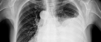
X-ray of the stomach
How to determine gastric bleeding? In many cases, an X-ray of the stomach using a contrast agent will be quite informative. Using this study, it is possible to assess the condition of the walls of this organ and identify various pathologies.
X-rays are performed exclusively on an empty stomach. If the stomach is filled with food, the contrast agent will not be able to distribute properly throughout its walls.
Research methodology:
- The person should drink a solution containing barium sulfate. A special feature of this substance is that it does not transmit x-rays.
- Several x-ray pictures are taken. In this case, the person must take different positions so that the study is as informative as possible.
- An analysis of x-rays is carried out, on which the stomach is clearly visible.
Angiography
Angiography or X-ray contrast examination is indicated when there is a suspicion that gastric bleeding is caused by vascular pathology. This may be caused by atherosclerosis or other serious disorders.
This study is carried out using a special contrast agent, which is injected into the vessel using a catheter. After this, an x-ray is taken. The resulting images clearly show the vessel. Based on the data obtained, it is possible to draw conclusions about the cause of gastric bleeding.
Radioisotope scanning
This study is carried out in cases where other diagnostic methods have failed to identify the causes of bleeding.
Before radioisotope scanning, a solution containing red blood cells, which are marked with a contrast agent, is injected into the blood of a sick person. After this, they accumulate in large quantities at the site of bleeding. It can be identified in photographs taken by a special device.
Magnetic resonance imaging
This study is carried out when the doctor needs additional data about the health status of a sick person. Magnetic resonance imaging allows you to obtain a 3D image of a specific organ or images with layer-by-layer sections of the necessary parts of the body.
This study is very informative and is carried out using a special installation - a tomograph.
Colonoscopy – indications and how it is performed
Colonoscopy is considered the most informative method for diagnosing defects of the large intestine. With its help, you can identify pathologies that caused heavy bleeding. The procedure is carried out with a special long probe - an endoscope. It is inserted through the anus into the intestines, after which the doctor sees an image of the mucous membrane on the screen. A significant disadvantage of the method is that a colonoscopy cannot be performed if a person has intense bleeding.
This procedure is quite complex and time-consuming. To avoid discomfort, you must strictly follow all the doctor’s instructions. Before the procedure, you need to completely cleanse the intestines. To do this, you need to take a special drug in the evening if the colonoscopy takes place in the morning. To reduce pain, antispasmodics are also prescribed. For the same purpose, immediately before inserting the probe into the intestines, the doctor injects the patient with an anesthetic.
The procedure does not last long. On average, 10-15 minutes are enough.
Capsule endoscopy is an innovative method for diagnosing gastrointestinal bleeding
In some cases, a doctor may decide to use a modern capsule endoscopy method to diagnose pathologies of the digestive system. It consists in the fact that the patient must swallow a special endocapsule.
This device transmits in real time an image of the mucous membranes of the digestive system. Capsule endoscopy allows you to identify pathologies of the stomach, esophagus, small and large intestines, duodenum, which may have gone unnoticed during other studies.
The duration of the procedure is 8-9 hours. The advantage of diagnosis is that the patient does not need to lie down during this time. A person can do his usual activities. There is also no need to specifically remove the endocapsule. When the device passes through the entire digestive tract, it comes out naturally on its own.
Symptoms of acute gastric bleeding
Internal bleeding of the stomach can be hidden or obvious
Symptoms of acute bleeding depend on a group of factors: the rate of blood loss, the volume of fluid lost, the person’s age, and the presence of additional diseases.
In the clinic of acute bleeding, two periods are distinguished: latent and obvious.
The first occurs from the moment blood enters the intestines from the stomach. The second is from the moment blood is detected in vomit, feces or gastric contents (with FGS).
The severity of the clinical manifestations directly depends on the rate of blood loss. If it is more than 750 ml per day, the patient feels weakness, sweating, and severe headache. As blood pressure drops, a person notices a noise in the head, “flickering of spots,” and fainting occurs. In some cases, the only sign of bleeding is sudden loss of consciousness.
Vomiting indicates the presence of bleeding; its color can indicate the massiveness of blood loss. “Coffee grounds” indicates slight bleeding, since hemoglobin has time to turn into hydrochloric acid hematin under the influence of hydrochloric acid.
With heavy bleeding, cherry-colored vomit. If the blood is bright red, the bleeding most likely comes from the esophagus or is very massive in the stomach. Vomiting is often one-time, its repetition within two hours indicates continuous bleeding.
Sigmoidoscopy (rectoscopy)
If necessary, an examination of the rectum is performed. Sigmoidoscopy (rectoscopy) is a method of endoscopic examination of the rectum and distal sigmoid colon by examining their internal surface using a sigmoidoscope inserted through the anus.
Sigmoidoscopy is the most accurate and reliable examination of the rectum and lower sigmoid colon. In the practice of a coloproctologist, sigmoidoscopy is an obligatory component of every proctological examination. The examination allows you to visually assess the internal surface of the rectum and distal third of the sigmoid colon to a level of 20-35 cm from the anus.
Symptoms of chronic bleeding
Internal stomach bleeding leads to severe anemia
Symptoms of chronic blood loss are vague and depend on the degree of iron deficiency anemia. Often the body compensates for losses so well that the patient walks independently and lives a normal life with a hemoglobin level of 30-40 g/l. The main symptoms of chronic gastric bleeding are signs of anemic syndrome. Severe weakness and fatigue appear, which increases even from slight physical activity.
The condition of the skin changes: the skin is pale, dry, flaky, and “stubs” appear in the corners of the mouth. Women immediately notice brittle nails and cracks in the skin of their hands.
With severe anemia, patients feel tachycardia even at rest, and shortness of breath appears. The condition of the oral cavity changes - there is a burning sensation in the tongue, difficulty swallowing, redness of the tip of the tongue. Sometimes there is hoarseness, coughing, redness and itching in the palate.
In especially severe cases, olfactory tastes change - patients may fall in love with the musty air of the basement, the smell of shoe polish or car tires. Sometimes there is a desire to eat sand or earth.
Peroxidase test to confirm the presence of guaiacol
This is the most commonly used stool blood test. The test available is called Hemoccult. In this study, a stool sample is distributed into a guaiacol-impregnated cardboard box, which, if the stool contains heme, turns blue (heme causes a reaction similar to peroxidase). The more blood in the stool, the higher the likelihood of a positive result.
The Hemoccult test gives a positive result in 50% of cases when 10 ml of blood enters the gastrointestinal tract during the day. Under physiological conditions, 0.5-1.5 ml of blood is normally extravasated into the lumen of the gastrointestinal tract during the day.
The sensitivity of the first test is estimated to be approximately 30%. Performing 3 tests (which is the standard) increases the confidence to 92%. False negative results are especially common in people taking medications or foods high in vitamin C.
Relatively often, the test gives false positive results when consuming large amounts of red meat and foods high in peroxidase (radish, radish, horseradish). Taking iron supplements does not give false positive results in the study.
Treatment
In the acute period, the following manipulations are performed to stop bleeding:
- Drug hemostasis: the patient is injected intramuscularly with hemostatic drugs (Vikasol, Etamzilat, Aminocaproic acid).
- Endoscopic hemostasis (vessel clipping, photocoagulation, sclerotherapy).
- Arterial embolization.
- Surgical intervention (vascular ligation).
At the same time, the volume of circulating blood is replenished: saline solutions are administered (Disol, Trisol, Ringer's solution), microcirculation in the vessels is improved (Reopoliglyukin).
In case of severe blood loss, blood is transfused and iron-containing drugs are administered. In the future, iron supplements are prescribed for 3-6 months.
After stopping the bleeding, treatment tactics will depend on the disease that caused the disease.
First aid
For stomach bleeding, there is little that can be done at home. If you suspect a pathology, you must:
- urgently call an ambulance;
- the patient should be placed with the leg end raised; if he loses consciousness, the head must be turned to the side to prevent aspiration of vomit;
- Ice should be placed on the patient’s stomach;
- You cannot give food or drink, or take medications on your own.
Folk remedies
Photo: oprostatite.info
If gastric bleeding develops, traditional methods cannot be used. In the absence of timely specialized care, this condition can lead to the development of hemorrhagic shock and death of the patient. It is necessary to immediately call an ambulance and not self-medicate. During the period of remission, the following remedies can be used:
- 1 tbsp. l. horsetail is steamed for an hour in 250 ml of boiling water. The broth is filtered and consumed half a glass each time after meals.
- A tablespoon of pink immortelle flowers is infused in 250 ml of boiling water. Then the infusion is filtered and given to the patient 1 tbsp. l. every 2 hours.
- A decoction of blueberries is brewed and drunk like tea three times a day.
Parsley helps prevent stomach bleeding. In addition, decoctions of chamomile, bearberry, and yarrow are used. All these herbs have hemostatic properties. Any folk remedies are allowed to be used only after the approval of a specialist.
The information is for reference only and is not a guide to action. Do not self-medicate. At the first symptoms of the disease, consult a doctor.
How do blood vessels bleed?
Bleeding can be mild, moderate or severe. His character may be:
- arterial - strong, jet;
- venous - blood loss occurs gradually;
- capillary - minor discharge due to damage to small vessels.
The problem itself can be stable/unstable, recurrent. Vessels of the mucosa, submucosal and intermuscular plexus, as well as those located outside the gastrointestinal tract, can bleed.
The expiration can last for several hours or days. In medicine there are:
- profuse blood loss - the patient loses more than 1 liter of blood in 1-3 hours and needs urgent medical attention;
- acute - less than 1 liter expires in 1-2 days, the patient’s vital signs are relatively stable;
- chronic - develops slowly, often over several weeks or even months, the intensity of symptoms gradually increases.
Infusion therapy
Begin infusion therapy with the introduction of balanced salt solutions.
Important! If there are signs of ongoing bleeding or unstable hemostasis has been achieved, blood pressure should be maintained at the minimum acceptable level (SBP 80-100 mm Hg), i.e. infusion therapy should not be too aggressive. Blood transfusions are carried out if adequate infusion therapy fails to stabilize the patient’s hemodynamics (blood pressure, heart rate). Consider the need for blood transfusion:
– when the hemoglobin level decreases below 70 g/l. when bleeding has stopped;
– with ongoing bleeding, when hemoglobin is below 90-110 g/l.
In case of massive blood loss (more than 50-100% of the blood volume), transfusion treatment is carried out in accordance with the principles of “Hemostatic resuscitation”. It is believed that each dose of packed red blood cells (250-300 ml) increases hemoglobin levels by 10 g/l. Fresh frozen plasma is prescribed for clinically significant coagulopathy, including drug-induced coagulopathy (for example, the patient is receiving warfarin). And in case of massive blood loss (>50% of blood volume). If reliable hemostasis is achieved, there is no need to administer FFP even with significant blood loss (more than 30% of the blood volume). Dextrans (polyglucin, rheopolyglucin), hydroxyethyl starch (HES) solutions may increase bleeding and their use is not recommended.
Sequence of assistance
If the patient received anticoagulants before bleeding occurred, they should, in most cases, be discontinued. Assess the severity of the condition and the estimated amount of blood loss based on clinical signs. Vomiting blood, loose stools with blood, melena, changes in hemodynamic parameters - these signs indicate ongoing bleeding. Arterial hypotension in the supine position indicates large blood loss (more than 20% of the blood volume). Orthostatic hypotension (a decrease in systolic blood pressure above 10 mm Hg and an increase in heart rate by more than 20 beats per minute when moving to a vertical position) indicates moderate blood loss (10-20% of blood volume);
In the most severe cases, tracheal intubation and mechanical ventilation may be required before endoscopic intervention. Provide venous access with a peripheral catheter of sufficient diameter (G14-18); in severe cases, install a second peripheral catheter or perform catheterization of the central vein.
Take a sufficient volume of blood (usually at least 20 ml) to determine the group and Rh factor, combine blood and conduct laboratory tests: general blood count, prothrombin and activated partial thromboplastin time, biochemical parameters.