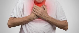
Chest pain
(chest pain) is a common symptom. In many cases, chest pain can be caused by heart disease, and this makes it especially serious. However, the list of causes of chest pain is much wider. This area contains the heart, lungs, esophagus, large vessels, bones and muscles - and any of these organs can be a source of pain. Chest pain can also be caused by the diaphragm and abdominal organs, primarily the stomach. Finally, pain can be caused by problems in other organs, and its localization in the chest is explained by the complex arrangement of nerves and muscles.
Possible nature of chest pain - what you should pay attention to
Chest pain can be aching, stabbing, shooting, squeezing, burning. In some cases it can be acute, intense, even intolerable.
It is important where exactly the pain is localized. Usually the pain can be quite clearly localized as follows:
- in one half of the chest - right or left. Sometimes the localization of pain can be more specific, for example, in the ribs;
- behind the sternum. Pain behind the sternum, especially when it hurts both behind the sternum and to the left of it, is characteristic of heart disease;
- just below the sternum (in the epigastric region). Such pain is typical for diseases of the digestive tract (stomach or esophagus).
Chest pain without a clear localization may indicate lung cancer or tuberculosis.
Chest pain may be accompanied by symptoms such as:
- shortness of breath, difficulty breathing;
- cough, increased body temperature;
- weakness, cold sweat;
- belching, heartburn, nausea, vomiting;
- changes in blood pressure.
The distribution of pain is also important for diagnosis. The pain may radiate to the arm. This type of pain is observed with myocardial infarction and osteochondrosis.
Spine pathologies
When chest pain appears, it is difficult to immediately suspect its cause in the development of spinal diseases. Nevertheless, osteochondrosis of the thoracic region and intercostal neuralgia can also make themselves felt in a similar way.
Osteochondrosis of the thoracic spine
Osteochondrosis is the process of dehydration of intervertebral discs and the occurrence of degenerative changes in them. Damage to the thoracic region is much less common than to the cervical or lumbar, and the symptoms are largely reminiscent of angina pectoris and gastrointestinal pathologies. Therefore, patients are often falsely diagnosed with other diseases, and treatment that does not bring results is prescribed.
The risk of developing osteochondrosis directly depends on lifestyle. Therefore, if previously it was mainly found in older people, today it is often detected in young people. Prolonged, regular sitting increases the likelihood of dehydration and progressive degeneration of the discs in the thoracic spine. Therefore, it is most often diagnosed in people who are forced to sit for a long time at a computer, behind the wheel of a car, at a machine, etc.
Osteochondrosis of the thoracic region can manifest itself:
- constant dull pain between the shoulder blades;
- girdle pain localized at the level of the armpits;
- pain in the heart and behind the sternum;
- burning sensation in the back.
A characteristic feature is the occurrence of painful sensations after prolonged sitting, at the end of the working day.
The main danger of osteochondrosis is its steady progression, especially in the absence of comprehensive treatment. As a result, over time, the affected disc, which is a dense cartilage with a jelly-like internal content, becomes deformed and bulges, usually towards the spinal canal. This is called protrusion. Subsequently, it can transform into an intervertebral hernia, which means the final rupture of the outer dense ring of the disc, through which the jelly-like internal contents (nucleus pulposus) can exit into the spinal canal. This is dangerous due to the infringement of the spinal roots passing here and even the spinal cord itself, which will lead to disruptions in the functioning of the internal organs innervated by them, shooting pains, numbness and other sensitivity disorders.
Intercostal neuralgia
Intercostal neuralgia is a consequence of damage to the peripheral nerves passing through the tissues in the area of the intercostal spaces. This may be due to their mechanical compression, herpetic infection, hypothermia, exposure to toxic substances, injuries and other factors.
With intercostal neuralgia, the pain is lumbago-like or burning. They spread along the ribs to the sternum. They can be either short-term or last for hours or even days. When touching the skin in the intercostal spaces, changing body position, walking, deep breathing, coughing, the pain intensifies.
Causes
Chest pain due to heart disease
Chest pain is a characteristic symptom of heart disease. It is observed, in particular, when:
- coronary heart disease (caused by insufficient oxygen supply to the heart muscle). The most common form of coronary artery disease is angina pectoris, which manifests itself in the form of discomfort, a feeling of heaviness or compressive pain behind the sternum and to the left of it. An attack of pain is usually provoked by physical activity or emotional stress. The duration of the attack usually ranges from several minutes to half an hour. Relief occurs after taking nitroglycerin. The pain may be accompanied by shortness of breath, radiating to the left arm, under the shoulder blade, and the left half of the lower jaw;
- acute myocardial infarction. Myocardial infarction is also a form of coronary heart disease that requires immediate medical attention. The pain during myocardial infarction is very intense and has a compressive, pressing or bursting character. It is observed behind the sternum and to the left of it, and can radiate to the left arm, under the shoulder blade, to the left side of the neck and lower jaw. The pain is accompanied by shortness of breath, fear of death, weakness, and cold sweat may appear. The pain lasts more than 15-30 minutes (i.e. longer than during an attack of angina) and is not relieved by nitroglycerin. If you suspect a myocardial infarction, you should immediately call an ambulance;
- pericarditis (inflammation of the outer lining of the heart - the pericardium). In this case, the pain may be constant or intermittent. It is usually localized behind the sternum. The pain increases when lying down, and decreases if you lean forward;
- myocarditis (inflammation of the heart muscle). Myocarditis occurs most often as a complication of an infectious disease. With myocarditis, chest pain is combined with fever and shortness of breath;
- mitral valve prolapse. In most cases, this disease is asymptomatic, but sometimes there is pain in the left side of the chest, which may be accompanied by a feeling of shortness of breath, a feeling of rapid or slow heartbeat, dizziness and fainting.
Chest pain due to respiratory diseases
Chest pain can occur with diseases such as:
- pleurisy (inflammation of the membrane of the lung - pleura). In case of pleurisy, the pain is usually acute and one-sided. Intensifies with deep breaths, laughter, and movement. The pain intensifies when bending to the healthy side. The pain decreases if you lie on the side where it hurts. Chest pain is combined with other symptoms - fever, chills, weakness, cough;
- pneumonia (pneumonia). Chest pain with pneumonia is a concomitant symptom that occurs against a background of cough and fever. The pain is usually one-sided - corresponds to the side on which inflammation develops. The nature of the pain is sharp or aching;
- bronchitis;
- tracheitis;
- pulmonary tuberculosis;
- lungs' cancer.
Chest pain due to diseases of the digestive tract
Pain in the chest area can be caused by diseases such as:
- stomach ulcer. With a stomach ulcer, the pain is usually described as “burning.” It is localized in the epigastric region and can radiate to the left half of the chest. Occurs after eating;
- gastroesophageal reflux disease (reflux of stomach contents back into the esophagus). Acidic or alkaline contents irritate the lining of the esophagus, causing bloating and severe pain in the epigastric region and the left side of the chest. An attack can be triggered by eating too much at night, strong coffee, or alcohol abuse. In addition to chest pain, heartburn and belching may occur;
- hiatal hernia. In most cases, the disease is asymptomatic. In some cases, aching or burning pain behind the sternum and in the epigastric region may be observed. Pain usually occurs after eating in a horizontal position. May be accompanied by hiccups, heartburn, belching, and sometimes vomiting (in obese women).
Other Possible Causes of Chest Pain
Chest pain can also be caused by:
- osteochondrosis of the cervical and thoracic spine. Pain with osteochondrosis can be similar to an angina attack and can radiate to the shoulder blade, arm, or shoulder. Sometimes there is numbness in the hand. Another option for pain with osteochondrosis is lumbago. It is provoked by turns of the body, movement of the arms, prolonged exposure to a lying position (during night sleep);
- intercostal neuralgia. In this case, the pain is usually limited to one intercostal space. The nature of the pain is “shooting”;
- shingles. The disease is caused by one of the varieties of the herpes virus. The pain is intense, burning. After 7-10 days, blistering rashes appear along the affected nerves;
- vegetative-vascular dystonia.
Diseases of the respiratory system
Pathologies of the pleura and pleural cavity, as well as the lungs themselves, can provoke pain in the chest. A distinctive feature of these diseases is the presence of a connection between pain and breathing: it increases with inhalation and decreases with exhalation. This causes an involuntary decrease in breathing rate - it becomes frequent and superficial. Most often, chest pain in such situations is caused by pneumonia, pleurisy, pleural empyema and tumors of the bronchopulmonary localization and pleura.
Pneumonia
Pneumonia is inflammation of the lungs that occurs as a result of damage to the lung tissue by a variety of bacteria, viruses or fungi. Most often, it is a consequence of acute respiratory infections, including infection with a new strain of coronavirus, which is accompanied by a deterioration in the patient’s condition after a temporary improvement.
As a result, pneumonia has typical symptoms characteristic of most respiratory infections:
- rapid increase in body temperature to 38 °C and above;
- chills;
- headache;
- decreased appetite;
- cough is initially dry, and then with sputum, less often hemoptysis;
- dyspnea;
- superficial or deep pain in the chest, which tends to intensify with coughing and breathing;
- muscle weakness, joint discomfort;
- general weakness, depression.
Today, diagnosing pneumonia is not difficult and is usually detected during the initial examination of the patient.
Pleurisy
Pleurisy is inflammation of the pleural layers covering the surface of the lungs and chest wall. This is accompanied by the formation of fibrinous deposits or the accumulation of liquid exudate in the cavity formed by two layers of the pleura. This may be a consequence of an infectious lung infection, in particular tuberculosis, although it also occurs with bacterial, viral and fungal infections of other origins. Also, pleurisy can develop against the background of rheumatism, after myocardial infarction, as well as with gastrointestinal pathologies and in some other cases.
There are dry and exudative pleurisy, and the second form is often a consequence of untimely treatment of dry pleurisy. In such cases the following are observed:
- intermittent chest pain, aggravated by deep breathing, coughing, swallowing, and in some cases, by tilting the body in the opposite direction;
- pain radiating to the stomach, shoulder;
- severe, prolonged hiccups;
- lag of the affected half of the chest when breathing;
- dry cough;
- dyspnea;
- weakness, decreased performance;
- increase in body temperature to subfebrile values;
With pleurisy, there is often a friction noise between the layers of the pleura, which resembles the creaking of snow underfoot. Moreover, if you place your palm on your chest, you can feel their friction.
With exudative pleurisy, pain is present at the beginning of the development of the disease and at the end. But after a sufficient volume of exudate accumulates in the pleural cavity, they disappear. The remaining symptoms are generally similar to those of dry pleurisy.
Empyema of the pleura
Pleural empyema, in fact, is one of the variants of the course of pleurisy, but unlike other forms, it is characterized by the accumulation of pus in the pleural cavity. It can be a consequence of pneumonia (pneumonia), the formation of tumors, cysts, bronchiectasis and other disorders of the lungs, less often in the abdominal organs.
The pathology is characterized by:
- chest pain;
- dyspnea;
- fever;
- chills;
- decreased appetite;
- forced position of the body (lying on the sore side).
The development of pleural empyema requires hospitalization in the surgical department.
Tumors of the lungs and pleura
There are many types of benign and malignant neoplasms that can affect the lungs, bronchi and pleura. They are often accompanied by recurrent pleurisy with constantly increasing pain on the affected side. But direct chest pain caused by a neoplasm, especially a malignant one, occurs after it reaches a large size. This significantly worsens the prognosis, although today there are methods for treating even large tumors that were previously considered inoperable.
Which doctor should I contact for chest pain?
If you have chest pain, you should first consult a general practitioner - family doctor or therapist. It is he (and not the patient himself) who should draw up the examination plan. Your GP can refer you to:
- to a cardiologist - if there is reason to believe that the pain is caused by heart disease (localized behind the sternum and to the left of it, relieved with nitroglycerin, accompanied by shortness of breath, etc.);
- see a pulmonologist if you suspect pneumonia or pleurisy (if pain is accompanied by cough and fever);
- to a gastroenterologist - if you suspect diseases of the esophagus and stomach (if the pain is accompanied by belching, heartburn, or occurs after eating);
- see a neurologist in case of “shooting” pain.
Treatment
Help before diagnosis
In the event of an angina attack, the person must be seated, his collar unfastened and freed from clothing that is compressing the chest. It is important to provide a flow of fresh air. The sternum with ischemic heart disease stops hurting after taking nitroglycerin, which patients should always carry with them. If the pain does not subside within half an hour, emergency help should be called.
To reduce the pain of GERD, it is recommended to eat small, frequent meals, and avoid physical activity and bending immediately after eating. It is recommended to sleep on a high pillow. If you have respiratory diseases, it is undesirable to stay in dusty rooms or rooms with low air humidity, so as not to provoke coughing and chest pain. If you experience severe pain in the center of your chest, you should consult a specialist to find out and eliminate the cause.
Conservative therapy
Medical tactics depend on the pathology against which the pain syndrome arose. For chest pain, etiotropic drugs are predominantly prescribed; specific analgesics are indicated for unbearable sensations with the threat of developing painful shock. To eliminate the etiological factor of unpleasant symptoms, the following medications are used:
- Antianginal drugs
. In complex therapy of angina, calcium channel antagonists, beta blockers, and vasodilators are used. Medicines dilate coronary vessels and reduce myocardial oxygen demand. They are combined with lipid-lowering drugs. - Nonsteroidal anti-inflammatory drugs
. Sharp pain in the chest caused by neuralgia or inflammation of the cartilage can be easily relieved with the help of selective COX-2 inhibitors. The drugs are taken in short courses during the acute period of inflammation to avoid side effects. - Antacids
. The drugs reduce the acidity of gastric juice and reduce its aggressive effect on the mucous membrane of the esophagus. If the sternum is very painful, antisecretory medications are additionally recommended, which provide a long-lasting effect. - Expectorants
. Effective in the treatment of bronchitis and tracheitis. They liquefy sputum and remove it along with pathogenic microorganisms, which reduces cough and chest pain and speeds up recovery.
What tests may be needed for chest pain?
If you complain of chest pain, the following may be prescribed to diagnose the disease:
- chest x-ray;
- computed tomography (MSCT chest);
- ECG;
- Holter monitoring (24-hour ECG monitoring);
- general blood test (allows you to determine the presence of inflammation);
- gastroscopy (if diseases of the esophagus or stomach are suspected).
stress tests (treadmill test);
Diagnostics
In case of pain in the chest on the right, a consultation with a therapist is indicated; in the future, the doctor can refer the patient to specialized specialists. A diagnostic search involves performing instrumental methods of visualizing the organs of the chest and abdominal cavities; to clarify the cause of thoracalgia, specific laboratory examination methods are performed. The most informative ones are:
- Radiography
. A plain radiograph of the lungs is recommended if pneumonia is suspected: attention is paid to focal heterogeneous darkening of the pulmonary parenchyma, expansion of the roots of the lungs and increased bronchial pattern. If a rounded shadow with a path to the root is detected at the apex of the lung on the right, a preliminary diagnosis of tuberculosis is made. - Ultrasonography
. If there is pain on the right side, not only in the chest, but also in the hypochondrium, an abdominal ultrasound is prescribed. During sonography, the anatomical position and structure of the gallbladder, liver, and bile ducts are assessed. The method also helps to identify gallstones and inflammation as possible causes of thoracic pain syndrome. - Endoscopy
. Bronchoscopy is indicated for severe respiratory diseases of unknown etiology, suspected malignant tumor of the bronchi. Wash water and areas of affected tissue are taken for laboratory diagnostics. Extensive inflammatory processes in the pleura are the basis for thoracoscopy - examination of the pleural cavity with an endoscope through a puncture in the chest wall. - ECG
. To exclude a cardiac cause of pain, an electrocardiogram is recorded in 12 standard leads to detect typical signs of “pulmonary heart”: deviation of the electrical axis of the heart to the right, high P wave, conduction disturbance of the right bundle branch. To clarify the diagnosis, echocardiography and duplex scanning of blood vessels are used. - Laboratory analysis
. To establish the etiological factor of pneumonia and bronchitis, morning sputum is collected for bacterial culture on enriched nutrient media. To confirm gallbladder damage, cholesterol, direct and indirect bilirubin, and alkaline phosphatase levels are measured. Additionally, a blood test is performed to determine the level of tumor markers.
X-ray of the chest organs
Gastrointestinal diseases
Major diseases that cause pain in the esophagus, or can spread from the abdomen to the chest area:
- Gastroesophageal reflux disease (GERD) is irritation of the esophageal mucosa due to the reflux of gastric contents.
- Dyskinesia or esophageal perforation is increased pressure in the esophagus due to slow movement of food or its rupture due to injury.
- Stomach ulcers.
Gastrointestinal diseases are characterized by pain that appears before, after or during meals; they may be accompanied by heartburn and other symptoms.
Preventive actions
In order to prevent the development of this unpleasant symptom, you need to take a number of measures:
- Treat inflammatory processes in a timely and correct manner;
- Eat right, minimize the consumption of salty, spicy, excessively fatty foods;
- To live an active lifestyle;
- Avoid stressful situations;
- Follow a daily routine, set aside enough time for sleep;
- Regularly undergo preventive examinations.
Don't forget: a burning sensation in the sternum can be a sign of serious illness. Its occurrence is a reason to seek professional medical help!
Make an appointment through the application or by calling +7 +7 We work every day:
- Monday—Friday: 8.00—20.00
- Saturday: 8.00–18.00
- Sunday is a day off
The nearest metro and MCC stations to the clinic:
- Highway of Enthusiasts or Perovo
- Partisan
- Enthusiast Highway
Driving directions