Kinds
Depending on the cause of development, primary idiopathic nephrotic syndrome and secondary glomerulopathies are distinguished. The following types of primary idiopathic nephrotic syndrome are distinguished:
- minimal change disease;
- membranous glomerulopathy;
- focal segmental glomerulosclerosis;
- membrane-proliferative (mesangiocapillary) glomerulonephritis;
- other proliferative glomerulonephritis.
Secondary nephrotic syndrome occurs within the framework of the following pathology:
- Infections (infectious endocarditis, syphilis, leprosy, hepatitis B and C, mononucleosis, cytomegalovirus infection, chickenpox, HIV, malaria, toxoplasmosis, schistosomiasis);
- Use of medications and narcotic drugs (gold preparations, penicillamine, bismuth preparations, lithium, probenecid, high doses of captopril, paramethadone, heroin);
- Systemic diseases (systemic lupus erythematosus, Sharpe's syndrome, rheumatoid arthritis, dermatomyositis, Henoch-Schönlein purpura, primary and secondary amyloidosis, polyarteritis, Takayasu's syndrome, Goodpasture's syndrome, dermatitis herpetiformis, Sjögren's syndrome, sarcoidosis, cryoglobulinemia, ulcerative colitis;
- Metabolic disorders (diabetes mellitus, hypothyroidism, familial Mediterranean fever);
- Malignant neoplasms (Hodgkin's disease, non-Hodgkin's lymphoma, chronic lymphocytic leukemia, multiple myeloma, malignant melanoma, carcinoma of the lung, colon, stomach, breast, cervix, ovary, thyroid and kidney);
- Allergic reactions (insect bites, hay fever, serum sickness);
- Congenital diseases (Alport syndrome, Fabry disease, sickle cell anemia, alpha1-antitrypsin deficiency).
Nephrotic syndrome can sometimes develop in patients suffering from preeclampsia, IgA nephropathy, and renal artery stenosis.
The morphological classification of nephrotic syndrome according to the ICD includes the following forms of kidney pathology:
- minor glomerular disorders;
- focal and segmental glomerular damage;
- membranous glomerulonephritis;
- mesangial proliferative glomerulonephritis;
- diffuse endocapillary proliferative glomerulonephritis;
- diffuse mesangiocapillary (membranoproliferative) glomerulonephritis;
- dense sediment disease;
- diffuse crescentic glomerulonephritis;
- Kidney amyloidosis.
According to the activity of the process, complete, partial remission and relapse are distinguished - newly emerging proteinuria after complete remission or an increase after partial remission.
Determination of the state of kidney function in nephrotic syndrome is based on 2 indicators: glomerular filtration rate and signs of renal damage. Kidney damage is structural and functional changes in the kidneys that are detected by blood tests, urine tests, or imaging tests.
3. Symptoms and diagnosis
In nephrology, the abbreviation MINS is used, meaning “minimal changes in nephrotic syndrome.”
However, already at this stage, characteristic swelling is observed, especially under the eyes, in the lumbar and genital areas.
As the pathology worsens, swelling spreads to the entire subcutaneous fat (this condition is called “anasarca”). Progressive hydrobalance disorders in the body, which initially affect local swelling, increasing deterioration of hair and skin (drying, cracking, then weeping cracks), over time can lead to very serious complications, such as:
- hypovolemia (sharp reduction in blood volume in the body);
- dystrophy of the liver and other organs;
- ascites, hydrothorax, hydropericardium (accumulation of large volumes of fluid, respectively, in the abdominal cavity, pleural space, cardiac sac);
- heart attack due to thrombosis;
- swelling of the brain, lungs.
On the part of the urinary system, there is a reduction in the daily volume of urine with an increase in its density and a specific change in composition.
Nephrotic syndrome can be suspected already during an external examination of the patient, collecting complaints and anamnesis. One of the most conclusive confirmations is the characteristic picture of laboratory analytical results: a sharp increase in protein in the urine with the appearance of abnormal cylindrical microclots from the glomerular system (proteinuria, cylindruria); a decrease in protein in the blood (hypoproteinemia, hypoalbuminemia) with a simultaneous increase in the blood concentration of fatty compounds (primarily cholesterol). In terms of diagnostics, various modifications of ultrasound of the kidneys, heart and blood vessels, nephroscintigraphy, as well as histological examination of renal tissue (biopsy) are also informative.
About our clinic Chistye Prudy metro station Medintercom page!
Causes
Some people substitute the concepts nephrotic and nephritic syndrome. In medicine, these concepts are divided. Thus, nephrotic syndrome is characterized by kidney damage. With nephritic syndrome, an inflammatory process and other associated symptoms develop.
The development of nephritic syndrome is associated with the penetration of antigens from the blood into the renal structures. After their penetration into the kidneys, immune mechanisms are activated, and an immune response to the foreign body occurs. Nephritic syndrome can develop due to the following reasons:
- viral or bacterial infection;
- autoimmune diseases;
- the effect of radiation;
- after administration of a vaccine or serum;
- tumor development;
- toxic effects after drinking alcohol or taking drugs;
- vascular damage due to diabetes;
- intoxication of the body with chemicals of various classes;
- chronic pathologies of the urinary tract;
- increased physical activity;
- sudden changes in air temperature.
Nephritic syndrome can develop both against the background of an inflammatory process and non-infectious disorders - depending on this, treatment tactics are developed.
The multidisciplinary Yusupov Hospital employs experienced specialists who accompany the patient during the treatment process. Thus, if this syndrome is of an autoimmune nature, consultation with a rheumatologist is necessary. Make an appointment
Symptoms
With nephritic syndrome, patients with typical complaints come to the Yusupov Hospital. Signs of nephritic syndrome are:
- the appearance of blood in the urine;
- change in the color of urine - it becomes purple;
- the formation of edema, the most pronounced swelling of the face, eyelids, legs in the evening;
- feeling of thirst and slow urine production.
- A pathological process in the kidneys can cause the following symptoms in patients:
- weakness, nausea;
- pain in the lumbar region that appears at night;
- headaches that appear when the body weakens;
- signs of infectious respiratory diseases;
- Body temperature rises extremely rarely.
Acute nephritic syndrome develops 7-10 days after exposure to a provoking factor or infection. When the first signs of pathological kidney function appear, you should use the services of specialists from the Yusupov Hospital and visit the diagnostic class=”aligncenter” width=”1024″ height=”682″[/img]
Diagnostics
During an outpatient examination of patients with nephrotic syndrome, doctors at the Yusupov Hospital carry out the following basic diagnostic measures:
- clinical blood test;
- general urine analysis;
- biochemical blood test (total protein, albumin, urea, creatinine, K, Na, Ca, glucose and cholesterol levels in blood serum);
- calculation of glomerular filtration rate using the Cockcroft-Gault formula;
- enzyme immunoassay for viral hepatitis B and C;
- blood test for HIV;
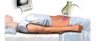
- Ultrasound examination of the kidneys and abdominal organs.
Additional diagnostic examinations that are carried out on an outpatient basis include daily proteinuria or protein/creatinine, albumin/creatinine ratios, immunological methods for studying blood serum, and determining the concentration of Cyclosporin-A in the blood serum.
If necessary, a computed tomography scan can be performed. Upon admission of a patient to the therapy clinic, nephrologists at the Yusupov Hospital prescribe the following mandatory diagnostic examinations:
- kidney biopsy;
- complete blood count (before and after kidney biopsy);
- general urinalysis (before and after kidney biopsy);
- daily proteinuria or protein/creatinine, albumin/creatinine ratios;
- ultrasound examination of the kidneys (after biopsy);
- coagulogram;
- morphological diagnosis of kidney tissue biopsy;
- immunohistochemistry.
Patients have the opportunity to undergo complex studies at partner clinics located in Moscow.
Additional diagnostic examinations that are carried out at the hospital level include electron microscopy of kidney tissue biopsy, determination of cryoglobulins in blood serum, electrophoresis of urine proteins, immunofixation of blood proteins for paraproteins, determination of the concentration of Cyclosporin-A in blood serum. If necessary, sternal puncture, fibroendogastroscopy, colonoscopy, and radiography of the flat bones of the skull can be performed. According to indications, computed tomography, Doppler ultrasound scanning of renal vessels is performed, and the level of tumor markers in biological fluids is determined. If a toxic origin of nephrotic syndrome is suspected, the presence and concentration of bismuth, lithium, paramethadone, and heroin in the blood are determined.
The patient is advised by specialized specialists depending on the suspected cause of nephrotic syndrome:
- oncologist - if paraneoplastic nephropathy is suspected;
- phthisiatrician – if a specific process is suspected;
- rheumatologist – if a systemic disease is suspected.
An ophthalmologist examines the fundus of the eye. The surgeon is invited for a consultation in case of nephrotic crises to exclude acute surgical pathology. A consultation with a neurologist is prescribed if neurolupus or systemic vasculitis is suspected. A hematologist examines patients with suspected multiple myeloma, a light chain disease. If a patient with nephrotic syndrome has a prolonged fever, he or she is consulted by an infectious disease specialist. Differential diagnosis of nephrotic syndrome is carried out with diseases that occur with severe edematous syndrome: liver cirrhosis, congestive chronic heart failure.
Nephrotic syndrome
Diagnosis is based on detected changes in blood and urine tests (proteinuria, hyperlipidemia, hypoproteinemia), and on clinical data. The MINS clinic develops gradually, and extrarenal symptoms predominate, especially edematous: increasing edema appears, first of the eyelids, face, lumbar region (later it can reach the degree of anasarca - widespread swelling of the subcutaneous tissue), genital organs, ascites, hydrothorax, less often - hydropericardium. Characterized by significant hepatomegaly due to liver dystrophy. The skin becomes pale (“pearly” pallor) in the absence of anemia, dry, signs of hypovitaminosis A, C, B1, B2, and degenerative changes appear. Brittleness and dullness of the hair may be observed, and cracks in the skin from which fluid leaks, striae distensae, may occur. The child is lethargic, eats poorly, develops shortness of breath, tachycardia, and systolic murmur at the apex (“hypoproteinemic cardiopathy”). A severe complication in patients with anasarca, i.e., severe hypoproteinemia, can be hypovolemic shock, which is preceded by anorexia, vomiting, and severe abdominal pain. In the observations of N. D. Savenkova and A. V. Papayan (1997), abdominal pain syndrome develops in 23.5% of children with hypoalbuminemia less than 15 g/l, and migrating erysipelas-like erythema in 33.3%, thrombotic episodes in 12.5 %, acute renal failure in 3.3% of children with the same severity of hypoalbuminemia, while nephrotic hypovolemic shock was noted only when the serum protein level was less than 10 g/l (in 5%). As the swelling subsides, the decrease in skeletal muscle mass becomes more and more noticeable. Blood pressure is usually normal, but up to 10% of children may have short-term hypertension. The serum albumin level in such children is less than 10 g/l.
The content of total protein in blood plasma (serum) is sometimes reduced to 40 g/l. The concentration of albumin and g-globulin is especially sharply reduced, while the level of a2-globulin is increased, i.e., severe disproteinemia is observed. The blood serum is milky in color and contains high levels of lipids, cholesterol, and fibrinogen. The level of nitrogenous wastes in the blood is usually normal, and the content of potassium and sodium is reduced. ESR is sharply increased (up to 50–70 mm/hour). Renal symptoms include oliguria with a high relative density (1.026–1.030) of urine and severe proteinuria. When studying glomerular filtration by endogenous creatinine, normal and even elevated values are obtained, but this is a false impression. If we take into account the degree of proteinuria, then glomerular filtration in MINS is always reduced.
The clinical picture, course and outcome of nephrotic syndrome, which complicated diffuse glomerulonephritis, differ from the MINS clinic. Urinary syndrome with MINS consists of the following symptoms: 1. proteinuria, 2. oliguria with high relative density of urine, 3. cylindruria.
Proteinuria in MINS is usually selective, i.e., blood plasma proteins with a molecular weight of less than 85,000 are found in the urine (albumin and its polymers, prealbumins, siderophilin, haptoglobin, transferrin, a1- and b-globulins, a1- and a2 -glycoproteins, etc.). In most cases, children with selective proteinuria have a better prognosis and are responsive to glucocorticoid therapy. In the genesis of proteinuria, impaired protein reabsorption in the renal tubules is also important. Nonselective proteinuria, when there are many large molecular proteins in the urine, is usually a consequence of the fibroplastic process, sclerosis, i.e., it is not typical for MINS. Let us remember that a healthy child over 4 years old can have up to 100–150 mg of protein in daily urine. Oliguria is associated with hypovolemia, hyperaldosteronism, and tubular damage. Due to proteinuria, the relative density of urine is increased, reaching 1.040. ADH is also highly active in the blood of patients.
Sometimes with nephrotic syndrome there is massive leukocyturia, caused by an immunopathological process in the kidneys. Leukocyturia is often short-term and is not associated with a bacterial infection, i.e., pyelonephritis. The frequency of detection of leukocyturia and erythrocyturia in MINS, according to various authors, does not exceed 10%.
If there is a large amount of protein in the urine, it can coagulate in the tubules, taking their shape; The fatty-degenerated renal epithelium is layered on this cast - this is how hyaline, granular and waxy cylinders are formed.
Edema. Massive and prolonged albuminuria in a patient with nephrotic syndrome ultimately inevitably causes hypoproteinemia, since protein loss exceeds the intensity of its synthesis. Hypoproteinemia leads to disruption of the Starling equilibrium between hydrodynamic, filtration and colloid-osmotic pressure. This leads to a predominance of fluid outflow from the arterial bed over inflow. Edema begins to appear when the albumin level decreases below 27 g/l of plasma and always develops if hypoalbuminemia reaches 18 g/l. Secondary hyperaldosteronism, typical of nephrotic syndrome, also plays an important role in the pathogenesis of edema. As a result, sodium is retained in the body, and therefore water, although there is hyponatremia in the blood.
Hypoproteinemia. The main cause of hypoproteinemia in patients with nephrotic syndrome is large losses of albumin in the urine and their movement into tissues. In addition, increased catabolism of albumin and disruption of the protein-synthesizing function of the liver are important. A decrease in the content of g-globulins in the blood of patients, primarily due to a violation of their synthesis. Hypoalbuminemia and hypovolemia, deficiency of anticoagulants - antithrombin III and proteins C and S, hyperfibrinogenemia, hyperlipidemia pose a threat to thrombotic disorders in patients with MINS.
Hyperlipidemia. Some authors associate an increase in the level of low and very low density lipoproteins, cholesterol and lipids (free fatty acids, triglycerides, phospholipids, etc.) in nephrotic syndrome with impaired liver function, others explain this phenomenon by a decrease in thyroid function. Due to the fact that intravenous administration of an albumin solution prevents the increase in hypercholesterolemia, it is assumed that the increase in cholesterol levels in the blood occurs compensatory due to a decrease in albumin content. Since lipidemia in the experiment can be obtained after ligation of the ureters, it is suggested that hypercholesterolemia and lipidemia in MINS are of renal origin and depend on damage to intermediary metabolism in the tubular enzyme system. In the genesis of hyperlipidemia, low blood levels of lecithin-cholesterol acetyltransferase, which is excreted in large quantities in the urine, and low lipoprotein lipase activity are also important. In MINS, types IIa and IIb of hyperlipidemia are usually diagnosed.
Disorders of phosphorus-calcium metabolism (hypocalcemia, osteoporosis, osteomalacia) are caused by impaired renal function, as well as the metabolism of vitamin D.
Disorders of the metabolism of iron and microelements with low levels of both iron and zinc, copper, and cobalt in the blood determine to a large extent the tendency of such patients to anemia, trophic skin disorders, growth retardation, and possibly immunodeficiency.
Blood viscosity in MINS is increased due to hyperlipidemia and increased platelet adhesiveness. At the same time, the levels of blood coagulation factors (procoagulants) and anticoagulation factors (antithrombin III, proteins C and S) are reduced, which explains the relatively low frequency of decompensated DIC syndrome in MINS.
Infections are one of the most common complications of MINS. Peritonitis was especially common, which in most cases was caused by pneumococci, but in 25–50% of cases by Escherichia coli.
The characteristic clinical and laboratory picture of MINS in the vast majority of cases (90–95%) in children 2–7 years of age makes it possible to make a diagnosis without a kidney biopsy. A good and rapid response to glucocorticoid therapy confirms the diagnosis. At the same time, it is advisable to determine the level of IgE in any child with nephrotic syndrome, to find out the presence of chronic persistent viral infections (hepatitis B, cytomegaly, herpes virus infections, etc.), since positive results significantly complement and modify therapy. The recurrent course of nephrotic syndrome is indicated by 2 relapses per year, and the frequently recurrent course is indicated by 3 or more relapses per year. Remission is stated in the absence of proteinuria or its value is less than 4 mg/m2 per hour and the serum albumin level reaches 35 g/l. A biopsy is indicated for children with nephrotic syndrome under the age of one year and over 12 years of age, because the incidence of MINS is very low in them.
Treatment
Treatment of nephritic syndrome has the following goals:
- achieving weakening or disappearance of signs of the disease;
- elimination of extrarenal symptoms (arterial hypertension, edema) and complications (electrolyte disturbances, infection, nephrotic crisis);
- slowing the progression of chronic kidney disease and delaying its terminal stage with the help of dialysis and kidney transplantation.
The patient, depending on the severity of nephrotic syndrome, is recommended free or bed rest (if the patient’s condition is serious and there are complications). Dosed physical activity – 30 minutes 5 times a week. Doctors recommend that patients stop smoking and drinking alcohol. The diet is balanced, with an adequate amount of protein (1.5-2g/kg), caloric intake according to age. In the presence of arterial hypertension and edema, limit the use of table salt (less than 1-2g per day).
Pathogenetic therapy after establishing a morphological diagnosis begins in the therapy clinic and continues on an outpatient basis. Significant edema is treated with diuretics. Diuretics are not prescribed for diarrhea, vomiting, or hypovolemia. For long-term edema, furosemide is prescribed intravenously as a single bolus or torasemide orally. To treat patients with refractory edema, a combination of loop and thiazide-like or potassium-sparing diuretics is used. If there is a high risk of developing nephrotic crisis, a combination of albumin and diuretics is used.
If the patient experiences resistant edema in combination with renal failure in the acute period of nephrotic syndrome, hemodialysis with ultrafiltration is performed until the critical state of renal failure is eliminated and diuresis appears.
Cardiologists at the Yusupov Hospital take a differentiated approach to the choice of treatment method for patients with nephritic syndrome. In the absence of symptoms of acute kidney injury, ACE inhibitors or angiotensin AT1 receptor blockers are prescribed for antihypertensive purposes, but not in combination. If high blood pressure persists, calcium channel blockers are used. If the patient has a rapid pulse or there are signs of chronic heart failure, beta blockers are additionally prescribed.
Pathogenetic therapy of nephritic syndrome depends on the morphological variant. It is prescribed only after a kidney biopsy and clarification of the morphological diagnosis.
When treating the disease, the disease that caused the nephrotic syndrome is also treated. If parasites or helminths are detected, antiparasitic (anthelminthic) drugs are prescribed. Patients with neoplasms undergo chemotherapy, radiation treatment, and surgery. For diabetic nephropathy, endocrinologists treat diabetes mellitus, nephrologists prescribe drugs that have a nephroprotective effect.
At the onset of nephrotic syndrome, patients are prescribed methylprednisolone or prednisolone. Upon achieving complete or partial remission, the dose of prednisolone is gradually reduced.
Patients who do not achieve complete or partial remission after taking a full dose of prednisolone for 16 weeks are defined as steroid-resistant. They are treated with combination therapy with cyclosporine-A and a minimal dose of prednisolone.
50-75% of patients with nephrotic syndrome who respond to steroid therapy experience relapses. If nephrotic syndrome reoccurs, prednisolone is prescribed. If relapses occur three or more times within one year or there is a steroid-dependent form, combination therapy with low doses of prednisolone in combination with one of the following groups of drugs is used:
- alkylating agents;
- calcineurin inhibitors;
- antimetabolites.
Patients with nephrotic syndrome and chronic kidney disease are prescribed statins. For resistant nephrotic syndrome, rituximab is used. Treatment of nephrotic syndrome in systemic lupus erythematosus is carried out depending on the class of lupus nephritis.
The pathogenetic treatment started in the therapy clinic is continued in the patient’s outpatient and home settings under monthly monitoring of the results of laboratory data (clinical blood test, general urine test, biochemical blood test). Proteinuria levels are monitored using test strips once every 1-2 weeks, and patients regularly measure blood pressure. If the amount of proteins in the urine increases, the protein/creatinine ratio is determined and immunosuppressive therapy is adjusted. If the patient does not respond to immunosuppressive therapy, a repeat kidney biopsy is performed in a therapy clinic.
If the cause of nephrotic syndrome is infectious diseases, antibiotic therapy is administered. Drugs are selected for each patient individually depending on the type of infectious agent. At a high risk of arterial and venous thrombosis, low molecular weight heparins are administered subcutaneously for a long time.
4.Treatment
Any therapeutic regimen for the treatment of nephrotic syndrome necessarily includes a strict diet with limited sodium intake (in particular, table salt); The emphasis in the diet is shifting towards potassium-containing and vitamin products. According to individual indications, which can be very different (or even opposite, which makes any attempts at self-medication dangerous), antibiotics, antihypertensive drugs, immunosuppressants, hormonal drugs, cytostatics, and means of normalizing protein levels in the blood are prescribed.


