Traumatologist-orthopedist (adults and children)
Bogatov
Victor Borisovich
22 years of experience
Highest qualification category. Doctor of Medical Sciences, Professor of the Department of Traumatology and Orthopedics, First Moscow Medical University. I.M. Sechenov.
Make an appointment
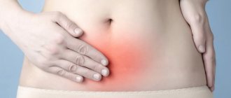
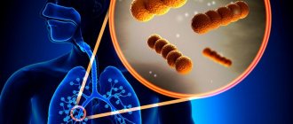
Tendonitis is a disease that manifests itself as an inflammatory process in the tendon area. Pathology can occur in acute or chronic forms. In the second case, degenerative processes develop in the affected tendon. In most cases, the source of inflammation is localized in the tissues adjacent to the bone. Symptoms of tendonitis are nonspecific: the patient suffers from pain when moving the limbs and local fever. The formation of mild edema and a zone of moderate hyperemia is possible. Treatment of pathology in the initial stages involves the use of conservative techniques. An advanced inflammatory process may require surgical intervention.
General information
Tendonitis occurs in patients of all ages. The risk group includes professional athletes and people engaged in monotonous physical labor. The source of inflammation is formed due to excessive stress to which the human tendons are exposed. Tissue microtraumas cause pain and increased local temperature.
Most often, the pathology affects the tendons located next to the elbow, knee and hip joints. Less commonly, inflammation develops in the ankle and wrist joints. Age-related changes in joint tissues lead to weakening of ligaments - the likelihood of developing inflammation increases. Patients over 60 years of age may suffer from the deposition of calcium salts, leading to the appearance of a calcifying form of the disease.
Reasons for the development of pathology
Symptoms of tendinitis identified by an orthopedist during an examination of the patient may indicate the causes of inflammation in the tendon. Often tissue microtraumas result from a high level of human motor activity. The pathology is common among professional tennis players, golfers, javelin throwers and skiers. The monotonous movements typical of gardeners, carpenters or painters often cause inflammation of the tendons.
A quarter of clinically diagnosed cases of tendonitis develop under the influence of other factors: rheumatic pathologies or diseases of the thyroid gland. Inflammation of the tendons can be a consequence of gonorrhea, intoxication of the body or abnormalities of the bone skeleton (different lengths of limbs, etc.).
Causes of tendonitis
Tendon tendinitis often becomes a consequence of pathological processes. The development of the disease is provoked by several factors.
Among the main ones:
- Infectious. Distribution through the bloodstream.
- Endocrine diseases. Malfunction of the thyroid gland.
- Physical. They can be post-traumatic.
- Chemical.
There are many more possible causes of the disease:
- immune imbalance;
- allergies to medications;
- infections caused by bacteria;
- frequent, excessive load on the muscle;
- anatomical features;
- joint diseases;
- rheumatic diseases;
- injuries;
- problems with posture;
- metabolic disorder.
The disease can strike at any age. But more often the diagnosis is given to people over forty years of age. As a rule, these are those who regularly experience heavy physical activity. The older a person is, the less elastic the tissues become, the greater the likelihood of developing the disease. Over the years, metabolic processes also change, which can lead to obesity, diabetes, and other diseases.
Pathogenesis
Tendons are formed in the form of dense, inelastic cords consisting of bundles of collagen fibers. Thanks to them, muscle tissue connects to bones. Tendons provide the transmission of movement from muscles to the skeleton and maintain stable joint function. Intense and monotonous movements interfere with the process of natural restoration of collagen fibers - the first signs of tendinitis appear.
The tendon structures swell and individual strands of collagen begin to break down. If the load remains high, foci of fatty degeneration, necrosis and deposition of calcium salts form in the tissues. Hardened calcifications lead to re-injury of the previously damaged area. The inflammatory process gradually spreads to the entire tendon.
Types of pathology
Tendinitis can affect tendons of any size - inflammatory processes develop in the tissues of the finger, hand, foot, elbow or knee. The classification of pathology used by orthopedists takes into account the localization of the source of pathology. Doctors distinguish the following forms of the disease:
- lateral,
- medial,
- inflammation of the patellar tendon,
- inflammation of the tendon of the shoulder joint.
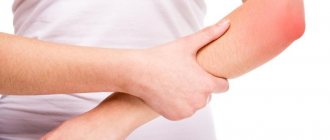
The lateral type of pathology develops in the tendons adjacent to the wrist muscles (extensor brevis and longus, brachioradialis muscle, etc.). The patient experiences pain spreading along the outer surface of the elbow joint. Over time, professional and amateur athletes develop hand weakness. A person begins to experience difficulty performing basic actions: twisting clothes, lifting a cup of drink, shaking hands.
The medial form of the disease affects the tendons adjacent to the flexor muscles of the forearm, elbow and palm. The pathology is diagnosed 7–10 times less frequently than lateral tendonitis and develops in individuals who regularly perform rotational movements of the arms. Those at risk include golfers, seamstresses, professional scorekeepers, gymnasts, tennis players and baseball players. The main symptom of the disease is acute pain localized on the inside of the elbow joint.
Inflammation of the patellar tendon is diagnosed in people who frequently perform jumps. Microtrauma is caused by short-term intense loads on the quadriceps muscle. In the initial stages of development of the pathology, pain occurs after the patient completes physical activity. Later, pain appears during exercise or at rest.
Inflammation of the shoulder tendons affects the tissue adjacent to the rotator cuff muscles. Pain occurs during any activity that requires hand mobility. Acute attacks can develop at night. An increase in the size of the focus of inflammation leads to the formation of pronounced edema.
Publications in the media
Tendonitis is an inflammation of tendon tissue, usually observed at the point of attachment to the bone or in the area of the musculotendinous junction; usually combined with inflammation of the tendon bursa or tendon sheath.
Etiology • Increased motor activity and microtraumatization • Diseases of a rheumatic nature •• Rheumatoid arthritis •• Gout •• Reactive arthritis.
Risk groups • Athletes • Manual workers.
Pathomorphology . Degenerative changes in tendons: presence of fibrinoid, mucoid or hyaline degeneration of connective tissue. Clinical picture
• Pain •• With active movements made with the participation of the affected tendon, while similar passive movements are painless •• With palpation along the affected tendon.
• Hyperemia, hyperthermia over the area of the affected tendon.
• Crepitus when the tendon moves, audible at a distance or only through a phonendoscope.
• Most common location •• Rotator cuff tendinitis, biceps tendonitis (see Periarthrosis humeroscapular) • • Lateral epicondylitis (tennis elbow) - tendinitis of the wrist extensor muscles (brachioradialis, extensor carpi radialis longus and brevis) ••• Pain when palpating the area of the lateral epicondyle of the humerus ••• Thomsen's test: when the patient tries to hold the hand clenched into a fist in the position of dorsiflexion, it lowers, moving to the position of palmar flexion ••• Belsh's test: the patient is given the command to simultaneously extend and supinate both forearms located at the level of the chin in a position of flexion and pronation, with the affected side lagging behind the healthy side • • Medial epicondylitis (“golfer’s elbow”) - tendinitis of the flexor and pronator muscles of the forearm (pronator teres, flexor carpi radialis and ulnaris, palmaris longus) ••• Pain on palpation of the medial epicondyle of the humerus ••• Pain with flexion and pronation of the forearm, radiating along its inner edge ••• Concomitant ulnar nerve neuropathy (25–50% of patients) • • Stenosing tenosynovitis of the extensor brevis and abductor longus muscles thumb (de Quervain's disease), accompanied by narrowing of the first canal of the dorsal ligament of the wrist ••• Pain when extending and abducting the thumb ••• Pain when palpating the styloid process of the radius ••• Elkin's test: pain when bringing the tip of the thumb together tips of the index finger and little finger • • Stenosing tenosynovitis of the extensor ulnaris (ulnar styloiditis) is accompanied by a narrowing of the VI canal of the dorsal ligament of the wrist ••• Pain in the area of the styloid process of the ulna ••• Swelling in the same area • • Tendonitis of the patellar ligament proper ••• Pain in the area of the tibial tuberosity when walking, running, going down stairs ••• Swelling in the area of the tibial tuberosity • • Tendinitis of the Achilles tendon and plantar tendons (talalgia) ••• Pain when stepping on the heel and when flexing the plantar ••• Local swelling - with concomitant achillobursitis and subcalcaneal bursitis.
• Children and teenagers. The most common form is patellar tendinitis associated with inflammation of the tibial apophysis (Osgood-Schlatter disease).
Research methods • Laboratory studies: changes are observed only with concomitant rheumatic pathology • X-ray examination •• Possible calcium deposits in the tendons •• Heel spurs - with tendonitis and tendobursitis of the Achilles tendon or plantaris tendon •• With tendonitis of the patellar ligament, signs of aseptic necrosis of the tuberosity are possible tibia (Osgood-Schlatter disease) • Special studies •• Echography of the tendon - contraction of the tendon, changes in its structure. It is necessary to ensure that the ultrasound wave does not cross the tendon along the oblique diameter. •• CT/MRI are informative for identifying tendon ruptures, but are not very informative in diagnosing stenosing tenosynovitis.
Differential diagnosis • Tendon avulsion • Bursitis (often combined with tendonitis should be remembered) • Infectious tenosynovitis (usually on the arm; pain on palpation and swelling are located along the tendon sheath, and not at the point of attachment to the bone).
Treatment • Management •• In the acute phase - rest, immobilization ••• Shoulder sling or splints for the upper extremities ••• Braces, cane and/or crutches for the lower extremities ••• Plasters placed tightly on the forearm slightly distal to the elbow joint - for epicondylitis •• Exercise therapy • Drug therapy •• NSAIDs ••• Piroxicam 10 mg/day ••• Indomethacin 25 or 50 mg 3 times a day ••• Ibuprofen 1800–2400 mg/day ••• Ointments with NSAIDs, for example ibuprofen, 3 times a day •• GK (injection into painful areas) ••• 40 mg of methylprednisolone with 4–6 ml of 1–2% lidocaine solution ••• 1–20 mg of hydrocortisone with the same volume of 1–2% solution of procaine. It is necessary to avoid insertion into the tendon sheath; in case of medial epicondylitis, the proximity of the ulnar nerve should be taken into account. After periarticular injections, despite a significant reduction in pain intensity, it is recommended to exclude physical activity due to the risk of tendon rupture • Surgical treatment - dissection of tendon aponeuroses, is used in the absence of the effect of conservative treatment, in the presence of signs of stenosing tendinitis, in Osgood-Schlatter disease.
Complication is tendon rupture.
The prognosis is favorable.
ICD-10 • M65 . 2 Calcific tendonitis • M75 . 2 Biceps tendinitis • M75 . 3 Calcific tendinitis of the shoulder • M76 . 0 Gluteal tendinitis • M76 . 1 Psoas tendinitis • M76 . 5 Patellar tendinitis • M76 . 6 Heel [Achilles] tendinitis • M76 . 7 Fibular tendinitis • M77 . 9 Enthesopathy, unspecified
Symptoms
Symptoms of the disease appear gradually. In the early stages, the patient experiences pain at the time of maximum physical exertion. The rest of the time, the injured tendon does not cause any discomfort to the child or adult. Over time, the pain syndrome becomes more intense: discomfort occurs with minimal physical activity. If left untreated, the patient may be unable to perform everyday activities such as washing dishes, putting on clothes, fastening buttons, tying shoelaces.
The skin next to the source of inflammation turns red and swelling forms. Local temperatures may rise. Palpation of the tendon increases the pain. With sudden movements of the injured limb, the patient may hear a crunching or crackling sound.
Achilles tendonitis
Achilles tendonitis (ICD 10 code in adults – M76.6) develops for the following reasons:
- Excessive load on the Achilles tendon (overstrain of the calf muscle, frequent running uphill or downhill, a sharp increase in the amount of physical activity);
- Using uncomfortable running shoes;
- Frequently wearing high-heeled shoes and changing heels to flat soles every night.
The patient experiences pain along the Achilles tendon, usually closer to the heel. Swelling and local redness of the skin appear in the tendon area. The pain intensifies when standing on your toes and jumping on your toes. Pain may occur within the first few minutes of walking in the morning. Mobility of the ankle joint is limited.
Diagnostics
Diagnosis of tendinitis is performed by an orthopedist. The doctor examines the patient and collects data for anamnesis. Confirmation of the primary diagnosis is carried out through radiography, ultrasound and magnetic resonance imaging of the injured tendon.
X-rays can reveal foci of calcification that have formed in collagen fibers. Ultrasound imaging of tendons demonstrates tissue thickening and decreased echogenicity. MRI is used to determine the location of the inflammation and its size.
Treatment
The treatment strategy for tendinitis is determined by the orthopedist, taking into account the data obtained during diagnostic procedures. In case of severe pain, the patient is advised to undergo short-term immobilization of the limb. Drug therapy includes anti-inflammatory drugs. Tissue swelling is eliminated through phonophoresis and electrophoresis.
Relief of the pain syndrome will allow the patient to begin therapeutic exercises. If acute pain persists, the child or adult receives glucocorticosteroid drugs by injection. The ineffectiveness of conservative therapy becomes an indication for surgical intervention.
During the operation, surgeons can perform complete or partial cutting of the tendons from the muscles for the purpose of subsequent lengthening. The postoperative rehabilitation period can last several months.
The treatment program for this disease at the Bone Clinic may include:
Shock wave therapy
More details
PRP therapy
More details
Acupuncture administration of ozone
More details
Teraquantum therapy
More details
Interference therapy
More details
Prognosis and prevention
Timely initiation of treatment for inflammatory processes in the tendons allows doctors to form a favorable prognosis for most patients. When the pathology is advanced, systematic relapses and severe injuries to the extremities are likely. Surgical treatment allows for complete restoration of limb mobility in 85–90% of cases.
Prevention of tendinitis is based on avoiding excessive stress on the tendons. Amateur and professional athletes should do a thorough warm-up before starting training or competition.