Morbidity statistics
Detection rate
Melanoma is quite rare - from 3% to 6% of the total number of malignant tumors. It is considered the most aggressive type of skin cancer, characterized by rapid growth and early metastasis. Of all deaths due to malignant skin tumors, it accounts for 80%. The average annual increase in the incidence of the disease in Russia is 3.9%.
Age and gender of patients
The tumor is mainly detected between the ages of 30 and 60 years, and also occurs in older people. Recently, the disease has become “younger” and is increasingly diagnosed in patients aged 24 to 40 years. The average age of patients is 52 years, more than a quarter of cases are detected in people under the age of 45.
Under the age of 40, neoplasms are more common in women. After 40 years, the incidence is higher in men and, as age increases, this trend becomes more pronounced - at the age of 65, men get sick twice as often, and at 80, three times more often than women.
Dependence on race and skin color
The risk of melanoma is highest in Caucasian people with fair skin. Hispanics, Asians and African Americans are less susceptible to the disease. At the same time, African Americans have the lowest survival rate.
Survival
The average mortality rate from melanoma in Russia is 2.13-2.52 per 100 thousand population. The average annual increase in the mortality rate is about 1.5%. Detecting a tumor in the early stages allows you to achieve almost 100% survival within 5-10 years.
How we treat
In addition to the enormous experience and professionalism of doctors, in addition to the latest technical base and the current range of drugs used, one of the significant advantages of treating melanoma in Euroonko clinics is the personal attitude of specialists towards this disease. Why for Andrei Lvovich Pylev the treatment of melanoma is an area of special scientific interest - in the following video:
Causes and risk factors
Melanoma develops due to malignancy (malignant degeneration) of melanocytes. No reliable causes of cell degeneration have been identified; every person is at risk of the disease. Factors that increase the risk of tumor development:
- hereditary predisposition;
- Phototypes I and II – fair skin, hair and eyes, pink freckles;
- multiple moles, age spots;
- excessive ultraviolet radiation - both natural and in a solarium;
- age over 50 years;
- endocrine diseases;
- previous melanoma.
The combination of any three of these factors is a reason for regular preventive visits to a dermatologist.
Symptoms
In most cases, melanoma develops from a mole (nevus). Early signs of a tumor include:
- horizontal increase in size of moles - growth in width over the surface of the skin;
- changes in the boundaries of the nevus - they become uneven, blurred, asymmetrical;
- change in the color of the mole, the appearance of heterogeneity (black, brown and other areas on one nevus), light spots in and around it.
These signs do not always indicate the development of skin melanoma. On the other hand, at a very early stage, the symptoms of the disease are easy to miss with the naked eye. Therefore, it is important to consult a doctor when the first signs or even suspicions appear. Timely treatment can save the patient's life.
Late symptoms:
- vertical growth - the formation rises above the surface of the skin;
- peeling, itching, pain, bleeding of the surface of the nevus;
- enlargement of regional and distant lymph nodes.
Sometimes people, especially those who are characterized by increased anxiety, begin to suspect melanoma, mistaking it for ordinary moles. There is another extreme - patients ignore the initial signs of melanoma, considering them a newly appeared or injured nevus, age-related pigmentation. It is important to distinguish a mole from a malignant formation.
Moles usually have a smooth surface and even, symmetrical edges. Their color varies from light brown to dark brown, it is uniform, without light or dark inclusions. The normal size of a mole is considered to be up to 6 mm in diameter (you can use a pencil eraser as a guide). Moles extremely rarely change their size, and they are not characterized by unpleasant or painful sensations.
General information
Melanoma (other designations used are melanoblastoma , melanosarcoma ) is a malignant disease, one of the types of skin cancer, which is formed as a result of mutational changes in cells that accumulate the pigment melanin .
This formation develops, as a rule, in open areas of the skin - on the skin of the extremities, back, etc., less often on the mucous membrane (oral cavity, rectum, vagina), and the retina of the eye. The ICD-10 code is C43 (malignant melanoma of the skin). Melanoblastoma is an aggressive formation that metastasizes . Experts note that this tumor can demonstrate a very wide variety of clinical courses. According to statistics, 89% of patients with this disease die in the first years after diagnosis. A favorable prognosis is observed only at the first stage of the disease. However, the outcome and prognosis of the disease largely depends on the condition of the patient’s body and the characteristics of the tumor. A pre-melanoma condition can be Dubreuil melanosis, as well as a number of other diseases - melanosis of the colon, sclera, etc.
The human melanoma gene was discovered in 1965 by US doctor R. Tuckington. But there are still many points regarding this disease that require further research.
Speaking about what skin melanoma is, it should be noted that this is a very serious disease that progresses rapidly. As evidenced by Wikipedia and other sources, how quickly this malignant process develops depends on a number of factors. This article will discuss how to recognize the symptoms of this terrible disease and what methods of treatment are currently practiced.
Types of skin melanoma
Superficial spreading
The most common type - about 70% of all cases of melanoma. Usually occurs against the background of a pigmented nevus. It is most often diagnosed in young and middle age (from 30 to 50 years), in 98% of cases it affects people of the Caucasian race. In women, superficial spreading melanoma is found more often than in men, and is mainly located on the legs. In men, the skin of the back is more often affected.
In the early stages, superficial spreading melanoma looks like a brownish spot of irregular shape with clear, even edges, slightly rising above the skin level. The color of the spot is uneven, with black and pinkish-gray splashes, and a reddish rim along the edge. The horizontal growth phase lasts from several months to several years. Then the neoplasm becomes more dense and convex, its surface turns black (depigmentation of the center is possible) and acquires a glossy shine. Foci of necrosis and bleeding appear on the surface of the spot. As the process spreads, small accompanying spots (satellites) appear around the spot, lymph nodes are involved in the process, and metastases occur.
If detected early, the prognosis is relatively favorable.
Nodular (nodal)
It ranks second in prevalence - from 14-15% to 30% of all cases of melanoma. More often it occurs on unchanged skin, without the presence of moles. Affects the scalp, neck and torso. The diagnosis of nodular melanoma of the skin is most often made between the ages of 40 and 60 years, and men predominate among patients.
The main difference between nodular melanoma and other types is the absence of horizontal growth. It grows immediately inward, vertically, penetrating deeply into the underlying tissue. The neoplasm is a dome-shaped exophytic node of dark brown, dark blue, black or grayish color with areas of ulceration or necrosis. It may look like a bleeding polyp on a stalk. Nodular melanoma quickly grows into the lower layers of the skin, metastasizes early to lymph nodes and organs, and has a less favorable prognosis than other types.
Acral-lentiginous (subungual)
The third most common type of melanoma. The only type of this tumor that most often affects people of the Negroid and Mongoloid races, located on non-pigmented surfaces: the nail bed, palms and soles. In Europeans, acral-lentiginous melanoma develops from complex nevi. Among them, people of phototypes I and II are more likely to get sick - fair skin, blond or red hair, freckles. In Asians and Africans, the tumor is localized under the nails in 50% of all cases of skin cancer, in Europeans - in 2%.
Acral lentiginous melanoma is diagnosed on average at 63-64 years in men, and at 67-68 years in women.
The characteristic difference between this type of tumor and others is that the provoking factor for it is not ultraviolet radiation, but other reasons: mechanical (injury), physical (frostbite, burns) or chemical (acids, alkalis, other aggressive substances) effects, as well as hereditary predisposition.
Subungual melanoma appears as a band of heterogeneous dark brown or black color, occupying more than a third of the nail plate. Pigmentation can spread to the nail fold, the skin of the phalanx at the free edge of the nail. Nail dystrophy develops, it becomes deformed, becomes thin, dull and fragile. Gradually, the nail cracks, revealing a lumpy, bleeding surface of brown or black color. The tumor can grow into the soft tissues of the hands, feet, and bones, causing severe pain.
The formation may be non-pigmented, which makes early diagnosis difficult. The survival prognosis is reduced by almost half when presenting at the second stage of tumor development.
Lentiginous (lentigo-melanoma)
Accounts for 5% to 10% of the total number of detected melanomas. It is more often diagnosed in women, but in them the tumor develops at a later age and is less malignant. In women, lentigo melanoma is detected on average at 60-70 years of age, in men - at 50-60 years.
In most cases, it affects exposed areas of the body: face, ears, neck, scalp, back of the hands. About 15% occur in other locations, mainly the back and lower extremities.
In the early stages, lentiginous melanoma looks like a pigment spot or freckle, not standing out on the skin. Its color can be different: from white, pink, yellowish to brown, more often it is heterogeneous. The color intensity increases as the tumor grows. The edges of the spot are uneven, clear, the surface is smooth and does not rise above the skin.
During the transition to the vertical growth phase, the boundaries of the formation are blurred and the color changes to black. The tumor rises above the skin, its surface begins to peel, crack, bleed, and itching appears.
Lentiginous melanoma has a long course - it can take from 2 to 20 years to reach the vertical growth phase; it metastasizes less often than others.
Pigmentless (achromatic)
It develops quite rarely, in 1-2% of cases. It is characterized by the absence of pigment, but can be light pink, reddish-bluish, bluish-pink. The lack of pigmentation makes diagnosis difficult. The tumor looks like a small, rough nodule on the skin, most often located on the toes, sole and heel. It quickly grows into the underlying tissues and, when disintegrated, forms an ulcer with hard, raised edges and a papillomatous bottom.
Spindle cell
A rare form of melanoma that most often develops in children and adolescents. Originates from spindle cells. It is a painless smooth or rough convex tubercle, small in size at an early stage, flesh-colored or pink. The absence of other signs of malignancy other than growth makes diagnosing this type of tumor difficult.
Etiology
The main causes of melanoma are based on two factors: uncontrolled exposure to solar radiation or artificial ultraviolet radiation through the use of tanning beds. Excessive passion for beach tanning or exposure to lamps with UV radiation entails a violation of the DNA structure of skin cells and increases the likelihood of cancer formation.
In addition, there are additional risk factors for skin cancer. Among them:
- Phenotype. People with light skin tones, blondes, and those with blue eyes are more likely to suffer from this disease.
- Weakened immunity increases the chances of developing skin cancer
- Having a large number of moles
- History of sunburn
- Xeroderma pigmentosum
- Age group over 50 years
- Heredity. If there were more than two relatives in the family suffering from a similar disease, this is a reason to turn to professionals for help.
- Presence of nevi (melanoform nevus is especially dangerous)
- Previously diagnosed melanoma
Pathogenesis
Medicine divides the development of melanoma into stages (according to Breslow):
- I – formation occurs only in the outer layers of the skin (in the epidermis, up to the basement membrane; thickness less than 2 mm).
- II – the tumor is found in the outer layers of the skin (thicker than 2 mm; may be characterized by the presence of an ulcer).
- III – the cancer process spreads to the lymph nodes near the affected area and/or extends beyond the primary tumor site (the border between the papillary and reticular layers of the dermis).
- IV, V – tumor cells are found in the reticular layer of the dermis, grow into fatty tissue, metastases spread throughout the body, with selective damage to organs.
Clark's microstage division includes thin, intermediate and thick (deep) forms of invasion.
Prognosis varies based on existing stages of melanoma.
If diagnosis and treatment occurred in the initial stages of the disease (I, II), then the prognosis is excellent and is 98.2% survival. Stage III leaves the chances of life within 61.7%. Stages IV, V (especially with metastasis) are characterized by a rapidly progressive and fatal course. As a result, less than 10% survive 5 years, and most patients die within a year of diagnosis.
Metastasis of melanoma occurs lymphogenously to the skin, lymph nodes and hematogenously to the liver, lungs, brain, bones, kidneys, adrenal glands. Trends in melanoma metastasis depend on the biological characteristics of the tumor. There are forms that metastasize for a long time only lymphogenously to regional lymph nodes. There are melanomas with a high malignant potential with a tendency to early hematogenous metastasis. Particular attention should be paid to such forms of skin metastases as satellite, nodular, erysipelas-like, and thrombophlebitis-like. Satellites are small multiple rashes near the primary lesion or at some distance from it in the form of spots that have retained the color of the primary tumor. The nodular form of skin metastases is manifested by multiple subcutaneous nodes of various sizes. The erysipelas-like form of cutaneous metastases appears as an area of swollen, bluish-red skin surrounding the tumor. The thrombophlebitis-like form of skin metastases is manifested by radially spreading painful compactions, dilated superficial veins and hyperemia of the skin around the melanoma.
Heredity of the disease
An independent study was conducted in Sweden, which involved 79,060 people with melanoma. Of these, only 9% turned out to be hereditary. Despite this low rate, the tumor is considered hereditary. It is assumed that a person inherits a violation of suppressors, which are responsible for suppressing oncological processes. Of the subjects with hereditary cancer, 92% were from families where there were already 2 cases of melanoma. Another 8% were among those with 3 or more cases in their family history. Scientists have also found that families with a history of melanoma have a significantly higher risk of other types of cancer.
Psychosomatics
Psychosomatics is a science that classifies all diseases as psychological. Conventionally, we can identify several mechanisms that influence the launch of the “self-destruction” mechanism - depression (primary and secondary), neurosis and trauma, situational psychosomatics (acute conflict, stress) and true (associated with our psychotype).
Stressful events
Why do we associate stress with cancer? Information about the “self-destruction” of the body is genetically embedded in us. When a person’s life begins to be dominated by various kinds of stress, conflicts, problems and seemingly minor troubles that do not find relief, quick resolution and compensation, sooner or later the person begins to be burdened by this situation psychologically, and physically his body constantly produces a stress hormone that significantly affects for immunity.
We live in a real physical body, in which real, sometimes independent of us, physical laws operate. Therefore, it is important to understand that in order for the disease to develop as it does, several factors must be combined. When we take a medical history and see in it a genetic predisposition to cancer, we additionally note the consumption of large quantities of food containing so-called carcinogens, the person lives in a certain environmentally unfavorable or radiation zone, when we observe other elements of self-destructive behavior: alcohol, smoking , self-medication with medications, a regime of stress (violence) on one’s body, and when we note problems of a psychological nature, only then can we say that the risk is really high. In this case, we consider the psychological factor as permissive. Indeed, in fact, in the body of each of us there are constantly those same immature, continuously dividing cells. But the principle of homeostasis is designed to prevent an increase in their number; every second our body works to maintain a healthy state.
Depression. Previously, depression was associated only with a reaction to the disease itself and treatment. However, patient stories have shown that often the disease can occur against the background of depression itself, or secondary, when a psychological disorder appears against the background of some disease.
So it is against the background of primary depression, when in the anamnesis of patients with oncology we see that they had previously been treated for depression. Moreover, experimental studies have shown that people suffering from depression have increased levels of a protein in their blood that is involved in the formation of cancer cells and the spread of metastases in the body. Moreover, one of the versions according to which oncology is classified as a so-called psychosomatosis is based precisely on the fact that often psychosomatic diseases are nothing more than a manifestation of somatized (hidden, masked) depression.
Neurosis and psychological trauma. Another option that we see in practice, although not in all patients, is also important and correlates with psychological trauma. More often, trauma that we remember but block on an emotional level manifests itself in organ neuroses, and here we would rather work not with oncology, but with cancerophobia.
Since we correlate true psychosomatics with constitutional characteristics (what is inherent in us by nature and does not change), more often this leads to the idea that oncology has a connection with some feelings, character traits, organs, etc. Indeed, we note that, for example, people with asthenic physique often have skin and lung cancer, but this is connected not so much with the person’s problems as with his personality. By the way, speaking about what decoding or significance this or that organ has in psychosomatics, we can immediately answer that most often there is none. In a hospital, people with the same diagnosis have completely different characters and psychological problems, any oncologist will confirm this to you. “The choice of tumor location” is more connected: with a constitutionally weak organ (where it’s thin, it tears – sometimes we talk about the risk of “breast cancer” for a woman whose mother had a tumor, but a woman can inherit her father’s constitution and our prognosis will not come true, and vice versa ); with the carcinogenic factors listed above (if a person smokes, then there is a higher probability of damage to the throat and lungs; if a person abuses medications and unhealthy food - to the stomach; sun/solarium - to the skin, but this is not the law and is considered with other components); with hormonal imbalance, in particular with the peculiarities of the production of neurotransmitters in a particular person at a particular point in time (each person needs a different amount of hormone to show a particular emotion and, by and large, although this depends on the constitution, it is also due to what happens in a person’s life) and even with age (each organ has its own history of development - renewal and destruction, so in different periods different cells can divide more intensively) or direct injury to the organ (often patients indicate that before the development of a tumor this area was injured (got a cold, hit, broke, broke), but we are talking about injury not as a cause of oncology, but as a localization, no need to be confused).
Is the disease contagious?
No type of cancer, including melanoma, can pass from a sick person to a healthy person. Only genetic predisposition is passed on in the family.
Stages of skin melanoma
There are four stages of melanoma development - 0, I, II, III, IV, of which I, II and III have substages A, B, C. The designations T is also used - the prevalence of the primary tumor, N - the absence, presence and prevalence of metastases in regional lymph nodes, M – absence or presence of metastasis to distant organs.
Staging is based on tumor size, thickness, rate of cell division, frequency and intensity of ulceration, and the absence or presence of metastases to lymph nodes and organs.
| Stage | Characteristic |
| 0 | Melanoma is only on the surface of the skin (in situ). Does not spread into underlying tissues. The lymph nodes are not affected, there are no distant metastases. |
| I (A, B) | At an early stage, the thickness of the tumor is up to 1 mm. There is no bleeding, ulcers or peeling. Low rate of cell division. There is no metastasis to lymph nodes and organs. |
| II (A, B, C) | Melanoma grows deep, its thickness is up to 2-4 mm. The surface of the tumor is hypertrophied, peels, ulcerated, and may bleed. There are no metastases in lymph nodes or distant organs. |
| III (A, B, C) | Melanoma of any thickness with growth deep into the tissue, with or without ulceration. Regional lymph nodes are affected and may be enlarged. There are no metastases in the organs. |
| IV | The tumor affects organs (brain, lungs, liver), distant areas of the skin and lymph nodes. |
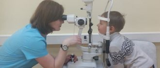
Dermatoscopy
Dermatoscopy is an examination in which a dermatologist uses a dermatoscope. This is an instrument that combines a magnifying glass 10 or 20 times, magnifying the fragment under study, equipped with a standardized light source. In such conditions, the doctor may initially assess skin changes.
Dermatoscopy
Videodermatoscopy is a study that, in addition to the techniques used in dermatoscopy, uses visualization on a computer monitor. This allows the clinician to store the displayed image in the device's memory, which can be useful for comparative purposes at a later stage in treatment. In practice, several dermoscopic methods are used to diagnose melanoma.
The doctor, based on the appearance of the lesion, will evaluate whether there is a reason to remove it along with a margin of healthy skin, and send the material after the procedure for histopathological examination.
The specialist studies the characteristic features of the mole, in particular:
- Color—uneven color, darker and lighter areas, or pink and red areas are abnormal;
- Ragged edges - moles with smooth edges are less dangerous to health;
- Asymmetrical shape;
- Size greater than 5 mm - smaller lesions are usually not a cause for concern, but if they are growing dynamically or show other abnormalities, they need to be removed;
- Bleeding, ulceration, presence of discharge.
Diagnosis that confirms or excludes a skin neoplasm, which is melanoma, is possible on the basis of a histopathological examination, which is carried out after removal of the entire lesion.
Diagnosis of skin melanoma
The first stage of diagnosis is an examination by a doctor, dermatologist or oncologist. The doctor performs dermatoscopy - examination of moles and other formations using a magnifying glass or a special device with multiple magnification to determine changes in nevi in the early stages.
If there is still a suspicion of malignancy, the patient is prescribed a biopsy and histological examination of a tumor tissue sample. Partial biopsy is practically not used to avoid the spread of cancer cells. Histology is performed after complete resection of the tumor.
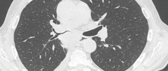
Computed tomography , magnetic resonance imaging , scintigraphy , and ultrasound are used to identify affected lymph nodes . The extent of the tumor process is determined by biopsy of the sentinel lymph node - located near the site of the lesion.
Treatment methods
The basis of treatment for both primary melanoma and its relapses is complete surgical removal with the capture of an area of unchanged skin. Depending on the stage, a section of healthy tissue ranging in size from 1 to 2-3 cm, subcutaneous tissue, and lymph nodes are removed if they contain metastases. Subsequently, if necessary, the removed part is restored with plastic surgery.
For metastatic melanomas, in addition to surgical treatment and for inoperable tumors, the following is used:
- polychemotherapy;
- radiation therapy;
- immunotherapy.
A relatively new method of treating metastatic melanoma of the skin is targeted therapy . Recommended for those patients whose tumor has a mutation in the BRAF gene. There are two types of targeted therapy: for patients with damage to regional lymph nodes and for patients with inoperable process and distant metastases.
The first group of patients is prescribed targeted therapy for prophylactic purposes. Therapy lasts a course of 1 year, is carried out in tablet form of drugs, and is combined with the main treatment.
For the second group, combined targeted therapy is used, acting directly on tumor cells. Treatment controls the process in 90% of patients, with a five-year survival rate of 34%. Others develop resistance and need immunotherapy.
Chemotherapy
Chemotherapy is usually prescribed in advanced stages, when surgical treatment of melanoma is not possible. Various chemotherapy drugs are used: dacarbazine, paclitaxel, carboplatin, cisplatin, vinblastine. Currently, chemotherapy is not used so often because new, more effective treatment methods have emerged:
- Targeted therapy.
They use drugs that block certain target molecules necessary for the survival and proliferation of tumor cells. Targeted therapy is more targeted than chemotherapy and causes fewer side effects. - Immunotherapy.
The most modern drugs for the treatment of melanoma are checkpoint inhibitors. They block molecules that cancer cells use to suppress immune activity. Checkpoint inhibitors are prescribed for advanced stages when other treatments are ineffective.
A modern type of treatment for melanoma of the arms and legs is isolated perfusion of the extremities. In this case, the arm or leg is, as it were, “disconnected” from the general circulatory system. A chemotherapy solution is supplied into the artery through a catheter, and the blood flows through the veins through the catheter into a special device. In order for the medicine to effectively destroy tumor cells, the solution is heated to approximately 42 °C. This type of chemotherapy is called hyperthermic.
Disease prognosis
Early detection of melanoma increases the likelihood of successful treatment and stable remission in more than 90% of patients. With timely diagnosis and a small thickness of the primary lesion, complete cure is likely in 90-95% of cases. Relapse with a tumor thickness of up to 1 mm and in the absence of metastases in regional lymph nodes occurs in approximately 8% of cases.
Overall survival of patients with cutaneous melanoma up to 5 years has increased due to early diagnosis and new treatment methods. Five-year survival data (if detected at the appropriate stage): I – up to 92%, II – 53-81%, III – 40-78%, IV – no more than 20%.
Prevention of melanoma
The main way to prevent melanoma is to protect the skin from UV radiation. Sunburn, even one-time and received many years ago, increases the risk of developing a tumor. Sunbathing should be done in the morning or afternoon, and always use sunscreen, wear a hat and dark glasses. Cosmetics with SPF in the spring and summer are mandatory even for residents of regions with low solar activity. Light skin, hair and eyes, a large number of moles on the body are reasons to completely give up tanning.
Solarium is always harmful to the skin. If it is not possible to exclude his visit, be sure to use sunscreen.
Periodically it is necessary to examine the skin, including those hidden under the hair, for the appearance of new moles or changes in existing ones. If a mole is injured or takes a long time to heal, there is a suspicion of uncharacteristic growth, or other changes, you should consult a doctor.
The information in this article is provided for reference purposes and does not replace advice from a qualified professional. Don't self-medicate! At the first signs of illness, you should consult a doctor.
Diet
Diet for cancer
- Efficacy: no data
- Duration: until recovery or lifelong
- Groceries cost: 2500 - 4800 per week
The diet of patients with melanoma should be light, low in calories, but still contain a sufficient amount of protein. The menu must include the following products:
- Lean meat - chicken, turkey, rabbit, beef, veal.
- Eggs – preferably quail.
- Vegetables – carrots, pumpkin, avocado, broccoli.
- Fruits – apples, citrus fruits.
- Cereals – buckwheat, lentils, wheat.
- Legumes - peas, beans.
- Dairy – cheese, milk, curds.
- Fish, seafood.
- Sesame seeds, flax.
- Nuts.
- Green tea.
It is better to cook by steaming, in the oven, using natural seasonings and spices. It is better to avoid fried foods. It is recommended to replace side dishes with boiled vegetables.
The following foods should be excluded from the diet:
- Margarine.
- Lard, animal fats.
- Sweets.
- Alcohol.
- Smoked meats, canned food.
- Pickles.