This oncopathology is usually registered in representatives of the stronger sex: men get sick 4 times more often than women. Seven out of ten cancer cases occur in people aged 65+.
Author:
- Sorokin Nikolai Ivanovich, head of the urological service of the university clinic of Moscow State University. M. V. Lomonosova
5.00 (Votes: 1)
Bladder cancer: diagnosis, treatment of stages 1, 2, 3. Symptoms, signs, metastases, prognosis.
Content:
- Overview of Bladder Cancer What is Bladder Cancer?
- Transitional cell carcinoma
- Cystoscopy is the main method for diagnosing bladder cancer
- Transurethral resection of the bladder
Disease prognosis
The survival prognosis depends on the stage at which the tumor is diagnosed and how timely and complete the treatment is. If a single epithelial lesion is detected and TUR is performed in conjunction with chemotherapy or immunotherapy, the chances of recovery are about 91%. In the second stage, without cancer spreading to other organs, timely transurethral resection and chemotherapy (less commonly, radiation) allow hope for recovery without relapse in 73-75% of patients. In the third stage and the process spreads to nearby organs, even after removal of the bladder and chemotherapy, there is a possibility of relapse; five-year survival is observed in 50% of patients. At the fourth stage, the process is considered inoperable, palliative methods are used, five-year survival rate is less than 7%.
Overview of Bladder Cancer
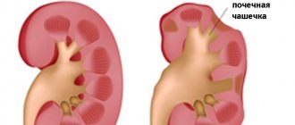
What is the bladder?
The bladder is a hollow organ located in the pelvic cavity, part of the urinary tract.
Urine, containing metabolic waste products, is produced by the kidneys and enters the bladder through the ureter. The bladder stores urine and then releases it through the urethra (urethra) when you urinate.
The bladder wall consists of three layers:
- the inner layer is called the mucosa. It is constantly in contact with urine accumulating in the bladder, providing tightness and protection against infection;
- the middle layer consists of muscle tissue. Due to this layer, the bladder is able to contract, expelling urine;
- The outer layer covering the bladder includes fibrous and fatty tissue, as well as blood vessels.
Prevalence.
RMP is the most common malignant tumor of the urinary tract and in terms of prevalence it ranks 7th in the structure of oncopathology in men and 17th in women. In the structure of cancer incidence in the Russian population, breast cancer ranks 9th among men and 16th among women. The incidence rate per 100 thousand population was 13.2 for men and 2.3 for women. There is a trend of increasing incidence for both sexes over the past 10 years, amounting to 28.3%. The standardized mortality rate for men and women was 4.7 and 0.5, respectively. The age composition is dominated by patients over 60 years of age, in Russia they account for 78.4%. BC occurs more often in men than in women (ratio 3:1), which is associated with a greater prevalence among men of smoking and occupations associated with carcinogenic substances, which increase the risk of developing the disease.
What types of bladder tumors are there?
Transitional cell carcinoma
Transitional cell carcinoma is the most common type of bladder cancer, accounting for 90% of all cases. About 70% of cases of transitional cell carcinoma are superficial tumors limited to the bladder mucosa.
These tumors rarely metastasize and in most cases are not life-threatening. In 30% of cases, invasive cancer develops, growing into the muscle layer of the bladder. Invasive tumors can grow into neighboring organs, metastasize and pose a threat to life.
Squamous cell carcinoma
Squamous cell carcinoma is rare and accounts for 1-2% of all bladder cancer cases. This type of bladder cancer is common in Africa and the Middle East. In 75% of cases, squamous cell carcinoma is caused by the parasite Schistosoma haematobium, which is widespread in these areas.
Adenocarcinoma
Adenocarcinoma (adenocarcinoma) - accounts for about 2% of cases and usually develops from the urachus, the urinary duct through which fetal urine is excreted into the amniotic fluid in utero).
Causes of tumor appearance
The mechanism of tumor development is active cell division. Such processes can be triggered by various factors that provoke mutation of the genetic cellular material. In the case of the bladder, smoking, unfavorable ecology and work associated with chemicals are especially dangerous. These factors can cause different types of bladder tumors:
- Benign: polyps, papillomas, fibroids, hemangiomas, fibromas.
- Malignant: solid, transitional cell, papillary and squamous cell carcinoma, adenocarcinoma, as well as superficial (non-invasive) and invasive cancer.
Benign tumors are often diagnosed in workers employed in the paint, paper, chemical and rubber industries. Stagnation of urine can provoke the disease. The higher its concentration and the longer it lingers in the bladder, the stronger the tumorigenic effect of the substances in its composition. In men, the causes of tumors are diseases of the genitourinary tract:
- BPH;
- prostatitis;
- stones in the urinary system;
- strictures and diverticula of the urethra.
Due to the peculiarities of the anatomical structure, men are more at risk of developing neoplasms. Malignant bladder tumors can develop for the same reasons:
- due to working in hazardous work;
- parasitic infections;
- prolonged urinary retention;
- infections and inflammation of the urogenital tract.
How common is bladder cancer?
Bladder cancer is one of the most common tumors, and is mainly detected at the age of 60-80 years. In the Republic of Belarus, more than 1000 new cases of bladder cancer are detected annually. Men get bladder cancer 4 times more often than women. This difference is explained by more frequent contact in men with external carcinogens (fuels and lubricants, petroleum products, metal processing), as well as a violation of the outflow of urine from the bladder due to an enlarged prostate gland. The prevalence of smoking is also observed to be higher in men than in women.
Prevention of bladder cancer
Quitting smoking and alcohol and eating plenty of fresh fruits and vegetables, which contain antioxidants that prevent cell degeneration, will help reduce the likelihood of developing a tumor. Bladder cancer is less likely to occur in those who drink enough water per day. It is believed that the risk of neoplasm is reduced by 7% for every 240 ml of added fluid.
It is necessary to be attentive to your health and consult a doctor when the first, even minor, signs of illness appear. It is important to monitor the regularity of urination, protect against genitourinary infections, and promptly diagnose and treat inflammatory processes in the urinary tract. Workers in hazardous industries should not neglect medical examinations. People over 40 years of age are recommended to undergo annual medical examination.
Sources
- Dolgikh D.V. Diagnosis of bladder cancer / D.V. Dolgikh [and others] // Siberian Medical Journal. - 2015. - No. 6. - P. 141-147.
- Dolgikh D.V. Bladder cancer (issues of etiology and pathogenesis) / D.V. Dolgikh [and others] // Siberian Medical Journal (Irkutsk). - 2015. - No. 7. - P. 26-30.
- Dunaev M.G. MRI diagnosis of bladder cancer / M.G. Dunaev // Remedium Volga region. - 2022. - No. 6 (156). — P. 22.
- Karyakin O.B. Bladder cancer: what's new in 2019-2020. / ABOUT. Karyakin // Oncourology. - 2022. - No. 4. - P. 147-154.
- Kulesh P.A. Study of clinical and morphological features of bladder cancer / P.A. Kulesh, A.A. Bagaeva // Eurasian Union of Scientists. — 2022.
- Nemtsova M.V. Molecular pathogenesis of bladder cancer / M.V. Nemtsova, N.E. Kushlinsky // Almanac of Clinical Medicine. - 2015. - No. 41. - P. 79-88.
- Startsev V.Yu. Modern possibilities of organ-preserving treatment of patients with muscle-invasive bladder cancer / V.Yu. Startsev [et al.] // Urological Gazette. - 2022. - T.9. - No. 1. — P. 29-38.
The information in this article is provided for reference purposes and does not replace advice from a qualified professional. Don't self-medicate! At the first signs of illness, you should consult a doctor.
What factors predispose to the development of bladder cancer?
Unfortunately, it is not always possible to answer the question of why one person gets bladder cancer and another does not. However, it is known that people with certain risk factors are more susceptible to this disease.
Research has identified the following risk factors for bladder cancer:
- smoking. Smoking has been shown to be the most important risk factor for bladder cancer. People who smoke for many years have a significantly higher risk than non-smokers or those who smoke for a short period of time. It has been proven that the likelihood of developing bladder cancer increases with the number of cigarettes smoked daily;
- chemical substances. Workers in the paint, chemical, metallurgical, textile, and leather industries have a higher risk of developing bladder cancer. Additionally, hairdressers, machinists, printers, artists and truck drivers are at increased risk;
- some cancer treatments. The risk of bladder development is increased in patients who have been treated for other cancers, such as after treatment with cyclophosphamide or after radiation therapy to the abdomen or pelvis.
Causes of the disease
There is no single and reliably proven cause of the tumor. Many risk factors contribute to the development of bladder cancer. Among them:
- Smoking. It is considered the main factor provoking bladder cancer. Approximately half of the detected cancer cases are associated with smoking; it is diagnosed in smokers 3 times more often than in non-smokers.
- Poor water quality and unfavorable environmental conditions. The chance of getting sick is increased by excess chlorine and fluorine in drinking water, and pollution of water and air by industrial waste.
- Abuse of alcohol, fried and fatty foods.
- Harmful working conditions – working with substances containing aromatic amines, phenols, phthalates. At risk are workers in the chemical, paint and varnish, rubber, oil, aluminum, leather and textile industries, artists, designers, and hairdressers. RMP often occurs in long-distance drivers due to prolonged sitting while driving, frequent hypothermia, and inability to urinate in a timely manner Source: Kulesh P.A. Study of clinical and morphological features of bladder cancer / P.A. Kulesh, A.A. Bagaeva // Eurasian Union of Scientists. — 2022..
- Non-infectious urogenital pathologies - chronic cystitis, urolithiasis, diverticulitis; genitourinary infections – chlamydia, mycoplasmosis, ureaplasmosis, trichomoniasis; parasitic diseases – schistosomiasis.
- Stagnation of urine or rare urination for various reasons - prostate adenoma, narrowing of the urethra, insufficient fluid intake.
- Radiation therapy for tumors of the cervix and ovaries, prostate gland, chemotherapy using Cyclophosphamide (increases the risk by 4.5 times), long-term use of phenacytin-containing analgesics (increases the risk by 2-6.5 times) Source: Kulesh P.A . Study of clinical and morphological features of bladder cancer / P.A. Kulesh, A.A. Bagaeva // Eurasian Union of Scientists. — 2022..
- In addition, the risk is significantly increased in people exposed to ionizing radiation Source: Kulesh P.A. Study of clinical and morphological features of bladder cancer / P.A. Kulesh, A.A. Bagaeva // Eurasian Union of Scientists. — 2022..
Main symptoms of bladder cancer:
- the presence of blood in the urine (hematuria) is the most typical, often the first and important symptom, especially if the blood appears without pain (urine may be rusty or dark red);
- feeling of urgent need to empty the bladder;
- having to strain when trying to empty your bladder;
- feeling of pain when trying to empty the bladder.
These symptoms may be associated not only with the presence of bladder cancer, but also with other diseases (infection, urolithiasis). If you have the above symptoms, you should immediately consult a doctor to identify the disease at an early stage and promptly begin treatment.
However, you should be aware that your urine may turn red if you eat beets, some berries, foods that contain food coloring, and some medications such as rifampicin (an antibiotic used to treat tuberculosis).
Symptoms of tumor diseases
Considering the symptoms of a bladder tumor, it is worth noting that at an early stage it does not manifest itself in any way. Signs occur when the tumor reaches such a size that it can irritate the walls of the organ. In such a situation, a person may have the following complaints:
- change in urine color;
- cramps and pain in the lower abdomen;
- feeling of a not completely emptied bladder;
- excretion of urine in small portions;
- frequent urge to urinate;
- disturbance of the menstrual cycle and discharge from the genital tract (in women);
- swelling in the legs, perineal area;
- disorders of the digestive system.
Diagnosis of bladder cancer
A tumor of the urinary tract can be suspected by the presence of an increased number of red blood cells during repeated urine tests. However, it must be remembered that such changes can be observed in other diseases of the genitourinary system, blood vessels, blood clotting disorders, etc.
A tumor in the bladder can be detected using ultrasound. A prerequisite for performing an ultrasound of the bladder is a well-filled bladder, for which you should gradually drink about 500 ml of liquid 30-60 minutes before the examination.
Cystoscopy is the main method for diagnosing bladder cancer
Cystoscopy is the main method for diagnosing bladder cancer. This is a procedure during which a urologist uses a special optical device to examine the inner surface of the bladder.
Cystoscopy allows the doctor to see the tumor and also take a piece of the tumor for histological examination. This test may not be very pleasant, since it is necessary to insert the cystoscope into the bladder through the urethra.
Local anesthesia may be required before the examination.
Questions and answers
How long do you live with bladder cancer?
Currently, over 95% of patients diagnosed with bladder cancer live longer than five years after treatment. The prognosis of treatment depends on the stage of tumor development and the type of cancer cells it consists of.
Is there a cure for bladder cancer?
Oncologists and urologists have become quite good at dealing with tumors in the bladder. After partial or complete removal of an organ, patients are able to return to normal life with certain restrictions.
How does bladder cancer manifest?
In three quarters of patients, the first symptom to suspect the presence of a tumor is hematuria - the presence of blood and urine. Blood turns the urine reddish, which forces a person to consult a urologist. Sometimes a blood clot blocks the urinary tract, preventing fluid from flowing out. For some patients, the first sign of the disease is enlarged lymph nodes in the groin area.
Attention! You can cure this disease for free and receive medical care at JSC "Medicine" (clinic of Academician Roitberg) under the State Guarantees program of Compulsory Medical Insurance (Compulsory Medical Insurance) and High-Tech Medical Care. To find out more, please call +7(495) 775-73-60, or on the VMP page for compulsory medical insurance
Surgical treatment of bladder cancer
There are several types of surgeries used to treat bladder cancer. In the early stages, it is possible to save the bladder by removing only the tumor and part of the bladder wall in the immediate vicinity of the tumor.
Transurethral resection of the bladder
This operation can be performed endoscopically (without an incision) - transurethral resection (TUR) or openly - open resection of the bladder. Transurethral resection of the bladder is used to treat early forms of bladder cancer.
In this case, no incisions are made on the body and the operation is carried out using a special instrument (resectoscope), which is inserted into the bladder through the urethra:
- Through the channel of the resectoscope, special optics are inserted into the cavity of the bladder and the image is displayed on the monitor screen.
- The tumor is removed using a wire loop at the end of the resectoscope through which a high-power electric current is passed.
Open resection of the bladder
Open bladder resection – may be performed for large superficial tumors or a single small invasive tumor:
- Open surgery involves making an incision in the lower abdomen, from the symphysis pubis to the navel, to access the bladder.
- During the operation, part of the bladder with the tumor and nearby lymph nodes are removed.
- Sometimes it is necessary to resect a section of the bladder at the junction with the ureter and transplant the ureter to another section of the bladder (ureterocystone anastomosis).
Radical cystectomy
For invasive cancer, the only radical treatment is to remove the entire bladder - a radical cystectomy. Radical cystectomy involves removing not only the bladder, but also the tissue adjacent to it.
In women, the uterus, ovaries, fallopian tubes, part of the vagina, and urethra are usually removed. In men, the prostate gland and sometimes the urethra.
After removing the bladder, the surgeon needs to create new pathways in the body to store and release urine. The operation to create a new pathway for urine to leave the body is called urinary diversion.
There are several ways to divert urine:
- One of the most preferred methods of urinary diversion from the point of view of restoring the function of “natural” urination is the creation of a urine reservoir from a section of the small intestine , which connects to the urethra and ureters and acts as a new bladder (ileocystoplasty);
- Sometimes, to drain urine , a section of the small intestine is used, one end of which is connected to the ureters, and the other is brought out onto the skin of the anterior abdominal wall in the form of a stoma, around which special flat urinals are glued for wearing under clothing - Bricker's operation (ileum conduit);
- The ureters can be brought out onto the skin of the abdomen, leaving tubes in them through which urine is released into a urine collection bag (urinal) - ureterocutaneostomy. The tubes can become clogged with mucus and blood clots, so you need to carefully monitor their condition and periodically rinse them. The tubes may also fall out of the ureter. In this case, you need to urgently contact a urologist.
Diagnostics
Programs for active detection (screening) of non-creatures. Diagnosis of bladder cancer includes
- Patient examination
- Physical examination
- Laboratory research
- Instrumental studies
- Ultrasound
- Cystoscopy
- MRI of the pelvis
- CT scan of the abdomen and chest
- Cytological examination of urine
- TUR biopsy.
- Morphological studies.
Cystoscopy can be performed as an outpatient procedure. Using a flexible cystoscope with transurethral injection of local anesthetic, better tolerability is achieved, especially in men. To prevent missing a tumor, a thorough examination of the entire epithelium lining the cavity of the bladder must be performed.
The use of fluorescence cystoscopy allows for a more accurate examination and a clearer determination of the boundaries of altered areas, especially in CIS (flat cancer)
Fluorescent cystoscopy is performed in violet light after intravesical exposure to a photosensitizer (aminolevulinic acid).
An alternative option may be narrow band imaging (NBI), which does not require the introduction of sensitizers.
Immunotherapy (BCG therapy)
For superficial bladder cancer, a treatment called immunotherapy is given. The essence of the treatment is to administer a solution of a specially created BCG vaccine into the bladder. BCG was originally developed as a vaccine against tuberculosis, but later activity was detected against bladder tumors.
The weakened bacteria included in the vaccine stimulate the body's own immune system, which destroys tumor cells. The use of the BCG vaccine reduces the likelihood of tumor recurrence by half.
Rehabilitation
Following organ removal surgery for bladder cancer, clinical recommendations include:
- special care for the area where the urinary catheter is inserted;
- enhanced drinking regime to flush out toxins and bacteria;
- avoiding alcohol, smoking, drinking drinks containing caffeine, as well as spicy and salty foods;
- exclusion of heavy physical activity;
- attending prescribed physical procedures, taking medications.
Chemotherapy
Chemotherapy is the use of drugs that kill cancer cells. For bladder cancer, several different ways of administering chemotherapy are used - intravesical chemotherapy, when the medicine is injected directly into the bladder, and systemic chemotherapy, when chemotherapy is administered intravenously:
- Intravesical chemotherapy targets cancer cells in the bladder and is used for superficial tumors to prevent tumor recurrence. When chemotherapy is injected into the bladder, side effects are usually minor. For several days after treatment, you may experience blood in your urine and more frequent and painful urination. These symptoms usually disappear after treatment.
- Systemic chemotherapy kills cancer cells throughout the body, not just the bladder, and is used for invasive or metastatic cancer. Chemotherapy is usually given in cycles. Each cycle has a treatment period and a rest period. Side effects depend on the chemotherapy drug and its individual tolerance.
Symptoms of bladder cancer
Signs of bladder cancer:
- Hematuria;
- Pain above the pubis;
- Dysuria.
At the initial stage, the disease is asymptomatic. The most characteristic early symptom of bladder cancer of all stages is blood in the urine. There are micro- and macrohematuria. Microhematuria is detected only by microscopic analysis; macrohematuria can be detected independently in the form of drops of blood in the urine. Doctors distinguish between terminal and total hematuria.
Terminal gross hematuria is detected at the end of urination and is observed in cervical cancer. Total gross hematuria is characterized by the release of blood clots throughout the entire act of urination. This type is determined by MP formations of any position. In this case, the color of the urine changes to bright red. In the final stages of formation of the neoplasm and its disintegration, urine takes the form of meat slop.
Bleeding often appears painlessly and suddenly, and can recur several times over several days. Blood clots can close the sphincter lumen and cause difficulty in the outflow of urine. Frequent hematuria leads to blood loss, anemia and weakness.
The course of bladder cancer is accompanied by urination disorders. Dysuria is the second most common symptom of bladder cancer. Patients complain of frequent, painful urination up to 10 times a day. As the volume of malignant growth increases, the capacity of the urinary organ and the number of bowel movements decreases, and the frequency of urge increases. When the lumen of the urethra is closed by a neoplasm or blood clots, urinary retention and an attack of renal colic are observed. Prolonged stagnation of urine causes the development of infections such as pyelonephritis and cystitis.
Pain in the first stages of the disease appears above the pubis and intensifies as the size of the tumor increases.
Pain from tumors in the bladder can radiate to:
- Crotch;
- Sacral area;
- glans penis;
- Anus;
- Lower limbs.
Common symptoms of bladder cancer:
- Persistent increase in temperature;
- Fast fatiguability;
- Exhaustion, weight loss;
- Sleep disturbance;
- The appearance of swelling of the legs, perineum, and scrotum in the later stages of the disease;
- Chronic pain in the suprapubic region;
- In the terminal stage, multiple organ failure syndrome occurs.
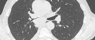
The following organs are affected by metastases in bladder cancer:
- Bone. Tumor cells lead to increased osteoclast activity;
- Lungs;
- Liver;
- Genital organs.
What kind of observation is required after treatment?
After treatment, recurrence of the tumor (relapse) may occur, the probability of which in superficial forms reaches 70%. A new tumor may appear in the same place as the primary one, and often relapses occur in another area of the bladder mucosa. Therefore, patients undergoing treatment for bladder cancer need to be examined every 3-6 months. One of the most important components of the examination is cystoscopy and urine cytology. The examination also includes an examination, blood and urine tests, and an ultrasound or computed tomography may be prescribed.
Diagnosis of bladder cancer
The most important factor in the successful treatment of such diseases is early diagnosis. The earlier the tumor is detected, the lower the risk of complications. BC can recur in 50% of cases, so not only diagnosis is important, but also complete removal of foci of cancer cells. The diagnosis is made based on anamnesis, physical examination, and the results of laboratory and instrumental diagnostics.
Diagnosis of bladder cancer includes:
- Analysis of patient complaints and subsequent physical examination. During the examination of the patient, the doctor palpates the bladder and possible areas of metastasis.
- Laboratory tests of blood and urine. General and biochemical blood tests do not provide the necessary information to establish a diagnosis of bladder cancer. A specific method is the determination of the UBS protein, the bladder cancer antigen. If the result is positive, the amount of protein is increased 15 times. However, false-positive results are possible in inflammatory diseases of the urinary system;
- Instrumental research. The most informative way to detect bladder cancer is cystoscopy. This invasive but highly informative method allows you to determine the size, location, shape and growth pattern of the pathological lesion. Cystoscopy provides the opportunity to take a biopsy - a piece of tissue for the differential diagnosis of cancer and a precancerous condition. If a biopsy is performed correctly, the procedure is not dangerous to the patient’s health and does not affect the rate of development of the tumor.
Using ultrasound of the pelvic organs, the shape, growth pattern, size of the pathological focus, as well as the presence of metastases in the lymph nodes are determined. The study is highly informative for tumor sizes greater than 5 mm.
Excretory urography displays the patency of the urinary tract, which may be impaired due to formation pressure or a blood clot.
CT and MRI for bladder cancer determines the presence of a tumor that has invaded nearby organs. CT helps determine the stage of bladder cancer formation according to TNM. To detect the spread of MP formation to neighboring organs, pelvic arteriography is used (study of the pelvic vessels after administration of a contrast agent).
Modern approach to cystectomy
In modern surgery, open cystectomy (through an incision in the abdominal cavity) is gradually fading into the background. Minimally invasive methods are becoming widespread: laparoscopic and robot-assisted cystectomy, which are performed through small punctures in the abdominal cavity. Minimally invasive methods can reduce the number of complications, blood loss and the risk of infection. The advantages also include: less intense pain, the absence of a rough postoperative scar and a short period of rehabilitation for the patient after surgery.
Among the minimally invasive methods, the high-tech robotic method should be highlighted. Additional benefits of da Vinci robotic cystectomy include5:
- No trembling of the surgeon’s hands during manipulations;
- Providing clear three-dimensional visualization of the surgical field, which allows for a clear view of the operated organ, neighboring structures and eliminates the risk of missing affected areas;
- Reducing the risk of bleeding due to effective hemostasis;
- The tools have 7 degrees of freedom of movement with the ability to rotate 90 degrees, which provides better access to hard-to-reach areas;
- The ability to operate on patients with severe obesity - in the deep pelvic areas in such patients, visualization is higher than with laparoscopy and open surgery.
Treatment of bladder cancer with robot-assisted method
– The robotic method is more convenient for cystectomy in general; it provides comfortable work in narrow spaces, such as the small pelvis. The robot helps to effectively perform orthotopic bladder surgery (creation of a new bladder): it should be noted the quality and convenience of applying an intracorporeal suture and tightness. This technology is also low-traumatic for the patient and ensures quick recovery. In obese patients, it makes it easier to cope with weight and provides better visualization, says Evgeniy Valerievich.
Reconstructive surgery after radical cystectomy
After removal of the bladder, it is necessary to create a new reservoir for the accumulation and removal of urine (diversion). For this purpose, reconstructive surgery is performed. There are several options for bladder reconstruction4:
- External urinary diversion (ureterocutaneostomy). The operation involves removing the ureters to the anterior abdominal wall with the formation of a stoma (an artificial opening between the organ cavity and the external environment). When choosing reconstructive surgery with the formation of a stoma, it is necessary to constantly wear a special bag on the anterior abdominal wall for collecting urine (urinal bag). Urine flows slowly, uncontrollably into the pouch, which the patient empties as it becomes full4.
- Diversion of urine into an isolated area of the intestine with the formation of a stoma. The most commonly used site for bladder replacement is the terminal ileum (a section of the small intestine). The ureters are connected to an isolated section of the intestine, which is brought to the anterior abdominal wall.
- Formation of a new reservoir for urine accumulation from a section of the intestine. To do this, the surgeon creates a new bladder from a piece of intestine, which is sutured to the urethra. This method allows the patient to urinate independently.
Reconstructive surgery with the formation of a new bladder from small intestinal loops
Evgeniy Valerievich explained what contraindications there are for performing reconstructive surgery.
– Reconstructive surgery is not indicated for everyone: we do not recommend this intervention for patients over 65 years of age due to the high risk of complications. Provided that the candidate is healthy (does not have diabetes, vascular diseases, etc.), we can perform the operation on a patient 65-70 years old. We also take into account the patient’s mental status: after reconstructive surgery, 6 months of regular training are required, and you need to wake up to an alarm clock at night. We must be sure that the patient can cope with the load.
Observation
In some patients, bladder cancer may disappear completely after treatment. Ending treatment can be both stressful and exciting. You may feel relieved to finish treatment, but you may find it difficult not to worry about the disease returning. This is very common among cancer patients. Life after bladder cancer means returning to some of the usual things as well as making new decisions.
Unfortunately, some patients will not be able to get rid of bladder cancer completely, and sometimes the disease may develop in another part of the body. Some patients may receive regular chemotherapy, immunotherapy, or other treatments to try to keep the cancer under control. Learning to live with terminal cancer can be difficult and very stressful.
After treatment, your doctors will continue to monitor you closely. People who have had bladder cancer have a high risk of developing bladder cancer again, so it is important to attend all follow-up appointments.
During follow-up visits, your doctors will ask questions about any problems you are experiencing and perform exams, laboratory tests, and tests to look for signs of cancer or side effects of treatment.
Your schedule for follow-up tests will depend on the stage and aggressiveness of the cancer, the treatment you received, and other factors. Be sure to follow your doctor's advice about follow-up tests.
The control examination includes:
- Doctor's examination every 3-6 months
- Cytological examination of urine
- Blood tests.
- Cystoscopy
- MRI of the pelvic organs.
- CT scan of the abdominal cavity and chest.
- Depending on your complaints, additional examinations and consultations with specialists may be prescribed.
If your bladder has not been removed, regular cystoscopy examinations will also be performed every 3 months for at least the first 2 years to determine whether the cancer has returned; there are no other tests that can replace cystoscopy.
If you have had your bladder removed and urine is being drained through a segment of bowel, you will be checked for signs of infection and changes in your kidneys. This may include urine tests, blood tests, and x-rays. Vitamin B12 levels will be checked at least once a year because urine drainage through the intestines may affect the absorption of vitamin B12
Storage of medical records
Even after treatment, it is very important to retain medical records. Tests and doctor's visits are very expensive, and even if no one wants to think about the cancer returning, it can happen.
At some point after cancer treatment, you may find yourself seeing a new doctor who doesn't know anything about your medical history. Having copies of earlier medical certificates will help the doctor better understand the course of the disease.
It is important to know that regular follow-up examinations are very important!
People who have had bladder cancer can still get other types of cancer. In fact, bladder cancer survivors are at higher risk of developing other types of cancer. Timely detection of tumor recurrence can significantly improve treatment results, reduce the manifestations of the disease, and preserve the body's reserves for treatment.
Immunotherapy
An important part of the immune system is its ability to avoid attacking normal cells in the body. To do this, the immune system uses “checkpoints”—proteins on immune cells that must be turned on (or off) to trigger an immune response. Cancer cells sometimes use these checkpoints to avoid being attacked by the immune system. But new drugs that target these checkpoints, called checkpoint inhibitors, may help restore the immune response against cancer cells. PD-1 and PD-L1 inhibitors Atezolizumab (Tecentriq) and avelumab (Bavencio) are drugs that target PD-L1, a protein on cells (including some cancer cells) that helps the immune system stop attacking them. By blocking PD-L1, these drugs enhance the immune system's response against cancer cells. This may shrink tumors or slow their growth. Nivolumab (Opdivo) and pembrolizumab (Keytruda) target PD-1, a protein on certain immune cells (called T cells) that normally helps these cells avoid attacking other cells in the body. Blocking PD-1 may allow the immune system to attack cancer cells, which can shrink some tumors or slow their growth. These drugs can be used in different situations to treat bladder cancer: Any of these checkpoint inhibitors can be used in people with advanced bladder cancer that has progressed after chemotherapy. Atezolizumab and pembrolizumab can be used in people who cannot receive chemotherapy (due to factors such as hearing loss, kidney failure, or heart failure). These drugs are given as intravenous infusions, usually every 2 to 6 weeks, depending on the drug.
Targeted therapy for bladder cancer
This is a new type of treatment for bladder cancer. Targeted drugs work differently than other treatments such as chemotherapy (chemotherapy). They may work in some cases where other treatments do not work. Targeted drugs also often have various side effects. FGFR Inhibitor Fibroblast growth factor receptors (FGFRs) are a group of proteins on bladder cancer cells that can help them grow. In some forms of bladder cancer, cells have changes in the FGFR genes (which control how much FGFR protein is made). Medicines that target cells with changes in the FGFR gene (called FGFR inhibitors) may help some people with bladder cancer. Erdafitinib (Balversa) This FGFR inhibitor can be used to treat locally advanced or metastatic bladder cancer that has certain changes in the FGFR2 or FGFR3 gene and continues to grow despite treatment with chemotherapy.
It is important to remember that treatment for bladder cancer depends on the stage, degree of differentiation of the cancer, and the size of the tumor.