Spasmophilia or infantile tetany occurs in young children between 3 and 24 months, with boys being more susceptible to the disease.
This pathological condition with increased neuromuscular excitability and the presence of clonic convulsive seizures is caused by a failure of mineral metabolism, namely calcium-phosphorus.
There are two forms of spasmophilia:
- hidden
- latent, especially noticeable by contraction of the muscles of the face, hand, and fleeting cessation of breathing during the period of application of external stimuli; - obvious
- manifest, very often arising from a latent form due to the minimum amount of vitamin D in the body, manifests itself as laryngospasm, carpopedal spasm and eclampsia.
This disease is directly related to rickets, as evidenced by the presence of hypocalcemia and hyperphosphatemia.
General description of the disease
The onset of the disease occurs at the same age as rickets, since these two diseases are etiologically and pathologically interrelated. The calciopenic state of tetany is accompanied by spasticity of the muscles of the face, larynx, and limbs.
In the modern world, 3.5–4% of children with obvious signs of rickets are susceptible to infantile tetany. Such a small percentage suggests that the number of severe cases of rickets has decreased.
In the case of an obvious form of spasmophilia, medical staff and parents must be fully prepared to assist the child in restoring vital functions.
The thetanoid state over a long period of time leads to disruption of the central nervous system, delayed mental development, and occasionally to death.
General clinical recommendations and prevention
With arthrosis of the temporomandibular joint, it is necessary to reduce the load on the joint. To do this, you need to restore the integrity of the dentition and periodically wear braces. If you are involved in (and cannot quit) contact sports (boxing, martial arts), be sure to wear sports mouthguards.
To restore blood circulation in the joint, it is recommended to slowly (!) open and close your mouth (without sudden or lateral movements).
You will also have to get rid of habits that create additional stress on the joint:
- chew gum vigorously;
- support your cheek with your palm;
- chew seeds, nuts, hard cartilage.
Osteoarthritis of the jaw joint is called a disease of suppressed emotions. The illness can be a consequence of divorce, dismissal, or critical life situations. The most severe forms develop in nice and non-conflict people who keep their own emotions to themselves. You need to learn to enjoy life and stop seeing the world in gray colors.
What triggers the development of the disorder?
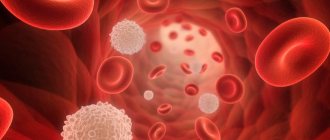
Due to a failure of calcium-phosphorus metabolism, a drop in the saturation of total and ionized calcium in the blood occurs, combined with alkalosis and hyperphosphatemia.
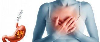
An increase in the amount of inorganic phosphorus is preceded by:
- feeding the baby with cow's milk, when excess phosphorus is partially excreted;
- overdose when taking vitamin D2 and D3 due to self-medication or due to uncontrolled prescription by a pediatrician;
- absorption of a large percentage of exposed skin from ultraviolet rays in the spring.
As a result of high levels of vitamin D metabolite:
- the quality of work of the parathyroid glands decreases;
- there is a change in the alkaline reserve of the blood, in advanced cases alkalosis occurs;
- the absorption of calcium and phosphorus in the intestine is stimulated or microelements are excreted in the urine in increased quantities, reabsorption of amino acids begins in the renal tubules;
- The baby’s bone tissue accumulates calcium above the limit, which is why its level in the blood decreases, and the amount of potassium increases, which leads to hyperkalemia.
Also, the causes of ultra-high muscular-nervous activity and convulsive readiness can be a decrease in the blood level of:
- magnesium;
- sodium;
- chlorides;
- vitamins B1 and B6.
In this case, any unfavorable factor - fear, screaming, crying, a viral disease, vomiting, a jump in temperature can provoke a convulsive attack.
Children who feed on mother's milk almost never suffer from spasmophilia; the risk group is artificial babies and premature babies.
Diagnostics
In the initial stages, arthrosis of the jaw is asymptomatic (more precisely, if there is pain, discomfort - they are attributed to a cold, problems with teeth, inflammation of the facial nerve, etc.). When constant pain appears, the face loses symmetry, it is impossible to chew - the patient begins to visit doctors.
Remember: at the slightest suspicion of TMJ arthrosis, you should consult a doctor; it is impossible to make a diagnosis yourself (if you are not an orthopedist or a healer).
In the clinic to confirm the diagnosis you will need:
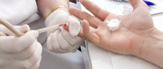
- take blood tests (clinical - to identify an infectious-inflammatory process, biochemistry - for arthrosis, biochemical parameters should be normal);
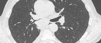
- take an x-ray in 2 projections (the image clearly shows the deformation of the osteophyte, the narrowing of the joint space, but the articular cartilage is not displayed in the image, and it is impossible to assess the degree of destruction of the TMJ in the early stages);
- undergo an MRI or computed tomography (MRI uses magnetic waves, and computer tomography uses X-rays, so in the early stages, MRI is an advantage).
Occasionally, an ultrasound of the joint is prescribed. In addition, a personal examination is required, because often it is necessary to treat not only arthrosis, but also to remove defects in the dentition, and to treat the accompanying inflammation of nearby tissues.
Clinical picture
Symptoms of spasmophilia will depend on the stage, form, course and type of disease.
Latent form
The latent form of the disease lasts from several weeks to several months; often these infants have all the symptoms of rickets:
- excessive sweating;
- tachycardia;
- anxiety at night;
- emotional lability;
- fear;
- malfunction of the gastrointestinal tract.
Manifest form
Eclamptic convulsions - begin with muscle twitching of the face, neck, limbs, and gradually take over the entire body. During the attack:
- unconscious infant;
- the face turns red;
- tachycardia, respiratory failure, general cyanosis occurs;
- uncontrolled urination and defecation;
- foaming from the mouth;
- due to increased intracranial pressure, the large fontanel is tense.
This situation lasts from several minutes to several hours.
Eclampsia is dangerous by stopping breathing or cardiac function.
Infantile tetany
Tetany is expressed in carpopedal spasm, lasting from 3–4 hours to several days, with:
- the child is overcome by severe pain;
- due to a long period of spasm, the limbs may swell;
- There is a bending of the arms to the maximum limits at the elbows and wrists, a shifting of the shoulders towards the body, and flexion of the hands.
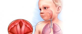
Development of laryngospasm
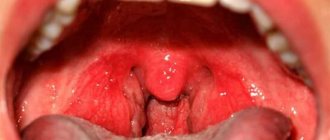
Laryngospasm or spasm of the glottis is a clear symptom of spasmophilia. It can develop during the process of crying or without obvious reasons; it happens up to several times a day. With moderate spasm, the child turns pale, a characteristic whistling breath appears, and when the gap is completely blocked, the following occurs:
- cold sweat;
- cyanosis;
- momentary unconsciousness;
- apnea;
- loud exhalation followed by noisy breathing;
- going to bed occurs.
The danger lies in possible death.
There are also local isolated spasms in the area:
- eye muscles – emerging strabismus;
- masticatory muscles – trismus, neck rigidity;
- smooth muscles - uncontrolled bowel movements and urination.
The obvious form of the disease overcomes the baby from a couple of days to 2 - 3 weeks.
Causes of arthrosis of the temporomandibular joint
Arthrosis can be triggered by a one-time injury (compression, blow, bruise), as a result of which cracks and erosions appear on the articular surfaces. The disease is caused by a fracture of the condyle and condylar process if the fusion is incorrect.
Other reasons:
- prolonged stress;
- consequence of acute traumatic arthritis;
- birth trauma (arthrosis develops due to improper application of forceps);
- underdevelopment of the jaw (microgenia);
- sudden removal of molars (accident, fight);
- errors during dental prosthetics;
- impaired coordination of muscle contractions during dislocation and subsequent sharp (jerky, zigzag, circular) movements of the jaw;
- complete absence of teeth;
- deep bite;
- introduction of drugs into the joint cavity (for example, hydrocortisone, glucose solutions, novocaine).
Structure of the TMJ
Etiological factors of arthrosis (without which the disease does not develop):
- infections;
- metabolic disease;
- injuries;
- atherosclerosis of the terminal branches of blood vessels;
- prolonged spastic contraction of the lateral pterygoid muscle (responsible for moving the jaw forward and to the side).
Even children are diagnosed with TMJ arthrosis. In newborns, the disease develops as a result of birth trauma. Dysfunction in the joint due to various malocclusions is observed in 40% of children from 4 to 14 years old, but in only 1% x-rays reveal coracoid (myogenic) arthrosis.
During menopause, the likelihood of developing arthrosis due to endocrine disorders increases. With age, it is possible to develop senile, i.e. invaluable arthrosis, when cartilage tissue cannot recover, dries out and collapses.
At risk are people whose professional activities involve inadequate load on the joint (violinists), or those suffering from spasms of the masticatory muscles (bruxism).
Diagnostics: instrumental methods and syndromes
Diagnosing manifest spasmophilia is not a complicated process; clinical and radiological signs of rachitic symptoms and confirmation of cases of convulsions and spasms are taken into account.
In case of latency, it is necessary to carry out mechanical or galvanic skin tests to determine the level of nervous and muscle activity, while identifying the presence of symptoms:
- Chvosteka
- when you tap with a finger or a hammer at the exit of the branches of the facial nerve - the cheekbones, a twitching occurs in the corner of the eye or mouth; - Trousseau's syndrome
- when the neurovascular bundle of the shoulder is compressed, the muscles of the arm involuntarily contract, bringing it into the “obstetrician’s hand” position; - Lyust's symptom
- tapping in the area of the head of the fibula, where the peroneal nerve exits, causes flexion of the foot with its abduction to the side; - Erba
- the impact of an electrical discharge on the median nerve of the elbow leads to compression of the fingers of the palm; - Maslov's phenomenon
is a reaction to a painful process, for example, an injection does not cause a long-term cessation of breathing; this does not happen to healthy children, since their breathing quickens at the moment of the injection.
How dangerous is the disease?
TMJ arthrosis is silent and unnoticeable; people live with the disease for years without even knowing about the problem. But in vain.
Degrees of TMJ arthrosis
In the Russian Federation, the Kosinskaya classification of arthrosis has been adopted, which takes into account both symptoms and radiographic data. However, the TMJ is an exception to the rule: the joint “hangs”, held by muscles and ligaments, and does not experience weight loads comparable to other joints.
When at stage 1 according to Kosinskaya the joint space narrows, the pressure on the jaw simultaneously increases, which leads to problems with the teeth, but maintains the distance. The process is gradual, so this moment can be recorded on an MRI, but since there are no symptoms characteristic of the disease in the initial stage, it cannot be said unequivocally that this is stage 1 arthrosis. Only at stage 2, when symptoms appear (pain, facial asymmetry, etc.), and the patient finally consults a doctor, is a diagnosis made.
Stage 3 according to Kosinskaya: absence of joint space, sclerosis, necrosis, inability to open the mouth, chew and speak.
Damage to the TMJ by arthrosis
Possible complications
Arthrosis is not only a problem of the joint. Compensatory, in an effort to maintain chewing function, the body redistributes the load, which leads to tooth loss and rapid wear.
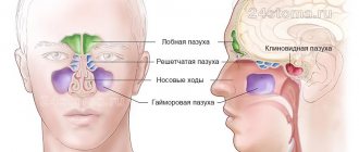
The previous diseases will be reflected in TMJ synovitis, and then the inflammatory process will affect the ear and nose (with decreased hearing, nasal congestion on one side), a headache will appear, which can radiate to the neck, back of the head and not stop.
The face will lose symmetry and become pasty (the skin appears loose, finely swollen, and grayish in color). Feeding is possible only through a tube; already at the second stage the ability to fully open the mouth is lost
Any localization and form of arthrosis has serious complications, so you should not delay treatment.
See how easily the disease can be cured in 10-12 sessions.
Exacerbations
Osteoarthritis is not arthritis; a chronic disease does not have periods of exacerbation. But this does not mean that the pain will be equally aching. The inflammatory process (cold, infection, virus) spreads to the joint with the development of synovitis. Swelling and pain appear, which can appear at any radial point (from the teeth to the back of the head). The source of inflammation expands, the oral cavity, ears, and breathing through the nose are at risk.
You need to understand that the brain is located nearby. And you shouldn’t wait for necrotic tissue to give rise to oncology.
Treatment of spasmophilia
If spasmophilia is at a latent stage, as certified by a specialist after diagnosis, emphasis is placed on changing the diet:
- a water break is arranged for 8–12 hours;
- infants are fed exclusively with mother's milk, if the baby is artificial they are looking for donor milk;
- you can add fermented milk liquid products;
- older children are prescribed a carbohydrate diet - pureed vegetables, porridge, tea with breadcrumbs, juices, vitamins;
- To completely get rid of the signs of the disease, children should be given calcium gluconate until its level in the blood is completely restored.
Antirachitic measures should also be carried out - gymnastics, long stays in the open air, massage of the whole body.
If spasmophilia is obvious, anticonvulsants are prescribed:
- Diazepam inside a muscle, into the root of the tongue or by injection into a vein;
- Sodium hydroxybutyrate intramuscularly;
- Phenobarbital - drink or in rectal suppositories;
- Chloral hydrate in an enema;
- 10% calcium gluconate solution intravenously;
- 25% magnesium sulfate solution.
To achieve a quick effect, you can use Seduxen or 20% gamma hydroxybutyric acid, the result is visible immediately, but the duration of its effect is only 30 minutes.
After normalization of the condition, barbiturate therapy is carried out for six months to a year.
In case of laryngospasm or eclampsia at the moment of respiratory or cardiac arrest, resuscitation should be carried out followed by mask inhalation of 100% oxygen.
To restore spontaneous breathing, you can spray your face with cool water, shake the baby, pat him on the bottom, and take him out of the room.
Types of arthrosis of the temporomandibular joint
For treatment to be effective, it is important to understand that there are several types of arthrosis of the lower jaw.
Deforming arthrosis
Osteoarthritis of the TMJ usually develops after injury. The clinical course depends on the nature of growth and the location of osteophyte proliferation (towards soft tissues or the articular cavity). If bone growth is directed to soft tissues, the disease is asymptomatic for a long time. If the osteophyte grows into the cavity of the glenoid cavity, local acute pain appears, which occurs with limited jaw movement. Clicking and crunching are dull, and sometimes popping sounds appear.
The joint becomes deformed with the growth of the condyle, changes occur in the synovial membrane and are accompanied by hemorrhagic synovitis. The reason for this is irritation of the TMJ, caused by the multiple presence of dead and rejected cartilage cells (intra-articular detritus). The synovial villi on the inner lining of the joint enlarge and fat is deposited in them. Occasionally, they degenerate, forming islands of bone and cartilage tissue (metaplasia), which are separated from the articular surface and form intra-articular free bodies.
Please note: this is not salt, it is osteochondral tissue. Therefore, folk remedies for arthrosis, which can still help with gout, do not work.
Viral and infectious diseases during this period inflame the joint membrane, accelerating the destruction of cartilage and bone.
Facial asymmetry does not appear in all patients diagnosed with arthrosis deformans. This depends on the compensatory capabilities of the neuromuscular complex and on the functional grinding of the articular surfaces.
Sclerosing arthrosis
Not only vessels can be sclerotic. With arthrosis, the 2 upper layers of bone become sclerotic (bone tissue is replaced by dense connective tissue). In this case, some compaction of the head occurs, followed by expansion. Since replacement is a slow process, the body manages to compensate for the changes. Therefore, the disease goes unnoticed in the initial stages.
Neoarthrosis (post-infectious arthrosis of the TMJ)
The disease is a consequence of an acute inflammatory process in the TMJ, with repeated acute respiratory viral infections and with the presence of dysfunctional jaw syndrome (luxation, neuromuscular, occlusal-articulatory). It is asymptomatic. With exacerbation of chronic inflammation, the following is noted:
- dull, aching pain that intensifies when moving the jaw;
- crunch;
- clicking in the HFNS.
X-rays show usuria (disappearance of osteochondral tissue), defects in the articulating surfaces of bones, and sometimes the complete absence of condyles.
Myogenic arthrosis of the TMJ
In orthopedics, there is a separate type of deforming arthrosis of the TMJ, myogenic. Its difference: a beak-shaped bone growth on the anterior surface of the condyle.
X-ray shows myogenic arthrosis, the contours of the articular surface due to osteophyte resemble a bird
Myogenic arthrosis occurs due to prolonged spastic tension of the lateral (lateral) pterygoid muscle. Its middle bundles are attached to the anterior-inner surface of the condyle and its process. Prolonged muscle spasm leads to a lack of coordination of muscle contractions, the bone beams change direction, stretch, positioned along the direction of the tendon traction. If the spastic contraction of the muscle continues, the bones that form the joint will begin to break down.
Differences from other forms:
- the condyle always has a beak-like shape;
- bone growth (osteophyte) is always localized in a specific place;
- no restrictions on jaw movement;
- the disease occurs without facial asymmetry.
The initial stages of the disease are asymptomatic. The osteophyte grows gradually on the anterior surface of the condyle, does not rub against hard tissues, and forms a bed in soft tissues. In the joint area, nutrition is disrupted, there may be a slight swelling on the face, spider veins - but very often this is explained by fatigue, overload, without paying attention to the TMJ. Painful symptoms occur at the moment of dislocation, subluxation of the lower jaw. Since the movement of the jaw in such cases is atypical, the osteophyte injures the soft tissues, irritating the nerve endings - severe pain appears (it hurts to chew hard food), severe swelling, clicking, mild swelling and paleness of the skin flap (pastyness). At the moment the mouth opens, the jaw begins to shift to the side.
Metabolic arthrosis
This is a rare type of disease that occurs when salt metabolism in the body is disrupted. The reason is needle-shaped crystals of uric acid settling in the TMJ. In patients, large joints are first affected; they suffer for a long time from metabolic polyarthritis, the visual manifestation of which is “gouty bumps” on the joints.
Symptoms:
- significant deformation of the head of the lower jaw, detected by palpation;
- asynchronous movement of the condyles when opening and closing the mouth;
- hinge movements on the side of the diseased temporomandibular joint;
- crunch;
- local dull pain;
- when opening the mouth, the jaw moves to the side;
- Lateral position of the head leads to facial asymmetry.
On radiographs with metabolic arthrosis, the condyle is covered with whitish needle-shaped curls of various shapes that are not permeable to x-rays.
Crunching in joints - when to worry
Intra-articular injections of hyaluronic acid
Senile arthrosis of the TMJ
Senile, or invaliable, arthrosis occurs with age. “Aging” of cartilage tissue occurs in 3 stages:
- cartilage tissue becomes soft and loose;
- loses some of the water, dries out, becomes denser;
- The smooth surface disappears, the cartilage becomes fragile and becomes covered with cracks.
After 60 years, bone exposure begins. Patients feel uncomfortable chewing and clicks are noted in the TMJ. The x-ray shows subtle changes.
Dangers and forecasts
Basically, the disease ends favorably. After 2–3 years, the pathology disappears due to the stabilization of the proportions of calcium and phosphorus in the blood.
In severe cases, not only proper treatment is important, but also compliance with a long prophylactic period. Death occurs extremely rarely due to suffocation during prolonged laryngospasm, cardiac or respiratory arrest.
With a long period of eclampsia, there is a deviation in the mental development of the baby.
To avoid such serious problems, it is important to provide the baby with the necessary amount of microelements in a timely manner, monitor his condition and behavior, and at the slightest sign of tetany, seek help from a pediatrician and pediatric neurologist.
Symptoms of TMJ arthrosis
Information about arthrosis of the temporomandibular joint on the Internet is 50% far-fetched descriptions of arthrosis of large joints, 30% is outdated data and obvious nonsense. And only 20% is true. Alas, texts are written by people without medical education, copying not from special educational literature or monographs, but from each other. Therefore, trust only trusted sources, and treat your health where there are no such ignorant things on the clinic websites.
First signs
A person may assume that he has arthrosis of the jaw when, after visiting doctors and following their recommendations, pain in the back of the head, ear, when chewing, hearing loss on one side, clicking, etc. does not go away.
Due to the structural features of the joint, the body manages to turn on the compensatory mechanism, so there is no long-term aching pain; due to the medications taken, it successfully disappears for a while.
Obvious symptoms
There are only 2 obvious symptoms (but it is also impossible to say 100% that this is arthrosis):
- displacement of the jaw to the side;
- pain when chewing.
You need to see a doctor immediately.
Frequently asked questions about the disease
Who treats arthrosis of the temporomandibular joint?
The treatment is complex. If there is no gnathologist in the medical institution, treatment is carried out by a surgeon or orthopedic traumatologist. In this case, a dentist, a neurologist, an otolaryngologist and, if necessary, a rheumatologist and an infectious disease specialist must be involved.
Is it possible to cure TMJ arthrosis?
If bone growths have begun, the process can be stopped, but it will not be possible to defeat the disease when the joint is young and healthy. But if you start treatment at least at stage 2 of the disease, you will be able to get rid of the symptoms, stop the destruction and even restore cartilage tissue.
Why is arthrosis of the TMJ dangerous?
Deformation in the joint leads to facial asymmetry, secondary inflammation spreads to the nasopharynx and ear. Due to spasmed muscles, teeth wear out and fall out. The skin on the face becomes pasty and ages quickly.
What is the difference between arthrosis and TMJ arthritis?
Arthritis is an inflammatory process in the temporomandibular joint of infectious-allergic, traumatic, autoimmune, etc. origin, which in advanced cases can lead to arthrosis. For example, a purulent infection (purulent otitis, boil in the ear canal, flu, sore throat, mumps, etc.) infects the joint fluid. The inflammatory process spreads to the joint capsule (the local temperature rises, the blood vessels of the heads of the bones grow and dilate). The purulent process then dissolves the cartilaginous surface and meniscus, and then destroys the bone tissue, leading to arthrosis. Arthrosis destroys the joint asymptomatically at the first stage and without an acute inflammatory process. The cartilage loses moisture, dries out, and cracks. The bone then grows, changing the structure of the joint.
Literature
- Evdokimenko P.V. Arthrosis
- Petrosov Yu. A., Kalpakyants O. Yu., Seferyan N. Yu. Diseases of the temporomandibular joint
Themes
Arthrosis, Joints, Pain, Treatment without surgery Date of publication: 10/08/2021 Date of update: 11/01/2021
Reader rating
Rating: 4.5 / 5 (2)