Despite the frequent use of the term “hypovolemia,” many doctors do not have an accurate idea of its meaning. Obviously, this is why they consider it possible not to use objective criteria when voicing this diagnosis; as a result, it turns into a subjective opinion. Moreover, slang expressions such as “underfilled” or “overfilled” are used in relation to the patient, referring to defects in infusion therapy.
Meanwhile, a violation of the patient’s volemic status is always a very serious symptom of trouble in the body, requiring objective confirmation and immediate therapeutic response, since volemia is the foundation of circulation, and therefore of exchange, that is, the very existence of the body. That is why a free interpretation of volemia can and does lead to great losses for the patient.
The terms “hypovolemia” and “hypervolemia” imply the presence of normovolemia, in comparison with which a decrease or increase in blood volume is determined. The search for the very concept of “normovolemia” continued for more than a hundred years, and now, finally, an almost generally accepted decision has appeared to consider normal only that volume of circulating blood (CBV), which in size corresponds to the capacity of the vascular bed. In this case, relaxed hemodynamics and breathing are observed, that is, there is no pronounced tachycardia, peripheral vasospasm, or increased respiratory movements.
Hypovolemia: a dangerous condition that requires immediate attention
What do we know about hypovolemia?
Hypovolemia is a condition when, as a result of fluid loss, the volume of blood that circulates in the human body decreases.
This violation poses a serious threat to the health and life of the patient. A severe degree can lead to hypovolemic shock and the development of multiple organ failure. Therefore, as a rule, the patient requires immediate medical attention: hospitalization and intensive care. Treatment is carried out in a hospital setting.
In rare cases, hypovolemia is caused not by the loss of fluid, but by its redistribution in the tissues of the body. At the same time, the filling of blood vessels with blood is reduced. This phenomenon is called relative hypovolemia. Blood loss and dehydration are absolute.
Reasons and mechanism of formation
The pathological hypovolemic process is based on severe dehydration.
Leads to it:
- Vomiting, characteristic of withdrawal syndrome after binge drinking.
- Diarrhea, with the help of which the body cleanses itself of alcohol toxins.
- Liver diseases characteristic of alcoholism (toxic hepatitis and cirrhosis). In this case, non-functional leakage of plasma into the intercellular space occurs due to a decrease in the oncotic pressure of the blood.
- Insufficient intake of fluid and protein into the body.
Hypovolemia is preceded by increased venous and hydrostatic pressure in small arteries, reflecting the negative influence of factors predisposing to a painful condition. Secondarily, the permeability of the vascular wall increases. Sweating of blood through arteries and veins leads to its general deficiency.
The body tries to compensate by:
- Producing the required amount of plasma.
- Slowing venous return.
- Spasms of small vessels.
The protective reaction allows you to maintain the activity of the brain and heart in a relatively normal state for some time and avoid circulatory complications.
What causes the condition
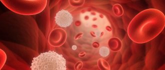
Most cases are associated with severe dehydration, blood loss, and large burns.
Dehydration can be caused by:
- repeated vomiting and diarrhea, excessive sweating
- insufficient fluid intake,
- taking medications, such as diuretics.
Hypovolemia that is caused by loss of plasma rather than blood is called polycythaemic.
Blood loss occurs as a result of injury, surgery, or internal bleeding. Blood loss associated with internal bleeding is considered especially dangerous, since it is not always possible to immediately determine where it is localized.
Massive blood loss is caused by:
- gastrointestinal bleeding,
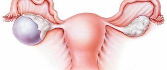
- rupture of the fallopian tube during ectopic pregnancy,
- rupture of an aortic aneurysm.
A general decrease in blood volume is called normocythemic hypovolemia.
Links[edit]
- Jump up
↑ McGee S (2018).
Evidence Based Physical Diagnosis
. Philadelphia, PA: Elsevier. ISBN 978-0-323-39276-1. OCLC 959371826. The term hypovolemia collectively refers to two distinct disorders: (1) volume depletion, which describes the loss of sodium from the extracellular space (ie, intravascular and interstitial fluid) that occurs during gastrointestinal bleeding, vomiting, diarrhea, and diuresis; and (2) dehydration, which refers to the loss of intracellular water (and all body water), which ultimately causes cells to dry out and increases plasma sodium concentration and osmolality. - "Definition of Hypovolemia - MedicineNet - Health and Medical Information Produced by Physicians". Medterms.com. 2012-03-19. Retrieved November 1, 2015.
- "Hypovolemia | definition of hypovolemia according to the medical dictionary." medical-dictionary.thefreedictionary.com. Retrieved November 1, 2015.
- Bhav G, Nelson EG (August 2011). "Volume depletion vs. dehydration: How understanding the difference can guide therapy". American Journal of Kidney Diseases
.
58
(2): 302–9. DOI: 10.1053/j.ajkd.2011.02.395. PMC 4096820. PMID 21705120. - ^ abc Jameson, J. Larry; Kasper, Dennis L.; Longo, Dan L.; Fauci, Anthony S.; Houser, Stephen L.; Loscalzo, Joseph, ed. (08/13/2018). Harrison's Principles of Internal Medicine
(20th ed.). New York: McGraw-Hill Education. ISBN 9781259644030. OCLC 1029074059. - "Hypovolemic Shock: MedlinePlus Medical Encyclopedia". medlineplus.gov
. Retrieved September 2, 2022. - Kolecki P (October 13, 2016). "Hypovolemic shock". Medscape
. - Danic B, Gouézec N, BigAnt E, Thomas T (June 2005). “[Cases of blood donation].” Transfusion Clinique et Biologique
(in French).
12
(2): 153–9. DOI: 10.1016/j.tracli.2005.04.003. PMID 15894504. - Taghavi S, Askari R (2019), "Hypovolemic shock", StatPearls
, StatPearls publishing house, PMID 30020669, retrieved 2019-09-02 - Carro A, El Hafny-Rahbi B, Matejuk A, Grillon C, Kieda C (June 2011). “Why is the partial pressure of oxygen in human tissue a critical parameter? Small molecules and hypoxia". Journal of Cellular and Molecular Medicine
.
15
(6): 1239–53. DOI: 10.1111/j.1582-4934.2011.01258.x. PMC 4373326. PMID 21251211. - Jump up
↑ Armstrong M, Moore RA (2019).
"Physiology, baroreceptors". StatPearls
. StatPearls Publishing. PMID 30844199. Retrieved September 2, 2019. - "Stage 3: Compensated Shock". Archived from the original on 2010-06-11.
- Alpert JS, Ewy GA (2002). Guide to Cardiovascular Diagnosis and Therapy. Lippincott Williams and Wilkins. paragraph 101. ISBN 978-0-7817-2803-4.
- ↑
Henry MS, Stapleton ER, Edgerly D (26 July 2011). Prehospital EMT Care. Jones and Bartlett Publishing. P. 471–. ISBN 978-0-323-08533-5. - Assuma Beevi (August 31, 2012). Child care plans. JP Medical Ltd. pp. 47–. ISBN 978-93-5025-868-2.
- ^ ab Clement I (20 May 2013). A Primer on First Aid and Emergency Medical Care. Published by Jaypee Brothers. P. 113–. ISBN 978-93-5025-987-0.
- Blaber A, Harris G (1 October 2011). Assessment skills for paramedics. McGraw-Hill Education. P. 83–. ISBN 978-0-335-24199-6.
- Hudson, Christy. "Hypovolemic shock - 1 Nursing CE". Archived from the original on 2009-06-06.
- "Stage 1: Waiting Stage (New Paradigm)". Archived from the original on 2010-01-16.
- Greaves I, Porter K, Hodgetts T, et al., eds. (2006). Emergency Care: A Textbook for Paramedics
. Elsevier Health Sciences. p. 229. ISBN 9780702025860. - ^ a b Agabeghi ED, Stephen SA. (2008). Step-Up to Medicine (Step-Up Series). Hagerstwan, MD: Lippincott Williams & Wilkins. ISBN 978-0-7817-7153-5.
- Kumar, Vinay; Abbas, Abul K.; Aster, John C., ed. (2015). Pathological basis of Robbins and Cotran's disease
. Illustrated by Perkins, James A. (9th ed.). Philadelphia, PA: Saunders. ISBN 9781455726134. OCLC 879416939. - Bulger, E.M.; and others. (2014). "Evidence-Based Prehospital Guidelines for the Control of External Bleeding: The American College of Surgeons Committee on Trauma." Prehospital emergency care
.
18
(2): 163–173. DOI: 10.3109/10903127.2014.896962. PMID 24641269. S2CID 15742568. - Takasu, A.; Prueckner, S.; Tisherman, S.A.; Stezoski, S. W.; Stezoski, J.; Safar, P. (2000). "Effects of increasing oxygen respiration in a volume-controlled outcome model of hemorrhagic shock in rats." Resuscitation
.
45
(3):209–220. DOI: 10.1016/s0300-9572(00)00183-0. PMID 10959021. - "Permissive hypotension". Trauma.Org. 1997-08-31. Archived from the original on 2013-11-27. Retrieved November 1, 2015.
- Kennamer WE, American Academy of Orthopedic Surgeons (AAOS) (September 30, 2013). Intravenous Therapy for Prehospital Providers. Jones and Bartlett Publishing. P. 63–. ISBN 978-1-4496-4204-4.
- ↑
de Franchis R, Dell'Era A (25 January 2014). Varicose bleeding. Springer Science & Business Media. P. 113–. ISBN 978-1-4939-0002-2. - Nordin, A. J.; Mäkisalo, H.; Höckerstedt, K. A. (1996-08-31). "Failure of Dobutamine to Improve Liver Oxygenation During Crystalloid Resuscitation Following Experimental Hemorrhagic Shock". European Journal of Surgery = Acta Chirurgica
.
Pubmed-NCBI. 162
(12):973–979. Retrieved November 21, 2022. - Geeraedts LM, Kaasjager HA, van Vugt AB, Frölke JP (January 2009). "Bleeding for trauma: a review of diagnostic and treatment options." Trauma
.
40
(1): 11–20. DOI: 10.1016/j.injury.2008.10.007. PMID 19135193.
Symptoms
The condition can be recognized by the following signs:
- dry mucous membranes
- pale skin
- decreased skin turgor
- rapid breathing
- lowering blood pressure levels
- tachycardia
- anxiety, confusion
- weak pulse (due to decreased cardiac output)
- thirst
- dizziness
- lack of urination for a long time.
Acute hypovolemia leads to an emergency condition - hypovolemic shock, which can result in coma.
4.Treatment
Depending on the situation, urgent resuscitation measures are taken. The primary objectives are to stop bleeding, restore breathing and replace blood loss through blood transfusion or transfusion of donor blood, and, if necessary, to relieve pain shock. In the future, measures are needed to normalize the acid-base balance, prevent thrombosis (DIC syndrome), prevent renal failure and secondary infections. Further management of the patient is determined by the results of the diagnostic examination and the dynamics of the condition.
Diagnostics
The main means of establishing a diagnosis is examination and interview (if possible) of the patient.
If hypovolemia is caused by visible trauma, large-area burns, its diagnosis is not difficult.
If a patient is suspected of dehydration or internal bleeding, the patient is sent for a clinical and biochemical blood test, a urine test, which will determine whether there is a change in the water-electrolyte balance.
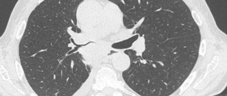
Also, to clarify the diagnosis, the doctor performs CT, X-ray, ultrasound and endoscopy of organs in which bleeding may develop.
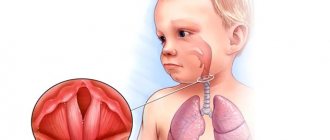
Hypovolemia in children
Danger of condition
The child's body is not sufficiently adapted to compensate for fluid deficiency. This feature should be taken into account when hypovolemia is suspected, since a state of shock in a child can occur much faster than in an adult. This is even more important the younger the child is. Thus, in children up to one year old, the ductus arteriosus may remain open, which, when fluid or blood is lost, actually deprives the lungs of blood supply. Therefore, the issue of hospitalization of the child is resolved immediately.
Causes
In newborns, hypovolemia can develop due to blood loss through the placenta, internal bleeding as a result of injury, or fluid accumulation in the abdominal or pleural cavity.
In older children, in addition to blood loss, the condition is caused by uncontrollable vomiting and diarrhea due to intestinal infections and intoxication; non-compliance with drinking regime.
Treatment
Treatment is prescribed based on the patient's condition.
Mild degrees require fluid replacement (drip, intravenous).
If the cause is injury, the first thing to do is stop the bleeding. Then restore normal hemodynamic parameters and breathing, while simultaneously treating the injury and pain relief for the patient.
Significant blood loss requires transfusion of plasma, red blood cells, platelets, and donor blood. Further therapy is aimed at preventing thrombosis, associated infections, and preventing renal dysfunction.
Signs of hypovolemic disorder
Symptoms of the disorder depend on its severity.
In the practice of narcology, the following stages of hypovolemia are distinguished:
- Easy. Minor fluid losses (up to 20% of bcc) are accompanied by a decrease in blood pressure, increased heart rate 10-15 beats higher than normal, and slight shortness of breath. The patient is pale, complains of weakness, dry mucous membranes, dizziness, and attacks of nausea.
- Average. The patient loses up to 40% of his total blood volume. In this case, the pressure is sharply reduced (below 90 mm Hg) and the pulse is above 120-130 beats per minute. Breathing is sharply rapid and intermittent. Externally, pallor and cyanosis of the skin in the area of the nasolabial triangle are revealed. There is severe sweating, lethargy, and decreased urine output due to severe thirst.
- Heavy. With a large-scale decrease in blood volume (above 40%), a critical drop in blood pressure is detected. The pulse is barely detectable and reaches 150 or more beats per minute. No urine is released. The patient's appearance indicates that he is in critical condition. There is deathly pale skin, a pointed face, sunken cheeks, and an unconscious state.
Loss of BCC is often accompanied by:
- Hypoglycemia is a serious complication of alcohol withdrawal syndrome. A harbinger of the development of hypoglycemia is any kind of liver disease, a decrease in glycogen reserves, and poor non-caloric nutrition.
- Hyponatremia is a disorder most often found in patients who abuse low-alcohol drinks (cocktails, beer). In this condition, peripheral edema occurs in combination with hypovolemia.
- Hypernatremia is a condition that occurs with encephalopathy, acute liver failure, and a clear deficiency in the amount of circulating blood.
- Hypomagnesemia is a deficiency of magnesium inside cells in people with alcoholism, regardless of its content in the plasma. The most common symptoms: general weakness of the body and drowsiness. The risk of seizures and heart problems (palpitations) may increase.
- Hypokalemia. In people suffering from alcoholism, as a rule, potassium deficiency is detected in the body, even if its content in the blood plasma is at normal levels. In combination with an overexcited nervous system, the lack of this element leads to irreparable disturbances in the functioning of the heart with a threat to life. Anyone suffering from alcohol withdrawal syndrome is prescribed potassium supplements.
This pathology manifests itself:
- Fatigue quickly.
- Depression.
- Attacks of tachycardia.
- Muscle weakness.
Alcohol withdrawal syndrome may be accompanied by metabolic acidosis, which occurs due to the accumulation in the body of breakdown products of alcohol and other substances of pathological metabolism, in the process of tissue hypoxia and respiratory alkalosis.
Drugs for hypovolemia
Treatment is carried out in a hospital, often in the intensive care unit. In most cases, infusion therapy is used.
The drugs of choice are solutions of colloids (solutions of gelatin and dextran, for example, rheopolyglucin) and crystalloids (Ringer's solution), blood substitutes (voluven, refortan).
To avoid infections, the patient is prescribed broad-spectrum antibiotics.
To raise the patient's blood pressure, norepinephrine and dobutamine are prescribed.
List of used literature
- Savelyev V.S. Comparative effectiveness of plasma substitutes for normovolemic hemodilution and correction of acute blood loss / V.S. Savelyev, N.A. Kuznetsov // Vestn. surgery. –1985.
- Bryusov P.G. Emergency infusion-transfusion therapy for massive blood loss / P.G. Bryusov // Hematology and transfusiology. –1991. – No. 2.
- Pshenisnov K.V., Aleksandrovich Yu.S. Massive blood loss in pediatric practice. Hematology and transfusiology. 2020;65(1):70-86
- Pleskov A.P. Invasive monitoring of central hemodynamics. // Intensive care, ed. V.D.Malysheva, M.: Medicine, 2002. P. 175–190.
Frequently asked questions about hypovolemia
What diseases can cause hypovolemia?
The condition occurs with gastrointestinal diseases, accompanied by uncontrollable vomiting and diarrhea, kidney disease, sharp drops in blood glucose levels, and some allergic reactions. However, hypovolemia is most often caused by blood loss or extensive burns.
How does hypovolemia manifest?
The patient has dry mucous membranes, tachycardia, thirst, and impaired skin turgor. As the condition worsens, a drop in blood pressure, weak pulse, and rapid breathing occur. In the acute stage - confusion.
How is hypovolemia treated?
The main method of treatment is fluid replacement. In most cases, intravenous administration of saline or electrolyte solutions is used.