Edited by: Kirgizov K.I., Deputy Director (Research Institute of Early Childhood Education, Federal State Budgetary Institution "N.N. Blokhin National Medical Research Center of Oncology" of the Ministry of Health of Russia) for scientific and educational work, pediatric oncologist, leading researcher, Ph.D. .
Childhood leukemia is a malignant disease of the blood system. One of the features of leukemia is that its manifestations at the initial stage can be mistaken for signs of other diseases. Today we will talk about how leukemia manifests itself in children, how to recognize it by the first symptoms and which specialists to contact so as not to waste precious time.
What is leukemia in children
Statistics show that leukemia is the most common cancer pathology in childhood (30%).
Most often, blood cancer is diagnosed in children aged 2 to 5 years. Classification of blood leukemia in children
Depending on the duration of the disease, there are:
- acute leukemia (up to two years);
- chronic leukemia (more than two years).
97% of cases in children are acute. A special type of it is congenital leukemia (observed in newborns or children in the first three months of life).
According to the criterion of morphological characteristics of tumor cells, acute leukemias are conventionally divided into:
1. Lymphoblastic. They arise as a result of the uncontrolled proliferation of immature lymphocytes (lymphoblasts). There are three types:
- with small lymphoblasts (L1) – the most common type in children;
- with polymorphic large lymphoblasts (L2);
- with polymorphic large lymphoblasts with vacuolization of the cytoplasm (L3).
According to antigenic markers, acute lymphoblastic leukemias are divided into:
- B-cell (1 to 3%);
- T-cell (15 to 25%);
- O-cell (70 to 80%).
2. Non-lymphoblastic (myeloblastic). Depending on the dominance of certain blast cells, types of acute myeloblastic leukemia are distinguished:
- undifferentiated (M0) – it is not possible to isolate a specific lineage of hematopoiesis modified by the tumor,
- poorly differentiated myeloblastic (M1);
- well-differentiated myeloblastic (M2);
- promyelocytic (M3);
- myelomonoblastic (M4);
- monoblastic (M5);
- erythromyelosis (M6);
- megakaryocytic (M7);
- eosinophilic (M8).
The clinical course of acute leukemia in childhood is described in three stages:
- Acute form (from the appearance of the first symptoms to a noticeable improvement in clinical and hematological parameters as a result of therapy).
- Incomplete/complete remission. In the first case, normalization of clinical parameters and hemogram is observed, the number of immature lymphocytes in the bone marrow puncture does not exceed 20%. With complete remission, this indicator is equal to up to 5% blasts.
- Relapse of the disease. Against the background of hematological well-being, extramedullary foci of leukemic infiltration are detected in the testicles, lungs, nervous system, etc.
LIST OF REFERENCES USED
1. Clinical guidelines of the Ministry of Health of the Russian Federation “Acute lymphoblastic leukemia”, 2022. 2. Bernbeck B., Wüller D., Janssen G., Wessalowski R., Göbel U., Schneider DT Symptoms of childhood acute lymphoblastic leukemia: red flags to recognize leukemia in daily practice. Klin Padiatr. Nov-Dec 2009;221(6):369–73. doi: 10.1055/s-0029-1239538. Epub 2009 Nov 4. PMID: 19890789 (https://pubmed.ncbi.nlm.nih.gov/19890789/). 3. Lyengar V., Shimanovsky A. Leukemia. StatPearls. Publishing LLC 2020. PMID: 32809325 (https://www.ncbi.nlm.nih.gov/books/NBK560490/). 4. Sokolova T.A., Kotlovsky Yu.V., Dubynina E.V., Ivanovskaya O.V., Veselova V.K., Kuznetsova E.Yu. Cytogenetic diagnostics for oncohematological diseases. Academy of Natural Sciences, 2012. ISBN: 978-5-91327-211-9. 5. Clinical guidelines of the Ministry of Health of the Russian Federation “Acute myeloid leukemia”, 2020. 6. Lee JW, Chung NG The treatment of pediatric chronic myelogenous leukemia in the imatinib era. Korean J Pediatr 2011 Mar;54(3):111–6. doi: 10.3345/kjp.2011.54.3.111. PMID: 21738540 (https://www.ncbi.nlm.nih.gov/pmc/articles/PMC3120996/). 7. Dvorak CC, Loh ML Juvenile Myelomonocytic Leukemia: Molecular Pathogenesis Informs Current Approaches to Therapy and Hematopoietic Cell Transplantation. Front Pediatr. 2014;2:25. doi: 10.3389/fped.2014.00025. PMID: 24734223 (https://www.ncbi.nlm.nih.gov/pmc/articles/PMC3975112/). 8. Davis AS, Viera AJ, Mead MD Leukemia: an overview for primary care. Am Fam Physician. 2014 May 1;89(9):731–8. PMID: 24784336 (https://pubmed.ncbi.nlm.nih.gov/24784336/). 9. National Cancer Institute, Advances in Leukemia Research, May 29, 2020 (https://www.cancer.gov/types/leukemia/research). 10. Whitehead TP, Metayer C, Wiemels JL, Singer AW, Miller MD Childhood Leukemia and Primary Prevention. Curr Probl Pediatr Adolesc Health Care. 2016 Oct;46(10):317–52. doi: 10.1016/j.cppeds.2016.08.004. PMID: 27968954 (https://www.ncbi.nlm.nih.gov/pmc/articles/PMC5161115/).
La Fondation La Roche-Posay and CCI (Childhood Cancer International) make every effort to ensure that the information provided is accurate and current at the time of publication. We are not responsible for information provided by third parties, incl. those that we refer to or highlight in this publication. The information in this publication should be used to supplement appropriate professional advice in your particular circumstances.
Causes of leukemia in children
It has been proven that acute leukemia is a “clonal” pathology. As a result of the mutation process occurring in the hematopoietic cell, its differentiation fails at the stage of immature forms (blasts) with their further growth (proliferation). A malignant tumor forms, replacing the bone marrow and preventing normal hematopoiesis. Blasts begin to leave the bone marrow, enter the blood and spread throughout the body. Leukemic infiltration of organs and tissues develops.
The origin of tumor cells from one mutated cell is proven by the fact that they all have identical morphological, biochemical and immunological characteristics. But what are the causes of leukemia in children and where does the mutant cell come from, scientists have not fully figured out.
It is believed that the following factors can trigger a pathological process in a child’s body:
- hereditary predisposition (if one of your relatives had leukemia, the disease may manifest itself in subsequent generations);
- genetic disorders (Down syndrome, Li-Fraumeni syndrome, neurofibromatosis, etc.);
- radioactive effects on the child’s body (man-made accidents, atomic explosions);
- increased solar insolation caused by ozone holes;
- poor environmental conditions;
- passive smoking (the child constantly inhales air containing smoking products);
- disruption of DNA structure caused by viral infections.
Why does malignant pathology appear?
Scientists are still unclear about some of the causes of leukemia in children. However, there are some theoretical and practical justifications for answering the question of why children suffer from leukemia. The following causes of leukemia in children are distinguished:
- Genetic predisposition. Pathological genes are formed as a result of intrauterine chromosomal changes, which produce substances that prevent the maturation of healthy cells.
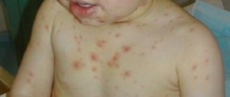
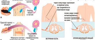
- Viral infection of the body. As a result of diseases of viral etiology suffered by a child, for example, chicken pox, mononucleosis, ARVI, etc., viruses are integrated into the cellular genome.
- Immunodeficiency. The immune system cannot cope with the destruction of foreign organisms and ceases to destroy its own pathological cells, including malignant ones.
- Radiation causes mutations in blood cells. Risk factors include maternal exposure to radiation (X-rays, tomography) during gestation, as well as living in a radioactive zone.
- Adverse habits of parents, especially mothers. Smoking, drinking alcohol and drug addiction.
- Secondary leukemia after radiation or chemotherapy for another cancer.
Leukemia also develops in children due to the formation of ozone holes as a result of active solar radiation. The causes of leukemia in children also lie in genetic pathologies, such as Down syndrome, Bloom syndrome, etc., as well as polycythemia.
Symptoms of leukemia in children
In most cases, the first signs of leukemia in children are veiled. The child sleeps poorly, gets tired quickly, does not want to eat, and for unknown reasons his body temperature rises. Parents mistakenly associate such symptoms with viral infections and colds. Sometimes the first sign of leukemia in children is:
- hemorrhagic syndrome (bleeding into the mucous membranes and skin);
- sudden general intoxication of the body (occurs with weakness, sweating, fever, anorexia, malnutrition, vomiting, nausea).
To recognize leukemia in a child, you should pay attention to:
- the color of the skin, mucous membranes - they become almost white, acquire a jaundiced/earthy tint (photos of patients with leukemia always show very pale children);
- condition of the mucous membranes – due to infiltration, tonsillitis, stomatitis, and gingivitis occur;
- the size of the lymph nodes - they increase, lymphadenopathy develops.
Symptoms of acute leukemia also include:
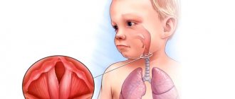
- sialadenopathy (pathological changes in the salivary glands);
- hepatosplenomegaly (simultaneous enlargement of the liver and spleen);
- hematuria (blood in the urine);
- uterine, gastrointestinal, pulmonary and nasal bleeding;
- hemorrhages into the joint cavity.
An invariable companion to acute leukemia in childhood is anemic syndrome. Its severity is determined by the degree of proliferation of blast cells in the bone marrow.
Symptoms of acute lymphoblastic leukemia can also be of a cardiac nature - arrhythmia, tachycardia, diffuse changes in the myocardium, expansion of the borders of the heart, and a decrease in ejection fraction are diagnosed.
Signs of an immunodeficiency state in leukemia are the addition of infectious and inflammatory processes to the underlying disease. Thus, death is often caused by pneumonia and sepsis.
If you notice similar symptoms, consult a doctor immediately. It is easier to prevent a disease than to deal with the consequences.
Treatment of leukemia in children and adults
The main types of treatment include: chemotherapy, radiation therapy, stem cell transplantation. Appointments are strictly individual. As additional treatment, detoxification and hemostatic therapy, transfusion of leukocyte and platelet mass, and prescription of antibiotics are used. Traditional medicine recipes can only be an addition to the main treatment and are used only after consultation with a specialist.
The prognosis for leukemia of different stages depends on many factors: the stage of the disease, the type of cancer cells, the patient’s age, his general condition, the presence of chronic pathology, etc. According to statistics, children and adult patients with early detection of the disease have a greater chance of recovery and remission.
Recurrent leukemia can occur only in adults. For children who received timely qualified help, cure occurs in 95%.
If you still have questions and need advice from a highly specialized specialist, make an appointment on the website or use the phone. Our doctors will help solve any health problem.
Related services: Chemotherapy course Consultation with an oncologist
How to identify leukemia in a child - diagnosis
Suspicions of leukemia in a child usually arise from a pediatrician. Further examination and treatment of the little patient is carried out by an oncohematologist. To make a diagnosis, it is necessary to carry out basic laboratory methods:
- bone marrow examination;
- peripheral blood examination.
A blood test for leukemia in children shows:
- anemia;
- reticulocytopenia (low content of young red blood cells);
- thrombocytopenia (low platelet count);
- high ESR;
- blastemia (release of blast cells into the peripheral blood);
- leukopenia (low white blood cell count);
- complete absence of eosinophils and basophils.
A classic symptom of leukemia in children is the phenomenon of “leukemic failure,” which is characterized by the absence of intermediate forms between mature and blast cells.
Mandatory procedures in the further diagnosis of leukemia:
- Sternal puncture. The procedure is performed under local anesthesia and is aimed at assessing the state of bone marrow hematopoiesis. The patient lies on his back. A Kassirsky needle with a special safety shield that prevents damage to the mediastinal organs is inserted at the level of 2-3 ribs in the midline.
- Myelogram study. To collect diagnostic material, a puncture of the sternum is performed at the level of the 2-3 intercostal space. The doctor uses a needle with a safety lock, so damage to internal organs is excluded. If the child is under two years old, a bone marrow sample is obtained by puncturing the tibia (the puncture is made in the upper third along the inner surface).
Additionally, when diagnosing leukemia:
- immunological, cytochemical and cytogenetic studies are carried out;
- consultations with an ophthalmologist and neurologist are prescribed;
- a lumbar puncture is performed (by inserting a puncture needle into the subarachnoid space of the spinal cord, the doctor takes cerebrospinal fluid (CSF) for further study);
- X-rays of the skull and ophthalmoscopy (examination of eye structures) are performed;
- Cerebrospinal fluid is examined.
According to indications, the following is performed: ultrasound of the liver, spleen, lymph nodes, salivary glands, scrotum in boys. In order to identify metastases, the child is prescribed a computed tomography scan.
During diagnostic procedures, it is important to differentiate leukemia from whooping cough, tuberculosis, cytomegalovirus infection, infectious mononucleosis, and sepsis. Full diagnostics can be performed in any modern hematology clinic
.
Medical Internet conferences
Early diagnosis of oncohematological diseases in children is extremely difficult due to the nonspecificity of primary symptoms, which are often hidden under the “masks” of other diseases [2, 27]. Oncological alertness and knowledge of probable symptoms will allow the practitioner to diagnose this pathology earlier and, therefore, significantly improve the prognosis for the patient [1, 3, 4, 5].
In most children, acute lymphoblastic leukemia (ALL) manifests rapidly and is characterized by clinical polymorphism. The preleukemia stage does not have characteristic clinical symptoms [6, 7], and the analysis of the symptoms of the initial period is carried out retrospectively [7], and therefore the diagnosis is not made on time. Despite the progress of laboratory diagnostics, it is difficult to detect leukemia in children in the early stages, because For characteristic changes in the hemogram to appear, the tumor must reach a critical mass [4], and the manifestation of ALL is associated with blast infiltration of various organs with disruption of their function [8]. Clinical symptoms in this case are ahead of laboratory changes, and the child is not hospitalized in a specialized hematology hospital [55].
ALL occurs predominantly in children aged 0–15 years, with a peak incidence observed at the age of 2–5 years [54]. A more favorable prognosis and effect of therapy is observed in children from 1 to 10 years of age, while in children younger than 1 year and older than 10 years, ALL has an unfavorable course and is difficult to treat [46].
Gender differences:
Studies aimed at studying gender differences in ALL showed that in B-ALL the gender distribution was the same: boys - 51%, girls - 49%, while the T-variant was found predominantly in boys (90%).
Constitutional features. There is data in the literature on the connection between the onset and course of the initial period of ALL with the constitutional type of the child (asthenoid, thoracic-muscular, digestive): the most severe course is observed in children with a digestive body type [55].
Predisposing factors. When diagnosing ALL, the doctor must take into account a wide range of predisposing factors that can lead to the development of ALL: socio-economic status of the child’s family, chromosomal abnormalities, Down syndrome, neurofibromatosis, variable immunodeficiency, Ig A deficiency, Fanconi anemia, Shwachman syndrome, congenital X- linked agammaglobulinemia [56].
In the clinical picture of acute lymphoblastic leukemia, 5 main syndromes are most common (Table 1):
- Intoxication syndrome. Most often, patients are concerned about prolonged fever of unknown origin, intoxication of varying degrees, weakness, fever, malaise, and weight loss. Fever may also be associated with the presence of a bacterial, viral, fungal or (less commonly) protozoal infection, especially in children with neutropenia [7].
- Osteoarticular syndrome. Manifestations from the musculoskeletal system are detected mainly in children with B-cell ALL, which is characterized by less involvement of extramedullary organs and unclear changes in the peripheral blood [18]. Osteoarticular syndrome is often the first manifestation of ALL in children [9, 10]. Studies have shown that in patients with ALL, osteoarticular syndrome was recorded in 54% of cases, with a higher frequency in children from 1 to 9 years of age. The presence of joint pain was noted in 16.2% of children with ALL, arthritis in 26.6%, and changes in gait in 32.8%. Large joints were most often affected: knees - 10.6%, ankles - 9.4%, elbows - 4.4%, shoulders - 3.6% [11]. Upon examination, swelling of the joints, the presence of effusion, and dysfunction of the affected limb were revealed [12]. Patients often complained of frequent and multiple fractures, which may be due to osteoporosis [13]. Clarifying diagnosis is also necessary if recurrent multifocal osteomyelitis is suspected [14]. Acute lymphoblastic leukemia in rare cases manifests itself as osteoarthritis, the incorrect interpretation of which can lead to errors in differential diagnosis with juvenile rheumatoid arthritis (JRA). Children with ALL have asymmetric oligoarthritis, which is manifested by fever, pallor, arthritis, night pain, and bone pain [19]. Damage to the skeletal system at the onset of ALL is characterized by polymorphism of radiological manifestations. In some cases they are uninformative (only a slight periosteal reaction is revealed) [15], in other cases osteolysis, osteopenia, osteosclerosis, pathological fractures, periosteal reactions and mixed lesions in the form of lysis-sclerosis were detected [16]. An important symptom is the discrepancy between clinical symptoms and radiological data. The combination of hyperthermia, intoxication and articular syndromes can mimic septic arthritis. However, the lack of an adequate response to antibacterial therapy allows one to suspect ALL. The pathological process in this case develops as a result of metaphyseal sclerosis [3]. Intractable pain and joint syndromes characteristic of juvenile idiopathic arthritis must be considered from the standpoint of differential diagnosis with ALL [16]. Similar clinical symptoms in combination with pancytopenia are characteristic of necrosis of the bone marrow substance [17]. These data highlight the importance of including ALL in the differential diagnosis of musculoskeletal disorders even in the presence of apparently normal peripheral blood findings.
- Lymphoproliferative syndrome. Enlarged lymph nodes are associated with leukemic tissue infiltration [57]. The most often enlarged cervical, inguinal, axillary lymph nodes, which have a dense elastic consistency, are painless on palpation, are not fused with the surrounding tissue, are mobile, the skin over them is not changed. Some children may experience Mikulicz's symptom complex (simultaneous enlargement of lymph nodes in the submandibular, parotid and periorbital region). There may be a significant increase in the lymph nodes of the mediastinum, up to compression of the superior vena cava (shortness of breath, cyanosis, puffiness of the face, pasty eyelids, bulging of the jugular veins). Along with lymphadenopathy, in 75% of cases hepatosplenomegaly is detected, which is not accompanied by pain [2, 6].
- Anemic syndrome is characterized by increasing pallor with worsening general condition, dizziness and headache, shortness of breath, tachycardia [2].
- Hemorrhagic syndrome is associated with both thrombocytopenia and intravascular thrombosis (especially with hyperleukocytosis) [46]. Patients may complain of polymorphic hemorrhages (from petechiae to large hemorrhages) on the skin and subcutaneous tissue of various locations. Bleeding from the mucous membranes (nasal, gingival, gastrointestinal, renal, uterine) is observed [2, 58].
, clinical manifestations from other organs may be observed, and in some cases predominate [27] (Table 1).
- Lesions of the mucous membranes. One of the early symptoms of ALL in children is lesions of the oral mucosa in the form of erythema, ulcers and swelling of the lips, tongue, palate and gums [21]. Often the diagnosis is accompanied by difficulties in differential diagnosis with surgical pathology of the face. Thus, a case of ALL was described in a girl with swelling of the nasolabial area, which was regarded as phlegmon. The changes described above were associated with infiltration of the mucosa and soft tissues by blast cells [22].
- Skin manifestations. ALL in children in some cases manifests itself in the form of various skin manifestations, which are usually based on a leukemic infiltrate with damage to the skin and subcutaneous tissues. Localization can be different: the head area is affected in the form of small nodules [23], or other areas of the skin [24]. A case of skin lesions in the external auditory canal is described, in which the child was initially diagnosed with diffuse external otitis media [25].
- Eye lesions. Ophthalmological manifestations in patients suffering from acute lymphoblastic leukemia may also be its first manifestation [26, 27]. Complaints and clinical manifestations differ in polymorphism [28]. The most common sign of eye damage in ALL is hemorrhages in various parts of the eyeball: retina, subconjunctival hemorrhages [29]. When examining the fundus, swelling of the optic nerve nipple and vascular infiltration may be detected.
- Abdominal syndrome. Abdominal syndrome manifests itself in the form of abdominal pain of unknown etiology, alternating diarrhea and constipation, which is associated with lymphoid infiltration of parenchymal organs and the intestinal wall. In rare cases, ALL can be hidden under the “mask” of acute pancreatitis, which is difficult to treat [30], as well as typhlitis [31], which requires additional differential diagnosis.
- Damage to the genitourinary system. Clinical manifestations of kidney disease caused by malignant infiltration may be the primary manifestation of the disease in patients with ALL and are represented by acute renal failure [32, 33], renal arterial hypertension [34], palpable mass syndrome and bilateral nephromegaly, which must be differentiated from Wilms tumor [ 35, 36]. A feature of the clinical picture of acute renal failure in such cases is the absence of oliguria against the background of severe nephromegaly [37, 38]. In addition, there is evidence of the primary manifestation of ALL in the form of nocturnal enuresis in a child [39].
- Neurological symptoms. Neurological symptoms are not only a sign of the development of neuroleukemia in children with ALL, but may also be the only early clinical manifestation of the disease. The clinical picture is predominantly associated with damage to the cranial nerves (CN). In one case, with damage to the motor branch of the trigeminal nerve in a child, the only symptom was trismus of the masticatory muscles [40], and in another, with damage to the sensory branch, sensory neuropathy (Numb chin syndrome) was noted [41]. The primary manifestation of ALL in a number of cases was an isolated lesion of the abducens nerve with corresponding oculomotor symptoms [42, 43]. In addition to lesions of the cranial nerve, manifestations of neurodegeneration (Louis-Bar ataxia-telangiectasia) with corresponding immune and hematological disorders can also be observed in the clinic [44]. ALL can debut as a paraneoplastic neurological syndrome (PNS), manifested by acute muscle weakness of the proximal muscles of the upper and lower extremities [20].
- Lung damage. Respiratory system disorders may be associated with enlarged mediastinal lymph nodes, characteristic of T-cell leukemia, leading to the development of superior vena cava syndrome or respiratory failure. There may be leukemic infiltration of the lung tissue and/or hemorrhages into it. It is sometimes difficult to differentiate these complications from an infectious process [45].
- Disorders of mineral metabolism. Disorders of mineral metabolism that develop in ALL are characterized by hypercalcemia, which can be the first symptom of the disease [50, 51] or its complication [52]. In patients with hypercalcemia, complaints from the gastrointestinal tract and osteoarticular system prevail [53].
- Infectious diseases. In some cases, the onset of ALL can occur under the “mask” of some infectious diseases, for example, acute respiratory viral infection, infectious mononucleosis, tonsillitis, whooping cough, parawhooping cough, pneumonia, mumps [59, 60, 61, 62].
Hematological changes. The most important diagnostic test to suggest ALL is a complete clinical blood test with a leukocyte count.
Hematological changes in ALL do not have clear patterns, and often their severity may not correspond to clinical symptoms; changes in blood tests characteristic of ALL may be absent, which indicates a comprehensive assessment of clinical and laboratory data.
Most often, a hemogram reveals pronounced leukocytosis, while the number of leukocytes varies widely and depends on the type of ALL. Thus, the average number of leukocytes in children with the B-variant ALL was 37.1 ± 12.2*109/l, and in the T-variant ALL – 123.3 ± 36.5*109/l [63]. It is noted that forms of ALL with hyperleukocytosis have a more unfavorable prognosis [46].
Along with leukocytosis, blastemia is observed up to 80-90% [27], and a small percentage of blast cells does not provide grounds for making a diagnosis of ALL, while their absence does not allow it to be completely excluded [47, 64].
In rare cases, persistent unexplained hypereosinophilia (up to 80%) is observed at the onset of the disease, which can be combined with skin manifestations [48], and in 25-30% of cases - lymphocytosis. The cytopenic variant of ALL is characterized by leukopenia and absolute neutropenia [4]. An increase in the leukocyte pool leads to inhibition of other hematopoietic germs, which is accompanied by a decrease in the number of red blood cells and hemoglobin, as well as thrombocytopenia [27, 46, 65, 66].
A number of studies contain data on the development of metabolic lactic acidosis in the initial period of ALL [49].
Thus, the primary clinical manifestations of acute lymphoblastic leukemia in children are characterized by pronounced polymorphism, which requires doctors of all specialties to be oncological vigilant, a comprehensive assessment of the anamnesis, consistent analysis of clinical data from all body systems, and the use of an adequate set of laboratory and instrumental studies. In doubtful cases, the child should be referred for consultation to a pediatric hematologist-oncologist.
Treatment of leukemia in children
Children diagnosed with leukemia undergo inpatient treatment in specialized oncohematological institutions. To avoid infectious complications, each child is in an isolated box, where conditions are created that are as close to sterile as possible. A lot of attention is paid to nutrition - it must be well-balanced and complete.
Treatment of leukemia involves the simultaneous use of several drugs (polychemotherapy) and is aimed at destroying the leukemic clone. Treatment regimens followed by oncologists for lymphoblastic and myeloblastic acute leukemia are different. Their difference lies in the combination of chemotherapy drugs, optimal doses and methods of drug administration.
Stage-by-stage treatment of leukemia:
- First, everything necessary is done to achieve clinical and hematological remission.
- Then the results obtained are consolidated (consolidation stage).
- Afterwards, maintenance therapy and prevention/treatment of complications are carried out.
In addition to chemotherapy, immunotherapy can be performed, the essence of which is the introduction of:
- BCG vaccine (against tuberculosis);
- leukemia cells;
- interferons;
- smallpox vaccine;
- immune lymphocytes, etc.
Among the most promising methods of treating childhood leukemia that doctors resort to today are transplantation of umbilical cord blood, bone marrow, and stem cells.
To eliminate unpleasant symptoms of the disease, the following can be done:
- transfusion of platelet and red blood cells;
- antibiotic therapy;
- hemostatic therapy;
- detoxification measures.
Forecast
Whether leukemia can be completely cured depends on:
- age at which the disease occurred;
- the stage at which it was diagnosed;
- individual characteristics of the body;
- presence of concomitant diseases;
- functioning of the immune system.
The worst prognosis is expected for children who:
- fell ill between birth and 2 years and from 10 years;
- who were diagnosed with neuroleukemia at the time of diagnosis;
- who suffer from lymphadenopathy and hepatosplenomegaly.
The earlier treatment is started, the better results can be achieved. Stable remission and complete recovery can often be achieved in patients (especially girls) who suffer from lymphoblastic leukemia type L1 from 2 to 10 years of age.
Treatment
Our clinic’s specialists will provide qualified assistance to young patients with oncology. The medical institution has all the necessary equipment, as well as extensive experience in treating children with various stages of leukemia.
To prescribe treatment, the patient requires a comprehensive examination. At the initial stage, a blood test should be done. Next, X-ray and ultrasound examinations, tomography, biopsy, puncture, etc. are prescribed.
Focusing only on symptoms, it is impossible to identify the problem.
For therapy, the child is prescribed a course of chemotherapy. The drugs can be administered intravenously or taken orally. At the end of the course, maintenance treatment is prescribed. If therapy fails, the patient undergoes a bone marrow transplant. Most often, cells from a related donor are used for this. If this does not produce results, TCT from an unrelated donor is indicated.
Diet for leukemia
There is no special diet for leukemia. Nutrition must be nutritious, since the pathological process occurring in the body leads to a weakening of the immune system. In order for the bone marrow to produce healthy blood cells, it needs large quantities of protein, carbohydrates, fats, vitamins, micro- and macroelements. Therefore, a child’s daily diet should include fruits, berries and vegetables. Freshly squeezed juices from pomegranate, carrots and beets are beneficial.
Due to anemia, you need to eat foods rich in iron. It is advisable to limit:
- fatty fish;
- fatty dairy products (can lead to metabolic disorders);
- baked goods, sweets;
- fried and smoked.
Replace pork with turkey or chicken.
The danger of leukemia - life prognosis in children
The life prognosis for children with acute leukemia, for whom adequate treatment has not been selected, is unfavorable - death occurs in 100% of cases. With the use of modern chemotherapy, the course of the disease without relapse is observed in 50-60% of children. We can talk about recovery after 6-7 years of life without relapses.
The most dangerous complication of leukemia is infiltration of the brain, nerve trunks and meninges. Signs of neuroleukemia are: nausea, headaches, dizziness, stiff neck, diplopia. Infiltration of the spinal cord substance is manifested by sensory disturbances, paraparesis of the legs, and pelvic disorders.
Classification of the disease
According to the course, acute and chronic forms of the disease are distinguished.
Spicy. The disease develops very quickly and tends to progress. The fight against acute leukemia is significantly different from the treatment of chronic leukemia.
Chronic. The disease is less aggressive and is often detected during diagnosis for another pathology. The chronic form is characterized by alternating periods of remission and exacerbation.
On our website Dobrobut.com you can sign up for a consultation with a specialist and learn about leukemia, what kind of disease it is, and its causes. At the clinic you can undergo a full examination of the body, including laboratory and instrumental studies.
Prevention of leukemia
Primary prevention of leukemia in children (that is, carried out before the disease develops) includes:
- strengthening the immune system in order to reduce the risk of contracting infectious diseases (proper nutrition, hardening, etc.);
- avoiding contact with carcinogens, sources of ionizing radiation, and potentially hazardous chemicals.
After the onset of leukemia, secondary prevention is possible. It involves undergoing regular preventive examinations in order to timely detect relapses.
This article is posted for educational purposes only and does not constitute scientific material or professional medical advice.