Pityriasis versicolor or pityriasis versicolor is a superficial skin lesion caused by a yeast-like fungus of the genus Malassezia.
These microorganisms are representatives of typical skin microflora, are found in most people and do not cause the development of the disease. But under the influence of external or internal factors, the pathogen can transform from a non-pathogenic form into a pathogenic one: the fungus penetrates the upper layers of the skin and damages melanocytes - the cellular structures responsible for the pigmentation of the skin.
As a result, the patient develops small spots of different colors: from pale pink to red-brown, with noticeable peeling in the center. When touched, the scabs flake off easily and resemble flakes, which is what gives pityriasis versicolor its name.
What is pityriasis versicolor?
Pityriasis versicolor is a dermatological disease, most often diagnosed in people under the age of 30, while it is rare in children and the elderly. A predisposing factor to the occurrence of infection is often excessive activity of the sweat glands, so the disease often develops in the summer, at resorts and in countries with a humid, hot climate.
Lichen versicolor develops against the background of:
- chronic gastrointestinal diseases (gastritis, ulcers);
- endocrine disorders;
- autoimmune diseases;
- hormonal fluctuations, including those caused by taking oral contraceptives, pregnancy, infertility treatment;
- genetic predisposition.
Ringworm can be triggered by taking certain medications, for example, immunosuppressants, glucocorticosteroids. Uncontrolled drug therapy also often causes lichen.
A decrease in the body's natural defenses can also trigger the growth of fungal colonies.
This can lead to:
- recent serious illnesses, injuries or surgeries;
- severe vitamin deficiency;
- hypothermia of the body;
- stress;
- prolonged exposure to adverse factors (for example, work in hazardous industries, contact with chemicals and reagents);
- food poisoning;
- period of increased mental and physical stress;
- sudden climate change;
- alcohol abuse.
Among the external factors that cause tinea versicolor, wearing tight synthetic clothing in the hot season stands out. Artificial materials disrupt natural skin respiration, increase sweating, and irritate the upper layers of the skin. As a result, colonies of the fungus penetrate the epidermis and begin to actively multiply.
Causes of pathology
Corynebacteria, which are the causative agents of the disease, are normally always present on human skin. Moreover, they provoke the development of pathology only under certain, favorable conditions. Corynebacteria do not penetrate deeper than the epidermis, and also do not affect nails and hair. Since the appearance of erythrasma is directly related to increased sweating, the disease most often manifests itself in the hot summer season.
Among the main reasons for the development of erythrasma are:
- hyperhidrosis;
- deviation of the normal pH of the skin to the alkaline side;
- diaper rash, constant friction and mechanical damage to the skin;
- dermatitis and other skin diseases;
- neglect of personal hygiene rules;
- wearing synthetic, overly warm clothing;
- the use of low-quality care products or the abuse of soap with an antibacterial effect, which destroys the natural protection of the skin.
Erythrasma is transmitted by contact, most often after the use of clothing, bedding and personal hygiene products of the patient. You can also become infected during sexual intercourse, when visiting a pool or bathhouse, and when walking barefoot on the ground or beach. At the same time, it is not always possible to accurately determine the source of infection, because the carrier may not have obvious external manifestations of the disease in the form of characteristic spots and peeling.
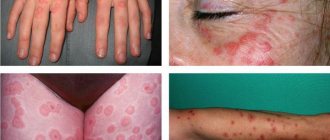
What does pityriasis versicolor look like?
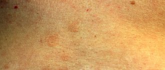
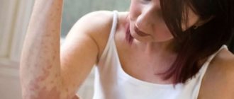
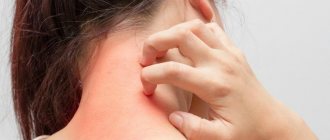
The first symptom of the disease is the appearance of small spots, most often localized on the torso, arms, scalp or external genitalia. The rash may be accompanied by mild itching or may not cause the patient any noticeable discomfort.
The rashes do not have a sharp outline, are not inflamed and do not swell. Initially they are pink in color, but gradually change shade: from pink-yellow to brown and red-brown. The number of spots increases: the fungus spreads across the skin, occupying more and more surface area of the body. The rash may appear on the hands, legs, face, but never affects the mucous membranes, palms or feet, which is due to the special structure of the epithelium in these areas.
The rashes can increase in diameter (up to several centimeters) and merge with each other. Peeling zones appear in the center - when you touch the spot, the scales easily peel off. An atypical form of pityriasis versicolor includes pityriasis alba, in which the affected areas completely lose their pigment (become white). In this case, there may be no peeling.
A distinctive feature of pityriasis versicolor is a disorder of skin pigmentation. Fungal colonies damage melanocytes, which are responsible for the production of melanin, the substance responsible for the color of the skin. Areas of hypopigmentation are especially noticeable during tanning: the epithelium damaged by yeast remains white. This effect can persist for a long time even after recovery.
Pityriasis versicolor is prone to a chronic course and frequent relapses.
To quickly recover and avoid the unpleasant consequences of the infection, it is recommended to consult a dermatologist at the first symptoms of the disease:
- the appearance of a pink or reddish rash;
- mild itching of the skin;
- a feeling of tightness and flaking in certain areas of the skin of the torso and arms.
Publications
Versicolor (pityriasis versicolor) (Tinea versicolor) is a mycotic skin infection characterized by widespread distribution and a chronic relapsing course. The disease affects 10% of the population. In hot countries, lichen versicolor is more common.
In mid-latitudes, most cases of the disease occur in the summer. Adults and young people get sick more often; The disease is rare in children and the elderly. Transmission of the pathogen from a patient or carrier: for example, through a shared bed or clothing (underwear), is possible, however, in most cases, the source of infection is endogenous. Therefore, lichen versicolor is not considered a contagious disease. Some authors consider iatrogenic immunodeficiencies, pregnancy, and hormonal contraception to be predisposing conditions. A hereditary predisposition to the disease cannot be ruled out.
In the International Classification of Diseases (ICD-10), lichen versicolor is included in the section “Other superficial mycoses” (B36.0) and has code B36.1. Its causative agents are opportunistic imperfect yeast lipophilic fungi. In recent years, the causative agents of pityriasis versicolor have been assigned to the genus Malassezia [2,4]. Currently, the genus Malassezia has 9 described species of fungi, among which the main causative agents of pityriasis versicolor are M. globosa, M. sympodialis, M. sloolliae |6, 7|. Fungi of the genus Malassezia are imperfect yeast fungi, basidiomycetes. A common characteristic of most Malassezia species is lipophilicity: (requirement of a source of lipids for growth). Malassezia are distinguished by polymorphism, consisting in different cell shapes (round or oval) in representatives of the same genus and even species, and the ability to form mycelial forms.
The disease begins with the appearance of small spots with a clear edge, or papules slightly raised above the surface of the skin. Small spots are often located around the hair follicles. The color of the spots ranges from yellowish-pink to brown in different shades. The spots often merge, forming large lesions with scalloped edges. On the surface of the spots you can notice a gentle “pityriasis-like” peeling. Peeling intensifies with scraping (symptom of shavings, or “nail strike” of Beignets). The color of the spots differs from the color of the surrounding skin: in people with fair skin, the lesions look darker, in people with dark or tanned skin - lighter, depigmented (pseudo-leukoderma, pityriasis versicolor alba). The same patient may have both hyperpigmented and hypopigmented macules. The color of the spots is mainly determined by exposure to ultraviolet radiation: unlike the color of the surrounding skin, it does not change after tanning. In rare cases, inflammatory lesions with erythema and slight infiltration occur. Typical localization of lesions in lichen versicolor is areas of the body rich in sebaceous glands: chest, back, neck, shoulders. Less commonly, the axillary, groin, forearms and lower legs are involved. In tropical climates, lesions on the face, abdomen, and scalp are more common. The inverse form (tinea versicolor inversa) is the lesion of large, usually inguinal, folds. Hair and nails are not affected. Changes in the skin, as a rule, are not accompanied by subjective sensations. The disease is characterized by a long-term relapsing course.
The variety of clinical manifestations makes timely diagnosis of mycosis difficult [6]. A long-term recurrent course, significant contamination of the skin and scalp with pathogens, and their penetration into the pilosebaceous follicles cause certain difficulties in the treatment of patients with lichen versicolor [1]. Until recently, keratolytic agents, which have low antifungal activity, are inconvenient to use and do not reduce the number of disease relapses, have been widely used in treatment.
N.V. Kungurov et al. developed an algorithm for the management of patients with lichen versicolor. When detected in patients on the skin in typical localizations (areas rich in sebaceous glands), spots of a round or oval shape, non-inflammatory in nature, not rising above the surface of the skin, with clear boundaries, scalloped edges, of various colors (from yellowish-pink, light brown to brown) and gentle pityriasis-like peeling on the surface for the purpose of diagnosis, a Balser test is performed (when the lesions are smeared with alcohol solutions of iodine or aniline dyes, the affected skin is colored much brighter than healthy), and the symptom of “chips” is revealed - with careful scraping of the spot, a barely noticeable tender pityriasis peeling intensifies, which is due to loosening of the stratum corneum of the epidermis.
The skin of the torso and extremities is examined under a Wood's lamp in order to identify non-visual, atypical forms of the disease. Lesions occupying up to 18% of the area are limited (localized) forms, more than 18% are widespread. The erythematous-squamous form (the most typical) is recorded in 98.5% of cases and is characterized by the clinical manifestations described above. The follicular variant, found in 7% of patients, is characterized by the appearance of perifollicular papules or pustules of a yellowish color, located on an erythematous background, while in the middle of the spots, point-shaped follicular openings are clearly visible. In the inverted variant (3.5%) of the clinical course, the lesions are localized in the inguinal-femoral folds, in the pubic area, on the thighs and legs. Less commonly, rashes of pityriasis versicolor rise above the skin and to the touch create the impression of nodules the size of a lentil or smaller, located in groups (pseudo-papular form). Certain difficulties in making a preliminary clinical diagnosis arise in the presence of hypopigmented lesions on the skin that appear in place of typical spots of pityriasis versicolor, usually after irradiation with ultraviolet rays or insolation. Differential diagnosis is carried out with diseases such as Zhiber's pityriasis rosea, syphilis (primary and secondary), seborrheic dermatitis, chloasma, vitiligo, as well as other skin dyspigmentations.
After making a preliminary clinical diagnosis of lichen versicolor, taking into account the course and extent of the process, it is confirmed in the laboratory. During microscopic examination, scales of the stratum corneum of the epidermis of the skin, obtained by scraping with a scalpel from the lesions, are treated with a 20% solution of potassium hydroxide and, after 30 minutes of exposure, are subjected to microscopic examination. The preparation shows short, slightly curved, septate, thick filaments of pseudomycelium and round spores with a double-circuited shell, located in clusters and singly.
To realize the pathogenic properties of the fungus, its transformation from a non-pathogenic form of blastospore into a pathogenic mycelial, certain conditions are necessary, therefore it is necessary to establish factors contributing to the occurrence and spread of fungal infection, analyzing anamnestic data, prescribing consultations and examinations with specialists. Pathology of the gastrointestinal tract, dysfunction of the endocrine system, diseases and functional disorders of the cardiovascular system, seborrheic processes, foci of acute and chronic infection - these conditions directly or indirectly contribute to hyperhidrosis, which results in changes in the physicochemical properties of sweat and sebum (in side of increasing the alkaline reaction), reducing the rate of physiological peeling of the stratum corneum or its loosening, which contributes to the spread of fungal colonies [3].
The choice of treatment tactics depends on the duration, prevalence and form of the fungal process, and identified concomitant diseases.
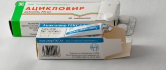
Most cases of lichen versicolor can be treated with external remedies. Patients with a limited form of lichen versicolor (the fungal process occupies less than 18% of the body skin area), whose disease duration is no more than 2 years, who have not previously been treated, as well as patients with widespread keratomycosis (affected area more than 18%), who have absolute contraindications for use systemic antimycotics, one of the external antimycotic drugs is recommended. The use of external forms that allow covering a significant surface of the body is of considerable importance in the treatment of lichen versicolor. Even if the lesion occurs in the form of separate small lesions, it is recommended to treat all areas where lichen versicolor usually develops. The shampoo is used once a day for 5 days.
Other imidazole antimycotics in the form of creams ( Fungazol "Farmakar") are used 2 times a day for at least 2 weeks. The use of modern antifungal aerosols is promising.
Lichen versicolor is characterized by relapses after treatment. As a rule, a year after treatment, a relapse is observed in every second patient, and within two years - in almost all. To reduce the likelihood of relapses, longer treatment and treatment of larger surfaces are recommended. For frequent relapses, systemic antimycotics, azole derivatives, are prescribed. Systemic antimycotics are prescribed to the following groups of patients: with the prevalence of the fungal process exceeding 18% of the skin area; with limited forms (less than 18% of the skin area), with a disease duration of more than 2 years, with relapses after treatment; with follicular and pseudopapular clinical variants of the course of keratomycosis, regardless of the area of the lesion; with the lightning-fast form of the fungal process.
Itraconazole ( Mikotrox "Farmakar") is an antimycotic with the widest spectrum of action, also effective in the treatment of lichen versicolor. It is prescribed at 200 mg/day for 1 week. Fluconazole ( Diflox "Farmakar") is prescribed once at a dose of 400 mg/day. These same drugs, with single or short-term use, can be used to prevent relapses.
We observed 24 patients with a common form of lichen versicolor, who were repeatedly treated with external antifungal drugs. To treat these patients, we chose the drug itraconazole ( Mikotrox ), which was prescribed at a dose of 100 mg 2 times a day for 1 week. All patients tolerated the therapy well, there were no side effects or cases of negative attitude of patients towards treatment. The criteria for achieving complete clinical and laboratory recovery are the absence of skin rashes, negative specific clinical tests, and the absence of fungal mycelium on microscopy.
Thus, despite the fact that lichen versicolor is a very common disease, it often causes difficulties in diagnosis and treatment, especially in recurrent forms. The use of the modern systemic antimycotic Mikotrox helps solve this problem and improve the quality of life of patients.
LITERATURE:
- Bragina E. E., Novoselov A. Yu., Stepanova Zh. V. // Esthet. honey. - 2002. - T. I, No. 5. - P. 447-453.
- Dmitriev G. A., Grammatikova N. E., Bibikova M. V. et al. // Vestn. dermatol. - 2002. - No. 2. - P. 7-9.
- Kungurov N.V., Skurikhina M.E., Budumyan T.M. etc. //Russian Journal of Dermatovenerol. - 2004. - No. 4. – P.49-51.
- Novoselov A. Yu., Bragina E. E., Stepanova Zh. V. // Immunopathol., allergol., infectol. - 2000. - No. 4. - P. 95-98.
- Potekaev II. P. // Ross. Journal dermatovenerol. - 2001. - No. 3. - P. 9-10.
- Sergeev L. Yu., Sergeev Yu. V. Fungal infections: A guide for doctors. - M., 2003. - 440 p.
- Crespo E, Ojeda M, Vera. A. et al. // JEADV. — 2000. -Vol. 14. - R. 47.
E.A. Levonchuk
Medical news, 2007 No. 13
How is pityriasis versicolor transmitted?
Pityriasis versicolor is transmitted through household contact, but this is rare: the disease is not very contagious. The causative agent of pityriasis versicolor, a fungus from the genus Malassezia, is present on the skin of most people, but does not penetrate the epithelium and does not lead to the appearance of a rash. However, under the influence of various factors, yeast transforms into a pathogenic form and begins to destroy skin cells. This process can be triggered by various factors: from excessive sweating to endocrine disorders.
Despite the low contagiousness of this type of lichen, patients are advised to adhere to some recommendations:
- do not share bed linen or towels;
- do not wear other people's clothes;
- do not visit baths, saunas, public swimming pools.
You should also take special care to disinfect the patient’s underwear, clothing and hats: wash at high temperatures (possibly boiling), iron with a hot iron, use steam treatment.
3. Signs and diagnosis of the disease
, asymmetrical spots up to 5 cm in size appear
, which merge over time, forming quite large foci. The edges are usually jagged and the color is yellow, pink, brown or white. Sun exposure often changes the color of the spots. Pityriasis-like peeling occurs on the surface, although inflammatory processes are rare. If papules and pustules appear, then most likely this is a consequence of other skin infections superimposing on the affected areas.
Foci of lichen are usually located on the back, neck, abdomen and shoulders, in the groin area.
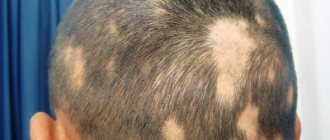
If the head is affected, the hair in these places does not change at all. Sometimes pityriasis versicolor causes itchy skin.
It is used to examine foci of affected skin.
Wood's lamp, microscopic analysis, Balser's iodine test.
About our clinic Chistye Prudy metro station Medintercom page!
Is it possible to cure pityriasis versicolor?
With proper therapy, pityriasis versicolor is easily treated and goes away within 2-3 weeks. To do this, you need to make an appointment with a dermatologist, who will conduct an examination and prescribe additional tests.
As a rule, the diagnosis is established in the presence of a typical clinical picture, but to confirm it, the following analyzes and tests are performed:
- examination under a Wood's lamp: fungal colonies are illuminated;
- microscopic examination of the scraping: shows the presence of yeast;
- Balzer test using iodine-containing solutions.
A dermatologist may also prescribe histological studies, general blood and urine tests. Based on the results of the examinations, treatment tactics are developed.
Depending on the severity and severity of the infection and the symptoms of lichen, therapy includes:
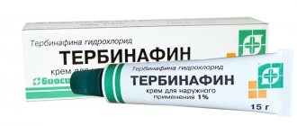
- the use of external agents (sprays, lotions, ointments) with antifungal components. If the scalp is affected, special shampoos are prescribed;
- antifungal drugs for internal use. Prescribed to patients prone to relapses of pityriasis versicolor, or to patients with atypical manifestations of infection.
If the patient suffers from severe itching (which is extremely rare), antihistamines may be prescribed.
Reasons for development
Versicolor versicolor is a type of fungal skin infection that affects the stratum corneum of the epidermis and hair follicles. Its causative agents are two types of fungi, and infection is possible only through prolonged and close contact with the patient. And in this case, provoking factors play a big role. These include:
- weakened immunity;
- hyperhidrosis;
- disruption of the sebaceous glands;
- diseases of the endocrine system (obesity, diabetes, Itsenko-Cushing syndrome, etc.);
- hormonal imbalance due to pregnancy, menopause or taking hormone-containing medications;
- vegetative-vascular dystonia;
- abuse of antibacterial personal hygiene products;
- excessive exposure to ultraviolet rays (intense tanning, frequent visits to the solarium) and regular overheating of the body.
It is noteworthy that patients with pityriasis versicolor over 60 years of age are extremely rare. This is due to natural age-related changes in the skin, which make it less susceptible to pathogens.
In children under 10 years of age, the main causes of pityriasis versicolor infection are neglect of personal hygiene rules or improper skin care. At this age, with the protective functions of the skin intact, the body independently copes with pathogenic microorganisms attacking it, so the development of the disease does not occur. But closer to adolescence, when hormonal changes begin, the body’s susceptibility to bacteria, viruses and fungi increases, so children over 10 years old become infected with pityriasis versicolor just like adults.
Is it possible to cure pityriasis versicolor permanently?
If you follow medical recommendations, pityriasis versicolor responds well to treatment: the rash completely disappears in 2-3 weeks. But the infection tends to become chronic: in this case, relapses occur every time sweating increases, for example, in the warm season, when traveling on vacation, or during intense physical activity.
To avoid this, you must:
- consult a dermatologist at the first signs of the disease. At the initial stage, pityriasis versicolor can be easily confused with other skin infections: tinea versicolor, seborrheic dermatitis, eczema, vitiligo, and so on. Each disease requires specific treatment, which is why it is so important to recognize the disease as early as possible;
- refuse self-medication. Uncontrolled use of medications and the use of “folk remedies” will not only not lead to a cure, but can also aggravate the course of the infection;
- follow your doctor's recommendations. This applies to both the use of prescribed medications and personal hygiene.
In this case, the patient can quickly recover from pityriasis versicolor and avoid its relapse.
Is it possible to sunbathe with pityriasis versicolor?
This dermatosis is popularly called “sun fungus”: during the beach season, the yeast is activated and the disease is diagnosed most often. At the same time, the areas of the skin affected by the colonies do not tan and remain white - the patient is covered with light spots, which bring him to the dermatologist’s office.
Since pityriasis versicolor can be diagnosed during rest, patients are often interested in: is it possible to sunbathe with pityriasis versicolor? Opinions differ on this matter. Previously, quartz treatment was prescribed for the treatment of lichen versicolor: under the targeted influence of ultraviolet light, colonies of the fungus died. But now many dermatologists dispute this method of treatment.
UV radiation can be dangerous for an organism weakened by infection. In addition, the occurrence of pityriasis versicolor indicates a decrease in immune strength. Therefore, experts do not recommend exposing the skin to additional stress.
If the patient doubts whether it is possible to sunbathe with pityriasis versicolor, he is recommended to consult a dermatologist. The doctor will give recommendations taking into account the individual characteristics of the body, the severity of the disease, its form and location. If you follow medical prescriptions, the patient will avoid the complications of lichen and will be able to recover in a short time.
It should also be remembered that if the patient decides to sunbathe with pityriasis versicolor, the areas of the skin affected by the colonies will remain light: it will not be possible to get an even and beautiful tan. Therefore, it is better to first get rid of the disease, and only after that return to the beach.
Signs of the disease
Externally, erythrasma manifests itself in the form of light brown, brick-red, brown or yellow-brown spots on the skin, most often round in shape and without signs of inflammation. The diameter of the lesions can reach several centimeters, and they tend to merge, forming large affected areas. First of all, erythrasma spots appear in the folds of the skin, where there is a favorable environment for the proliferation of bacteria.
In addition to spots, erythrasma is characterized by:
- Peeling of the skin on the affected areas, aggravated by touch. Usually this is where the development of the disease begins.
- Mild, irregular itching. It intensifies and begins to cause significant discomfort only if a secondary infection is added to the primary disease.
- Absence of fever, wounds, ulcers and ulcers with copious discharge. This distinguishes erythrasma from most bacterial skin pathologies.
- Getting wet. An optional symptom, the manifestation of which depends on the amount of sweating and the quality of skin care.
It is noteworthy that in children, symptoms of erythrasma appear extremely rarely. The risk group includes adults, primarily men, who are predominantly overweight and prone to excessive sweating. In this case, in men, the skin in the groin, navel and inner thighs is usually affected, and in women, the entire abdomen, armpits and areas under the breasts are affected.
Can pityriasis versicolor be cured with iodine?
Iodine-containing products are used in the diagnosis of lichen versicolor. Yeast colonies are sensitive to iodine, causing them to absorb it to a much greater extent than surrounding healthy skin. Therefore, if you apply an iodine solution to the epidermis, the affected areas will be much darker.
But many patients believe that iodine is suitable not only for diagnosis, but also for self-treatment of pityriasis. An alcohol solution of iodine is a popular antiseptic that is often used to disinfect the skin. Fungi from the genus Malassezia, which cause tinea versicolor, are sensitive to it and are partially killed upon contact with this substance, but it is impossible to completely destroy them with iodine.
In addition, an alcohol solution of iodine greatly dries already damaged skin, which can lead to various complications of dermatosis.
As a result of self-medication:
- the disease drags on - recovery does not occur, and the rash spreads to new areas of the body;
- the likelihood of relapse increases - after the rash disappears, it appears again when the immune system is weakened and is chronic;
- new symptoms are added to the symptoms of the disease: tightness and dryness of the skin treated with iodine, itching may occur.
Pityriasis versicolor can be cured with antifungal medications prescribed by a dermatologist. Special creams and ointments affect only colonies of the pathogen and do not affect healthy epithelium. Also, many products contain care components that additionally moisturize the skin, accelerate its regeneration and promote the resumption of melanin synthesis.