Home — For the public
- Map of medical organizations
- Vaccination
- Clinical examination
- Fluorography
- Addresses and opening hours of clinics
- Emergency rooms
- Oncology
- Where to take an HIV test
- Healthy child's office
- Services
- Prevention of CVD
- Disease Prevention
- World Patient Safety Day
- Newspaper "Medical News"
- specialist
- School of Health
— Disease prevention
- HIV infection
- All about vaccination
- All about proper nutrition
- Hepatitis
- Flu
- Dementia
- Schoolchildren's health
- STD
- Tick-borne encephalitis
- Whooping cough
- Measles
- Legionellosis
- Meningococcal infection
- Oncology
- Acute intestinal infection
- Pediculosis
- First aid
- Pneumococcal infection
- Pneumonia
- Prevention of rabies
- Dependency Prevention
- Rotavirus infection
- Diabetes
- Cardiovascular diseases
- Injuries
- Tuberculosis
- Tularemia
- Physical activity
- Obstructive pulmonary disease
- Exotic infections
- Ecology
- Why is swimming in ponds dangerous?
— Flu — Flu symptoms and treatment
Influenza is an infectious disease affecting the respiratory tract of a viral nature. Symptoms arise acutely; a person can often indicate the hour when he felt unwell: the temperature rises sharply, chills occur, pain in the joints and muscles, and cough. Diagnosis: influenza can be confirmed using PCR diagnostics.
Routes of transmission
In the transmission of infection, the main role is played by airborne droplets: when coughing or sneezing, small drops of saliva or sputum enter the air and spread over a distance of up to 1.5 meters. In the room, the droplets break up into smaller fragments and remain suspended. And the virus itself remains viable for up to several hours. Infection occurs by inhaling this aerosol.
The second most important is the contact route of spread: a sick person, sneezing, covers his face with his hand and then spreads the virus by touching objects in the environment. Therefore, using a handkerchief and washing your hands; actually reduce the risk of spreading infection.
The average incubation period is up to 2 days.
Main routes of infection
Influenza is transmitted by airborne droplets. An invisible drop (0.0001 ml) of secretion from the nasopharynx is enough to become infected. The virus is also transmitted through household contact. If there is a sick person in the house, he should have individual hygiene items, dishes, and bed linen. You can also become infected through contact. For example, a person sneezed while covering his mouth with his hand. Hands are rarely washed after this. By touching various objects, a person expands the area of potential infection. Crowds of people and closed types of premises are places where the flu spreads with lightning speed.
Diagnosis of influenza
Typically, infectious disease specialists rely on characteristic clinical manifestations and weekly monitoring of morbidity. Certain clinical difficulties are presented by the similarity of the disease with other viral infections “walking” in the population:
- respiratory syncytial virus (or RSV);
- adenovirus;
- parainfluenza;
- rhinovirus.
Laboratory diagnostics help detect the influenza virus. The fastest method is the polymerase chain reaction (PCR). The method allows you to detect not only influenza, but also viruses with a similar clinical picture.
According to the instructions of the Ministry of Health of the Russian Federation, the diagnosis of influenza is confirmed based on 2 criteria:
- Clinical signs;
- positive PCR result.
A general blood test for influenza does not help establish a diagnosis. It reflects only the presence of inflammatory changes of a viral nature: relative lymphocytosis and neutropenia.
WHO activities
WHO, through the WHO GISRS system and in collaboration with other partners, monitors influenza activity globally, makes recommendations on seasonal influenza vaccine formulations twice a year for the northern and southern hemispheres, and assists countries with tropical and subtropical climates in the selection of vaccine products (for northern and southern hemispheres) and decisions regarding the timing of vaccination campaigns and supports Member States in developing prevention and control strategies.
WHO works to strengthen national, regional and global capacity to respond to influenza (including diagnosis, antiviral susceptibility monitoring, disease surveillance and outbreak response), increase vaccination coverage in high-risk populations and ensure preparedness for next influenza pandemic.
(1) Estimates of US influenza-associated deaths made using four different methods. Thompson WW, Weintraub E, Dhankhar P, Cheng OY, Brammer L, Meltzer MI, et al. Influenza Other Respi Viruses. 2009;3:37-49
(2) Global burden of respiratory infections due to seasonal influenza in young children: a systematic review and meta-analysis. Nair H, Abdullah Brooks W, Katz M et al. Lancet 2011; 378: 1917–3 (3) WHO recommended surveillance standards, Second edition.
Signs of influenza and other viral infections
A person cannot always figure out which virus caused the deterioration and loss of performance. What manifestations of the disease should you pay attention to?
Typical flu symptoms
- One of the first signs may be chills, a sharp rise in temperature.
- Severe discomfort in muscles and joints – “the body aches and hurts”;
- Headache, feeling of weakness, “overwhelmed.”
This condition persists for up to 3-5 days. Discharge from the nasal passages is unusual. On the contrary, the patient may complain of a feeling of dryness, nasal congestion, pain, and sore throat. Often accompanied by a dry cough with chest pain.
When examining such a person, the therapist will identify signs of infection such as:
- hyperemia (redness) of the face, neck;
- congestion of scleral vessels;
- redness and characteristic “graininess” of the back wall of the pharynx.
Causes of occurrence and development
The cause of the disease is seasonal viruses (autumn and winter). There are different types of influenza and there are three types: A, B and C. Mild forms of the disease are caused by type C. The reason for exceeding the epidemic threshold is A and B from year to year. The source of the disease is an infected person. Viral molecules are resistant to survival. The room where the patient was may remain potentially dangerous for healthy people for 9 hours. On plastic surfaces, the activity of pathogenic molecules persists for up to 2 days, so prevention of influenza and ARVI in premises (wet cleaning, disinfection) is important.
Are you experiencing flu symptoms?
Only a doctor can accurately diagnose the disease. Don't delay your consultation - call
Complications of influenza: what are the dangers and how to recognize them
The most vulnerable groups of the population include children under 6 years of age, the elderly, pregnant women and people with chronic diseases (diabetes, cardiovascular and respiratory diseases). It is they who often experience the flu with complications.
The most common complications include pneumonia. Complications such as viral encephalitis, meningitis, myocarditis are also possible.
If you notice any of the following symptoms, you should immediately consult a doctor:
- breathing problems (frequent, noisy, a person can only breathe in one body position);
- pain in the ribs, intensifying or appearing when coughing;
- headaches with nausea, loss of consciousness, behavioral disturbances;
- the temperature does not drop after taking various antipyretic drugs (according to the scheme);
- convulsions due to high fever.
In addition, you should call a doctor at home if the body temperature remains high on the 4th day of illness or there is a decrease to 37-37.5℃ with a subsequent rise to 38℃.
The first thing a person should do when sick is stay home. You should not work “heroically” or study while barely standing. Such behavior will not bring any benefit, but will only undermine your health and those around you. Right:
- Call a doctor at home.
- Follow the doctor's recommendations and do not self-medicate.
Pathogen
There are 4 types of seasonal influenza viruses - types A, B, C and D. Influenza A and B viruses circulate and cause seasonal epidemics of illness.
Influenza A viruses are divided into subtypes according to combinations of hemagglutinin (HA) and neuraminidase (NA), proteins on the surface of the virus. Currently, influenza viruses of subtypes A(H1N1) and A(H3N2) are circulating among people. A(H1N1) is also referred to as A(H1N1)pdm09 because it caused the 2009 pandemic and subsequently replaced the seasonal influenza A(H1N1) virus that circulated before 2009. Only influenza A viruses are known to cause pandemics.
Influenza B viruses are not divided into subtypes, but can be divided into lineages. Currently circulating influenza B viruses belong to the B/Yamagata and B/Victoria lineages.
Influenza C virus is less common and usually causes mild infections. Therefore, it does not pose a public health problem.
Group D viruses mainly infect cattle. According to available data, they do not infect people or cause disease in them.
Prevention of influenza and ARVI
With the onset of the autumn-winter period, people spend more and more time indoors: at home, in educational institutions, clinics, entertainment centers, and shopping centers. Effective prevention of ARVI should work in different directions:
- limiting visits to crowded places, especially at the first signs of ill health;
- washing hands after places, using handkerchiefs when coughing and sneezing;
- regular ventilation of premises, air humidification;
- taking care of your health: proper nutrition, protection from hypothermia, avoiding stress.
Timely annual vaccination is considered the most effective way of prevention. Specific prevention has been developed to combat seasonal influenza. Vaccination is primarily important for vulnerable groups of the population: preschool children, pensioners and people with chronic diseases, the course of which may worsen after an infection.
When refusing vaccination “out of conviction,” you should remember that even in the absence of a clear clinical picture, a person who has contracted the flu spreads the virus among family and friends. Including those who have medical contraindications to vaccination.
Methods of nonspecific prevention include:
- regular hand washing;
- avoiding crowded places during seasonal epidemics;
- maintaining a healthy lifestyle;
- hardening;
- playing sports;
- prolonged exposure to fresh air;
- proper nutrition;
- full sleep.
Treatment of influenza in adults
When you have an influenza infection, bed rest is very important. Attempting to carry the disease “on your feet” will only make you feel worse and will contribute to the infection of others. During an influenza infection, doctors recommend drinking a lot - the heat causes sweating, and, as a result, greater loss of moisture. Contrary to popular belief, very hot drinks are contraindicated - burns to the mucous membranes of the larynx make them vulnerable to bacterial infections. The drink should be warm. It is not advisable to drink coffee - it increases heart rate and can worsen the condition. Maintain a comfortable temperature in the room, humidify the air and do not forget about regular ventilation.[vi]
The following medications are used to treat influenza:
- symptomatic remedies - antipyretics and antihistamines, drops for the common cold.
- etiotropic drugs are drugs that act on the causative agent of the disease. This group includes antiviral agents that fight the virus at different stages of its life cycle and prevent its spread.
Use of the drug VIFERON for influenza
One of the antiviral drugs that helps restore the immune system and has a wide spectrum of antiviral activity is the drug VIFERON. Recombinant interferon alpha-2b, which is part of this drug, prevents the synthesis of viral DNA and blocks the reproduction of the virus, and also helps restore immunity. It is identical to human interferon alpha-2b, but is produced using modern technology without the use of donor blood.
The drug, which is available in the form of suppositories, gel and ointment, was developed as a result of fundamental research in the field of immunology, which has proven that in the presence of antioxidants (vitamins C, E), the antiviral effect of interferon is enhanced. The drug in the form of suppositories (suppositories) can be used during pregnancy (from the 14th week of gestation), as well as during breastfeeding, for the treatment of newborns, starting from the first days of a child’s life, and the elderly.
The inclusion of the drug VIFERON rectal suppositories in the complex therapy of influenza in adults contributes to:
- a decrease in body temperature to normal 24 hours after the start of use in 48.6% of patients;
- a decrease in body temperature to normal on the second day after the start of use in 82.9% of patients;
- reducing the total duration of intoxication symptoms (by an average of 2 times);
- shortening the duration of the febrile period to 1.53 days (control group 3.55 days).1
The antiviral drug VIFERON Gel can also be used for the treatment and prevention of ARVI and influenza for pregnant and lactating women, as well as newborn children, starting from the first days of life, and the elderly. When applied externally and locally, systemic absorption (the physiological process of absorption, that is, the penetration of medicinal substances into the blood and lymph) is low and the drug has an effect only at the site of the lesion.
Use of the drug VIFERON for the prevention of influenza
In order to protect yourself from acute respiratory viral infections and influenza infections, you need to apply a strip of gel approximately 0.5 cm long to the nasal mucosa 2 times a day as a preventive measure. Course duration is 2-4 weeks. For the treatment of ARVI, including influenza infection, including those complicated by a bacterial infection, the application of VIFERON gel must be repeated 3-5 times a day for 5 days.
VIFERON Ointment for the treatment of influenza and other acute respiratory viral infections is applied in a thin layer to the mucous membrane of the nasal passages 3 times a day. The amount of the drug is one pea with a diameter of 0.5 cm, the approximate course of treatment is 5 days.
Home remedies
At home you need to follow a diet. The diet should include not only vegetables and fruits, but also juices from them, fruit drinks, still mineral water, herbal teas (mint, chamomile, raspberries). You can eat fish, lean meat, dairy products, eggs. It is worth giving up heavy fatty and fried foods, alcohol, cocoa, and fresh baked goods.
A popular means is an inhaler. Put your cold and flu pills aside and try breathing in sage, eucalyptus or oak bark. Don't forget about the phytoncides that are contained in onions and garlic. Eat them fresh. Home therapy should include tea with ginger and milk with honey. Remember also to constantly ventilate the room.
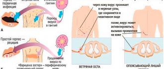
Influenza: history, clinical picture, pathogenesis
The term "flu" is derived from it. greifen and french. gripper (grab, paralyze), becoming widespread in Europe during the epidemic of 1742–1743. In Russia, the term “flu” began to be used only at the beginning of the 19th century - for example, in the first volume of L. N. Tolstoy’s novel “War and Peace” we find: “Anna Pavlovna coughed for several days, she had the flu, as she said (the flu was then a new word, used only by rare people).
Etiology of influenza
Influenza is an acute respiratory viral disease, etiologically associated with representatives of three genera - Influenza A virus (influenza A viruses), Influenza B virus (influenza B viruses) and Influenza C virus (influenza C viruses) - from the family Orthomyxoviridae [2, 26].
Influenza A virus was first isolated from pigs by American virologist Richard Shoup (1901–1966) in 1930; from people - three years later by a group of English scientists: Wilson Smith (1897–1965), Christopher Andrews (1896–1987) and Patrick Laidlaw (1881–1940) [26].
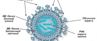
| Rice. 1. Structure of the influenza A virus virion (Orthomyxoviridae, Influenza A virus) |
On the surface of the virion (viral particle) of the influenza A virus there are two functionally important molecules (Fig. 1): hemagglutinin (with the help of which the virion attaches to the surface of the target cell); neuraminidase (which destroys the cellular receptor, which is necessary during the budding of daughter virions, as well as to correct errors due to incorrect binding to the receptor) [2, 24, 26].
Currently, 16 types of hemagglutinin (designated as H1, H2, ..., H16) and 9 types of neuraminidase (N1, N2, ..., N9) are known. The combination of hemagglutinin type and neuraminidase (for example, H1N1, H3N2, H5N1, etc.) is called a subtype: out of 144 (16 × 9) theoretically possible subtypes, 115 are known today [24].
The natural reservoir of the influenza A virus is wild birds of the aquatic ecological complex (primarily dabbling ducks, gulls and terns), but the virus is able to overcome the interspecies barrier, adapt to new hosts and circulate in their populations for a long time [9–12]. Epidemic variants of the influenza A virus cause an annual increase in incidence and dangerous pandemics every 10–50 years [1, 11, 16].
The influenza B virus was discovered in 1940 by American virologist Thomas Francis Jr. (1900–1969). Influenza B virus does not cause a pandemic, but is the causative agent of large epidemic outbreaks [26].
Influenza C virus was discovered in 1947 by American virologist Richard Taylor (1887–1981). Influenza C virus causes local epidemic outbreaks in children's groups. The infection is most severe in young children [26].
Influenza viruses occupy an important place in the structure of the incidence of acute respiratory viral infections (ARVI), which account for up to 90% of all other infectious diseases. According to the World Health Organization (WHO), 3–5 million people worldwide fall ill with severe forms of influenza alone every year. Every year, 25–35 million people fall ill with influenza and other acute respiratory viral infections in the Russian Federation, of which 45–60% are children. The economic damage to the Russian Federation from seasonal epidemic influenza amounts to up to 100 billion rubles/year, or about 85% of economic losses from infectious diseases [2–8, 20–23].
History of influenza pandemics
Hippocrates' (460–377 BC) description of influenza as the "Perinthian cough" is considered the first scientific description of the disease (412 BC). In the 9th–18th centuries, influenza was known as “peasant fever”; warm foot baths and heated red wine with spices were recommended as treatment. From the beginning of the 16th century, with an increase in population density, epidemic “waves” began to roll across all of Europe. In the 17th century, five major epidemics were recorded in Europe, and three in the 18th century. During the epidemic of 1675, the famous English physician Thomas Sydenham (1624–1689) suggested that the “English prickly heat” was of an infectious nature and described a version of this disease, which he called febris comatosa, which was accompanied by the development of symptoms from the central nervous system (CNS).
In 1889–1892 The first documented influenza A(H2N2) pandemic occurred. The Spanish flu (H1N1) pandemic (1918–1919) sickened 600 million and killed 50–100 million1 (i.e., 30% and 5% of the world's population, respectively). The Asian influenza (H2N2) pandemic (1957–1959) killed more than 1 million; Hong Kong flu pandemic (1968–1970) - about 1 million; major epidemic of “Russian flu” (H1N1) (1977–1978) - about 300 thousand people.
A feature of the pandemics of 1889–1892. and the Spanish Flu of 1918–1919. there was a lack of information about the etiological agent of the disease (doctors of that time considered the Afanasyev-Pfeiffer bacillus, Haemophilus influenzae, as the causative agent of the infection). The number of severe and fatal cases of infection sharply declined during subsequent pandemics, which is associated primarily with the advent of influenza vaccines, the strain composition of which is determined annually by WHO based on data from the Global Influenza Monitoring Program (operating since 1947), etiotropic antiviral drugs Amantadine and Rimantadine (1963), as well as antibiotics (1941) for the treatment of secondary pneumonia.
The modern pandemic of “swine flu” (H1N1) swl2 (2009–2010) began as a result of the emergence of epidemic potential in one of the variants of the swine influenza A (H1N1) virus [16]: in mid-March 2009 there was a sharp jump in incidence in the city La Gloria (Mexico, Veracruz state), at the end of March the first laboratory-confirmed cases appeared in the United States, at the end of April - in Canada, and then in Europe. In the Russian Federation, the identification of the first case of the disease and isolation of the strain were carried out by employees of the Federal State Institution Research Institute of Virology named after. D.I. Ivanovsky” Ministry of Health and Social Development of Russia May 21, 2009 [13, 15, 25]. The WHO announced the beginning of the next influenza pandemic on June 11, 2009, and its end on August 10, 2010. During this period, influenza caused the death of 18.5 thousand people worldwide.
In the first post-pandemic epidemic season of 2010–2011. pandemic influenza A(H1N1) swl caused more than 70% of ARVI cases in the world, influenza A(H3N2) - 1-5%, influenza B - 10-20%.
Composition of influenza vaccines in the 2011–2012 epidemic season. (same as 2010–2011): A/California/07/2009 (H1N1) swl; A/Perth/16/2009 (H3N2); B/Brisbane/60/2008.
The pandemic influenza A(H1N1) swl virus is resistant to Remantadine and Amantadine, but sensitive to Tamiflu, Relenza, Ingavirin, Arbidol and Ribavirin [13–16, 18, 19, 23].
Highly virulent avian influenza A(H5N1) is a possible causative agent of the next pandemic.
The likelihood of the influenza A virus crossing the interspecies barrier and entering the human population with dangerous consequences increases sharply during epizootic periods3. Therefore, the highly virulent avian influenza A(H5N1) virus, which has caused a modern large-scale epizootic among wild and domestic birds of the Old World and has an increased ability to reproduce in mammalian cells, is considered as the most likely causative agent of the next influenza pandemic [10, 11, 17]. Further spread of this virus could have catastrophic consequences if it develops epidemic potential (the ability to be transmitted from person to person), since, firstly, humanity does not have collective immunity to influenza A (H5) viruses, and secondly, because 563 laboratory-confirmed cases of human disease in 15 countries as a result of infection with the influenza A(H5N1) virus of avian origin during 2003–2011. 330 died, i.e., the mortality rate is approaching 60% [11, 24].
In the Russian Federation, no human cases of influenza A(H5N1) virus of avian origin have been detected. However, Russian researchers continue to regularly monitor this dangerous infection (si vis pacem, para bellum) and have made a significant contribution to reducing the likelihood of unfavorable developments: the main features of the development of the epizootic process were predicted, prototype viral strains were deposited in the State Collection of Viruses of the Russian Federation, their biological properties were studied (in particular, it has been shown that strains of highly virulent avian influenza A(H5N1) virus are sensitive to commercial anti-influenza chemotherapy drugs: Remantadine, Amantadine, Tamiflu, Relenza, Ingavirin, Arbidol, Ribavirin), at the Federal State Institution "Research Institute of Virology named after. D.I. Ivanovsky" Ministry of Health and Social Development of Russia received Russian patents for the vaccine strain, and developed domestic veterinary (which is actively used) and medical (which is waiting in the wings) vaccines [10, 11, 17, 24].
Pathogenesis of influenza
In humans, influenza viruses infect epithelial cells of the mucous membrane of the respiratory tract, as well as goblet cells (secreting mucus), alveolocytes and macrophages [3, 4, 7]. All these cells have on their surface a receptor to which the viral hemagglutinin binds (Fig. 1), the terminal residue of sialic, or N-acetylneuraminic acid (Neu5Ac) (Fig. 2), as part of the polysaccharide chains that are part of gangliosides and glycoproteins. The terminal sialic acid residue can bind to the next monosaccharide in two ways: via an alpha2-3 or alpha2-6 linkage (Fig. 2) [14, 18].
| Rice. 2. Structural formulas of sialic or N-acetylneuraminic acid (Neu5Ac) and two ways of covalent bonding with the following monosaccharide (in this case galactose, Gal): alpha2-3- or alpha2-6-bond |
Epithelial cells of the human upper respiratory tract contain mainly alpha2-6-sialosides; lower sections - alpha2-3-sialosides (Fig. 3). Therefore, epidemic strains of influenza viruses, having alpha2-6 specificity, easily reproduce in the upper parts of the human respiratory tract, are actively released into the environment when speaking, sneezing, coughing, and effectively infect other people through airborne droplets.
Variants of influenza A virus adapted to birds have alpha2-3 specificity (Fig. 3). Terminal alpha2-3-sialosides are found in birds mainly on the surface of epithelial cells of the intestinal mucosa, therefore in birds influenza occurs in the form of enteritis; the virus is released into the external environment with feces, and infection occurs through the nutritional route. The alpha2-3 specificity of avian variants of influenza A virus explains their inability to effectively infect the epithelium of the upper parts of the human respiratory tract and, as a result, be transmitted by airborne droplets in the human population. At the same time, if a highly virulent avian influenza A virus somehow manages to cause a productive infection in the human body, then it will effectively infect the lower parts of the respiratory tract, causing severe primary viral pneumonia (according to WHO, in 60% of cases - fatal) .
Epithelial cells of pigs simultaneously contain both alpha2-6- and alpha2-3-sialosides (Fig. 3), therefore, both epidemic and avian variants of the influenza A virus can simultaneously circulate in the body. As a result, in pig populations, firstly, the formation of reassortants4 of human and avian strains with new biological properties; secondly, strains with mixed alpha2-6/alpha2-3 specificity are selected. It is precisely this mixed alpha2-6/alpha2-3 specificity that strains of the pandemic influenza A(H1N1) swl virus possess, and, as a result, they have the ability to spread by droplet airborne routes and cause severe pneumonia [13–15, 18, 19, 23 ].
| Rice. 3. Sialoside receptors of influenza A viruses on the surface of epithelial cells of humans (alpha2-6 - on the mucous membrane of the upper, alpha2-3 - on the mucous membrane of the lower parts of the respiratory tract), pigs (alpha2-6 / alpha2-3 mixture on the mucous membrane of the respiratory tract) and birds (alpha2-3 - on the intestinal mucosa) |
Infection of epithelial cells results in a rapid increase in the viral load, apoptosis, degeneration and necrosis of this type of cell with the subsequent development of toxic and toxic-allergic reactions. In humans, damage to the columnar epithelial cells of the trachea and bronchi is typical. The main link in the pathogenesis of influenza A is damage to the vascular and nervous systems that occurs as a result of the toxic effect of the virus. At the same time, one of the main mechanisms of the influence of the influenza A virus on the vascular system is the formation of reactive oxygen species that interact with phospholipids of cell membranes, causing lipid peroxidation in them, disruption of membrane transport and barrier functions, contributing to the further development of viral infection. Lysosomal enzymes additionally damage the capillary epithelium and the basement membrane of cells, which contributes to the spread of influenza infection and viremia. Increased vascular permeability, fragility of their walls, and impaired microcirculation are the cause of hemorrhagic manifestations - from nosebleeds to hemorrhagic pulmonary edema and hemorrhages in the brain. Circulatory disorders, in turn, cause damage to the central nervous system: the pathomorphological picture is characterized by the presence of lymphomonocytic infiltrates around small and medium veins, hyperplasia of glial elements and focal demyelination, which indicates the toxic-allergic nature of the pathological process in the central nervous system during influenza [3–8, 23] .
An important factor in the pathogenesis of influenza is the production of the viral protein PB1-F2, which causes apoptosis of tissue macrophages of the lungs and thereby contributes to the development of secondary bacterial pneumonia (in the modern pandemic variant of the influenza A(H1N1) swl virus, the production of PB1-F2, fortunately, is absent, which reduces - but does not eliminate! - the likelihood of developing secondary pneumonia, leaving the danger of primary viral pneumonia intact - see below) [2, 26].
Clinical picture of influenza in humans
The onset is acute, with chills, a rapid rise in temperature to high numbers, and a sharp increase in symptoms of intoxication. The temperature reaches maximum values (39.0–40.0 °C) on the first day of the disease. During the same period, signs of intoxication increase: chills, severe headache, dizziness, myalgia, arthralgia, severe weakness. On external examination: the face is hyperemic, puffy, the vessels of the sclera are injected, hyperemia of the conjunctiva, cyanosis of the lips and mucous membrane of the oropharynx are detected, pinpoint hemorrhages on the soft palate are possible. Cyanosis in general is an important symptom of influenza: you should pay attention not only to cyanosis of the lips, but also to the cyanotic tint of the uvula, tonsils, palatine arches against the background of bright hyperemia of the oropharyngeal mucosa; the mucous membrane of the soft palate also has a cyanotic tint, fine granularity, vascular injection and pinpoint hemorrhagic elements are clearly visible; on the posterior wall of the pharynx - moderate hyperplasia of lymphoid tissue.
Localization of headache: in the frontotemporal region and in the eyeballs (with light pressure on them or when they move). Meningeal signs are often detected, which gradually disappear with a decrease in intoxication and a decrease in body temperature. The range of clinical manifestations from the nervous system is quite wide: from functional disorders to serous meningitis and severe meningoencephalitis.
Damage to the respiratory tract during influenza is the leading one: catarrhal symptoms in the form of nasal congestion or slight rhinitis are observed in all patients; cough (dry, painful, with pronounced rumbles and “roars”, dry wheezing in the lungs), which develops by the end of the first day of illness, is very characteristic of influenza A - due to tracheitis common with influenza - and can serve as a differentiating clinical sign. In the pandemic (2009–2010) and post-pandemic (2010–2011) periods, a long (8–10 days) cough was recorded, with symptoms of bronchitis, which in some patients persisted for several weeks (which indicated involvement in the pathological process small airways); During auscultation of the lungs, hard breathing was heard, often dry scattered wheezing. In addition, in 2009–2011. Some patients experienced diarrhea in the first two days of the disease: 5% of moderate and 10% of severe patients [8, 18, 23].
Pneumonia is one of the most dangerous complications of influenza. There are three types of pneumonia: 1) primary viral; 2) secondary viral-bacterial; 3) secondary bacterial (or “day 14 pneumonia”) [3–8, 23].
Primary viral pneumonia develops in the first days of the disease (2–5 days) and is characterized by severe intoxication, often hemorrhagic manifestations (nosebleeds, streaks of blood in the sputum), a rapid increase in respiratory failure (feeling of lack of air, shortness of breath, increased cyanosis). Auscultation during inspiration reveals characteristic crepitating rales. Bilateral lung damage and severe respiratory failure are consistent with acute lung injury (ALI). Developing acute respiratory distress syndrome (ARDS) is characterized by significant arterial hypoxemia and diffuse alveolar infiltrates. Computed tomographic examination is characterized by uneven thickening of the interalveolar and interlobular septa (“reticular lung”) and a decrease in the transparency of the lung fields like “ground glass”. Signs of active bacterial infection, as a rule, cannot be detected. Most patients have leukopenia, thrombocytopenia, high levels of lactate dehydrogenase, creatine phosphokinase.
With the development of secondary pneumonia against the background of a viral infection, signs of bacterial exposure are determined, confirmed by the detection of bacteria Streptococcus pneumoniae, Staphylococcus aureus, etc. in the sputum. As a rule, secondary pneumonia develops after 5–7 days of influenza and is characterized by a repeated rise in temperature to febrile values, increased cough, the appearance of mucopurulent sputum, often streaked with blood, radiologically - focal and focal-confluent infiltrates, often with signs of destruction and abscess formation. After 10 days, pneumonia usually has a bacterial etiology and is most often associated with gram-negative microflora.
One of the main factors contributing to the severe course of influenza is concomitant pathology. In particular, in patients who died during the last two epidemic seasons of 2009–2011, heart and vascular diseases, diabetes mellitus, metabolic syndrome (obesity), alcoholism and tobacco smoking predominated. A special risk group is pregnant women, in whom pneumonia can develop rapidly, and therefore they require special attention from clinicians and immediate treatment.
An analysis of fatal outcomes showed that outpatient doctors are gradually losing their wariness regarding pandemic influenza A(H1N1) swl: there were cases of late hospitalization due to underestimation of the severity of the patient’s condition and the risk of developing rapidly progressing pneumonia. All patients require urgent etiotropic therapy according to the principle “the sooner the better”: the first 36–48 hours of illness are optimal. The use of antiviral drugs within the specified time frame significantly reduces the duration and severity of the disease, as well as the frequency of complications. In future epidemic seasons, lessons need to be learned from previous experiences.
Literature
- Gendon Yu. Z. Influenza pandemic: is it possible to fight it // Questions of Virology. 1998. No. 1. P. 43–46.
- Kaverin N.V., Lvov D.K. Orthomyxoviridae. In the book: Medical virology. Hand-in. Ed.: Academician of the Russian Academy of Medical Sciences D.K. Lvov. M.: MIA, 2008. pp. 176–183.
- Ketiladze B. S., Ivanova L. A., Eliseeva I. Ya. et al. The importance of various respiratory viruses in the development of chronic nonspecific bronchopulmonary processes // Questions of Virology. 1986. No. 3. P. 310–314.
- Kolobukhina L.V. Viral infections of the respiratory tract // Breast Cancer. 2000. T. 8. No. 13–14. pp. 559–564.
- Kolobukhina L.V. Clinic and treatment of influenza // Breast Cancer. 2001. T. 9. No. 16–17. pp. 710–713.
- Kolobukhina L.V. Viral infections of the respiratory tract. In the book: Respiratory medicine: a guide. Ed.: Academician of the Russian Academy of Medical Sciences A. G. Chuchalin. M.: GEOTAR-Media, 2007. T. 1. P. 449–474.
- Kolobukhina L.V., Lvov D.K., Burtseva E.I. Gripp. In the book: Medical virology. Hand-in. Ed.: Academician of the Russian Academy of Medical Sciences D.K. Lvov. M.: MIA, 2008. pp. 382–393.
- Kolobukhina L.V., Shchelkanov M.Yu., Merkulova L.N. et al. Etiotropic therapy of influenza: lessons from the last pandemic // Bulletin of the Russian Academy of Medical Sciences. 2011. No. 5. pp. 35–40.
- Lvov D.K. Possible significance of natural biocenoses in the variability of influenza A virus // Questions of Virology. 1974. No. 6. P. 740–744.
- Lvov D.K. Population interactions in a biological system: influenza A virus - wild and domestic animals - humans; causes and consequences of the penetration of the highly pathogenic influenza virus A/H5N1 into the territory of Russia // ZhMEI. 2006. No. 3. P. 96–100.
- Lvov D.K. Ecology of viruses. In the book: Medical virology. Hand-in. Ed.: Academician of the Russian Academy of Medical Sciences D.K. Lvov. M.: MIA, 2008. pp. 101–118.
- Lvov D.K., Ilyichev V.D. Migrations of birds and transfer of infectious agents. M.: Nauka, 1979. 270 p.
- Lvov D.K., Burtseva E.I., Prilipov A.G. et al. Isolation on May 24, 2009 and depositing in the State Collection of Viruses (GKV No. 2452 dated May 24, 2009) of the first strain A/IIV-Moscow/01/2009 (H1N1) swl, similar to swine virus A (H1N1) from the first patient identified on May 21, 2009 in Moscow // Questions of Virology. 2009. T. 54. No. 5. P. 10–14.
- Lvov D.K., Burtseva E.I., Prilipov A.G. et al. Possible connection of lethal pneumonia with mutations of the pandemic influenza virus A/H1N1 swl in the receptor-binding site of the HA1 subunit of hemagglutinin // Questions of Virology. 2010. T. 55. No. 4. P. 4–9.
- Lvov D.K., Burtseva E.I., Shchelkanov M.Yu., Prilipov A.G., Kolobukhina L.V., Malyshev N.A., Bazarova M.V., Merkulova L.N., Deryabin P. G., Kuzmichev A. G., Fedyakina I. T., Grebennikova T. V., Usachev E. V., Sadykova G. K., Shevchenko E. S., Trushakova S. V., Lavrishcheva V. V. ., Alkhovsky S. V., Samokhvalov E. I., Belyakova N. V., Ivanova V. T., Oskerko T. A., Latyshev O. E., Belyaev A. M., Belyaev A. L., Feodoritova E. L. Spread of the new pandemic influenza virus A(H1N1) v in Russia // Questions of Virology. 2010. T. 55. No. 3. 4–9.
- Lvov D.K., Malyshev N.A., Kolobukhina L.V. et al. Influenza caused by the new pandemic virus A/H1N1 swl: clinical picture, diagnosis, treatment. Guidelines. M.: Moscow Department of Health, 2009. 18 p.
- Lvov D.K., Fedyakina I.T., Shchelkanov M.Yu. et al. Effect of in vitro antiviral drugs on the reproduction of highly pathogenic strains of influenza A/H5N1 virus that caused an epizootic among poultry in the summer of 2005 // Questions of Virology. 2006. T. 51. No. 2. P. 20–22.
- Lvov D.K., Shchelkanov M.Yu., Bovin N.V. et al. Correlation between the receptor specificity of strains of the pandemic influenza A(H1N1) pdm09 virus isolated in 2009–2011, the structure of the receptor-binding site and the likelihood of development lethal primary viral pneumonia // Questions of virology. 2012. No. 1.
- Lvov D.K., Yashkulov K.B., Prilipov A.G. et al. Detection of amino acid substitutions of aspartic acid with glycine and asparagine in the receptor-binding site of hemagglutinin in variants of the pandemic influenza A(H1N1) swl virus from patients with a fatal outcome and with a moderate form of the disease // Questions of Virology. 2010. T. 55. No. 3. P. 15–18.
- Sadov A. A. Epidemic influenza. L., 1927. 60 p.
- Smorodintsev A. A. Influenza and its prevention. M.: Medicine, 1984. 384 p.
- Supotnitsky M.V. The Spanish Flu pandemic of 1918–1920. in the context of other influenza pandemics and “bird flu” // Medical records. 2006. No. 11. P. 31–34; 2006. No. 12. P. 15–22; 2007. No. 1. pp. 16–22.
- Chuchalin A. G. Influenza: lessons from the pandemic (clinical aspects) // Pulmonology. 2010. Appendix 1. pp. 3–8.
- Shchelkanov M. Yu., Lvov D. K. Genotypic structure of the genus Influenza A virus // Bulletin of the Russian Academy of Medical Sciences. 2011. No. 5. pp. 19–23.
- Shchelkanov M. Yu., Lvov D. N., Fedyakina I. T. et al. Dynamics of the spread of pandemic influenza A(H1N1) swl in the Far East in 2009 // Questions of Virology. 2010. T. 55. No. 3. P. 10–15.
- Shchelkanov M. Yu., Fedyakina I. T., Proshina E. S. et al. Taxonomic structure of Orthomyxoviridae: current state and immediate prospects // Bulletin of the Russian Academy of Medical Sciences. 2011. No. 5. pp. 12–19.
M. Yu. Shchelkanov, Doctor of Biological Sciences, Associate Professor L. V. Kolobukhina, Doctor of Medical Sciences, Professor D. K. Lvov, Doctor of Medical Sciences, Professor, Academician of the Russian Academy of Medical Sciences
Federal State Institution "Research Institute of Virology named after. D. I. Ivanovsky", Moscow
Contact information for authors for correspondence
1 For comparison, 8.3 million people died as a result of hostilities during the 5 years of the First World War (1914–1918).
2 The abbreviation “swl” refers to the English. the phrase “swine-like”, i.e. strains “similar to swine”.
3 Epizootic is the process of spreading an infectious disease in animal populations.
4 The influenza A virus has a genome consisting of 8 individual RNA molecules. Reassortment is the formation of a strain in which the source of different genetic segments was different parental strains that simultaneously infected the same cell.
How is the flu different from a cold?
- Onset of the disease . Flu symptoms develop rapidly. And it progresses quickly in the body.
The source of influenza is sick people during the incubation period or the height of the disease. The disease is transmitted by airborne droplets when sneezing, coughing, shaking hands, and also through dirty food (it has not been thermally processed enough). The infection begins to manifest itself in the period from 4 hours to 3 days. In general, the disease goes away in 5-7 days.
A cold is a disease of the body associated with hypothermia. A cold develops gradually, progressively, and painful symptoms appear within a day or two. It is usually easy to treat. But in advanced forms it can develop into other more severe diseases.
- Temperature . With the flu, there is a high body temperature, which can reach 39-40 degrees. With a cold, the temperature usually remains at the standard level.
- Weakness . People with a cold often ignore symptoms such as cough and runny nose, thinking that it will “go away on its own” with time. A cold can be carried on your feet, although it is undesirable.
But during the flu, the patient immediately realizes that he is sick; often he cannot even have the strength to get out of bed. Aches all over the body, severe headache, fever - these symptoms cannot be confused with anything else.
- Cough and runny nose . During the flu, these symptoms do not appear immediately (only after 5 days), and may not appear at all. With a cold, a debilitating cough, sore throat when swallowing and a runny nose are noted.
- Red eyes, itching and watery eyes . The picture of the flu is complemented by red eyes, severe weakness, fever, and bursting blood vessels in the nose. And in severe forms, even convulsions, vomiting, rapid heartbeat, and lack of air are possible.
When you have a cold, your eyes do not turn red or water, and if this happens, it is more likely to indicate a bacterial infection.
Myths and dangerous misconceptions in treatment
The question of whether to get a flu vaccine in the midst of a mass disease of people has a clear answer - no. During an epidemic, a person may already have the virus, which will create a double infectious load for the body. The flu vaccine should be given earlier. Be careful with aspirin. This is an excellent antipyretic, but not for the flu. It thins the blood and increases the penetration of viruses into blood vessels and cells, aggravating the health condition. Hot foot baths are also famous. They help relieve swelling of the mucous membrane of the upper respiratory tract - this is true. They can only be done during periods when there is no fever. Otherwise, the load on the heart increases significantly. Hot inhalation with potatoes is a traditional method, but it is also dangerous due to the risk of getting a steam burn to the mucous membrane.
Diagnosis and treatment of the virus
Influenza is diagnosed by clinical manifestations. If identification of the etiological factor is required, rapid tests are used to detect the viral antigen, tests of samples from the throat, nose, and the RT-PCR method. They are not mandatory for everyone, but they are necessary for patients at risk of developing complications. At JSC “Medicine” (clinic of Academician Roitberg) near the Mayakovskaya metro station, modern technologies and our own clinical diagnostic laboratory allow us to accurately diagnose, prescribe therapy, or, if necessary, hospitalize.
If a person is not in a high-risk group, influenza is treated symptomatically. Prescribe bed rest, plenty of warm drinks, antipyretic and antitussive medications, isotonic water for rinsing the nasal cavity and vasoconstrictor drops. Any cold and flu remedy must be prescribed by a doctor. In severe conditions and complications, treatment is carried out in a hospital.