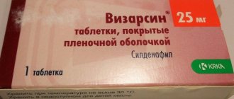
Thiolepta 600 mg 60 pcs. film-coated tablets
pharmachologic effect
The drug is an endogenous antioxidant that binds free radicals. Thioctic (α-lipoic) acid is involved in the mitochondrial metabolism of the cell; it functions as a coenzyme in the complex for the transformation of substances that have a pronounced antitoxic effect. They protect the cell from reactive radicals arising during intermediate metabolism or during the breakdown of exogenous foreign substances, and from heavy metals. Thioctic acid exhibits synergism with insulin, which is associated with increased glucose utilization. In patients with diabetes mellitus, thioctic acid leads to a change in the concentration of pyruvic acid in the blood.
Composition and release form Thiolept 600 mg 60 pcs. film-coated tablets
Tablets - 1 tablet: thioctic acid 600 mg.
10 pieces. — contour cell packaging (6) — cardboard packs.
15 pcs. — contour cell packaging (4) — cardboard packs.
Directions for use and doses
When taken orally, a single dose is 600 mg.
300-600 mg/day is administered intravenously (slow stream or drip).
Indications for use Thiolept 600 mg 60 pcs. film-coated tablets
Diabetic polyneuropathy.
Contraindications
Pregnancy, lactation (breastfeeding), hypersensitivity to thioctic acid.
Application of Thiolept 600 mg 60 pcs. film-coated tablets during pregnancy and breastfeeding
Contraindicated during pregnancy. Breastfeeding should be stopped during treatment (there is no sufficient experience with the drug).
special instructions
With simultaneous use, it is possible to enhance the hypoglycemic effect of insulin and oral hypoglycemic agents.
When used simultaneously with cisplatin, its effectiveness may be reduced.
Side effects Thiolept 600 mg 60 pcs. film-coated tablets
After IV administration, diplopia, convulsions, pinpoint hemorrhages in the mucous membranes and skin, and impaired platelet function are possible; with rapid administration - increased intracranial pressure.
When taken orally, dyspeptic symptoms are possible (including nausea, vomiting, heartburn).
When taken orally or intravenously - allergic reactions (urticaria, anaphylactic shock); hypoglycemia.
Drug interactions
Thioctic acid reacts with ionic metal complexes (for example, with cisplatin), therefore, when used simultaneously, Thiolept® may reduce the effect of cisplatin. After taking Thiolept® in the morning, it is recommended to take iron and magnesium supplements, and also consume dairy products (due to their calcium content) in the afternoon or evening. With simultaneous use, Thiolept® enhances the effect of insulin and oral hypoglycemic agents. Alcohol (ethanol) reduces the therapeutic activity of thioctic acid.
Guidelines for the management of patients with acute facial neuropathy (2020)
Fieux M, et al. French Society of ENT (SFORL) guidelines. Management of acute Bell's palsy. European Annals of Otorhinolaryngology, Head and Neck diseases (2020)
Introduction
Bell's palsy is the most common form of facial neuropathy.
at least in 8–12% of cases the cause is the presence of a space-occupying formation [1,2] (level
evidence 4). In more than 70% of cases, Bell's palsy has a favorable prognosis with
complete recovery within 6 months [3,4] (level of evidence: 3); practically
complete recovery (to grade I-II on the House-Braakman scale) reaches more than 80%
patients [5] (level of evidence: 3). Thus, the prognosis of the disease is favorable,
however, the lack of complete recovery disrupts social adaptation and worsens the quality of
patients' lives.
Clinical examination
Bell's palsy is a form of facial neuropathy that
characterized by rapid development (within 24–48 hours) of weakness of the upper and lower groups
facial muscles of half the face and autonomic disorders. The onset of the disease may
preceded by pain in the ear or in the postauricular area, taste disturbance, hyperacusis. Diagnosis
determined by excluding other causes. Clinical examination should include
assessment of the strength of facial muscles, primarily to confirm peripheral
level of lesion with detailed neurological and otolaryngological examination [5,9, 10] (level of evidence: 2).
Criteria for peripheral level of damage:
- involvement of the upper (limited mobility of the eyebrow, Bell's sign when closing the eye/or smoothness of the nasolabial fold on the affected side) and lower (drooping corner card or inability to hold air when puffing out the cheeks on the affected side) groups of facial muscles of half the face;
- absence of dissociation in the presence of motor and autonomic disorders;
- absence of other focal neurological symptoms (motor, sensory disorders or signs of damage to other cranial nerves).
Confirmation of the peripheral level of facial nerve damage:
(Class B)
- In patients with a clinical picture of peripheral neuropathy of the facial nerve, a complete neurological and otorhinolaryngological examination should be performed, including otoscopy, palpation of the parotid salivary gland and neck area.
- The weakness of both the upper and lower groups of facial muscles of the half-face in the absence of dissociation between motor and autonomic disorders, as well as the absence of other focal neurological symptoms, suggests a peripheral level of damage to the facial nerve.
Assessment of the rate of development of symptoms suggests a possible etiology of damage to the facial nerve, for example, a space-occupying lesion. True Bell's palsy develops quite quickly, unlike neuropathy.
Most often, a differential diagnosis should be made [11] (level of evidence: 2).
According to the recommendations of the American Academy of Otolaryngology (2013), the maximum time
development of symptoms – 72 hours [6] (level of evidence: 1). Despite the fact that 6%
cases, Bell's palsy may recur [9] (level of evidence: 2), history of
patient with facial neuropathy questions the diagnosis of idiopathic paralysis
Bell [12] (level of evidence 4).
Isolated facial nerve neuropathy should not be accompanied by ipsilateral
hearing loss, and therefore audiometry is mandatory to exclude
conductive hearing loss, which suggests the presence of a tumor of the facial nerve in
pyramid of the temporal bone, or sensorineural hearing loss, the presence of which allows us to make
assumption of compression of the facial nerve by a space-occupying formation located in the area
cerebellopontine angle. Tympanometry allows you to evaluate the acoustic reflex of the stapedius
muscles, which provides additional information for topical diagnosis [13,14]
(level of evidence 4).
For clinical assessment of the severity of acute facial neuropathy, it is recommended
application of the House-Brackmann facial nerve grading system [15],
whereas for subsequent dynamic monitoring of the patient and assessment of the degree
restoration, the Sunnybrook classification is the most convenient.
- If the clinical symptoms of facial neuropathy progress >72 hours from the onset of the disease or there is a fluctuation in the severity of symptoms, involvement of the muscles of both halves of the face, the diagnosis of Bell's palsy should be questioned with the exclusion of other causes, including the presence of a space-occupying lesion (Class A ).
- If a patient has signs of facial neuropathy in combination with ipsilateral hearing loss, dizziness or other focal neurological symptoms, as well as in combination with pathology identified by otoscopy or palpation of the cervical and parotid lymph nodes, the diagnosis of Bell's palsy should be questioned. even if symptoms develop rapidly (within ≤72 hours) (Class B).
- All patients with a clinical picture of Bell's palsy are recommended to undergo audiometry (Expert opinion).
- All patients with a clinical picture of Bell's palsy are recommended to study the acoustic reflex of the stapedius muscle. In cases of severe facial neuropathy and intact stapedius reflex, the diagnosis of Bell's palsy should be questioned with a detailed examination of the parotid and neck region (Expert Opinion).
- All patients with newly diagnosed Bell's palsy are recommended to assess the severity of symptoms using the House-Braakman scale and enter the assessment results into the medical documentation. (Expert opinion).
Laboratory examination
In the American (2013) and Canadian (2014) guidelines for the management of patients with paralysis
Bell [6,7] (level of evidence 1) it is not recommended to carry out any
laboratory examinations.
- However, in patients with diabetes, assessing the degree of its compensation by examining the level of glycated hemoglobin is advisable due to the need to prescribe high doses of glucocorticoids to patients [16] (level of evidence, 4).
- Similarly, an elevated neutrophil/lymphocyte ratio in a complete blood count indicates a poor prognosis for recovery [17] (Level of Evidence: 4).
- In patients with Bell's palsy, serological diagnosis of HIV infection [18] (level of evidence 4) and Lyme disease [1,19] (level of evidence 4) is recommended.
- All patients with Bell's palsy are recommended to have a complete blood count and fasting blood glucose tested. In patients suffering from diabetes, it is advisable to further examine the level of glycated hemoglobin (Expert opinion).
- Serologic testing for Lyme disease is recommended for all patients with Bell's palsy. In some cases, a diagnosis of HIV infection and herpes viral infection (varicella zoster virus and herpes simplex virus) should be carried out (Expert opinion).
Neuroimaging examination
According to American and Canadian guidelines for the management of patients with paralysis
Bell [6,7] (level of evidence 4), conducting a neuroimaging examination in
patients with no other focal neurological symptoms and with normal
audiometric indicators are inappropriate. However, in recently published works
It is reported that in 8–12% of cases, neuroimaging examination of patients with typical
The clinical picture of Bell's palsy revealed the presence of voluminous neoplasms, 30% of
which were malignant [2] (level of evidence: 4). MRI of the brain with
contrast enhancement is the method of choice because it allows detailed evaluation
the state of the cerebral structures along the nerve, as well as the structure of the parotid salivary gland.
Bell's palsy is characterized, but not specific, by accumulation of contrast material in the facial
nerve on the side of clinical symptoms [20–22] (level of evidence: 4). If available
in a patient with a typical picture of Bell's palsy, an emergency neuroimaging examination is performed
examination is not practical [23] (level of evidence: 4), diagnosis should be
carried out within 1 month from the onset of symptoms.
- Patients with a clinical picture of Bell's palsy are recommended to undergo a contrast-enhanced MRI of the brain within 1 month from the onset of symptoms to assess the condition of the cerebral structures along the nerve and the nerve itself along its entire length, including the portion passing through the parotid gland.
- An emergency MRI of the brain is indicated only in cases where the clinical picture is atypical for Bell's palsy.
- Carrying out MRI of the brain in DWI mode excludes the presence of a stroke, but is insufficient when examining patients with the clinical picture of Bell's palsy.
- If the patient does not have a favorable recovery within 6 months from the onset of symptoms, a neuroimaging examination is recommended.
- Performing a CT scan of the brain and temporal bones in patients with Bell's palsy is inappropriate (Expert opinion).
Severity rating
Electroneuromyography (ENMG) is the main instrumental method for assessing
severity and prognosis of the disease, but it should not be carried out in the early stages [24] (level
evidence 4). Absence of motor unit potential (MUP) during voluntary
muscle contraction has no prognostic value until the 15th day from onset
symptoms [25] (level of evidence: 4).
- To assess the prognosis of the disease in patients with Bell's palsy, ENMG should not be performed earlier than 8 days from the onset of symptoms, due to the low prognostic significance of the results of the study conducted in the early stages (Expert opinion).
In accordance with expert opinion and literature data, based on the results
ENMG identifies the following variants of neuropathy of the facial nerve in Bell's palsy and its
forecast:
— myelinopathy with a favorable prognosis;
- a combination of myelino- and axonopathy with a relatively favorable prognosis;
- severe axonopathy with a poor prognosis [26] (level of evidence: 4).
In case of severe neuropathy of the facial nerve (grade V-VI on the House-Braakman scale), ENMG should be performed 9–20 days from the onset of symptoms to assess the prognosis of recovery (Expert Opinion).
For the greatest prognostic value of the results, the ENMG protocol should include:
- Bilateral facial nerve motor conduction study recorded in multiple muscles, including at least 1 perioral muscle (nasolabial groove, orbicularis oris, mentalis). Recording values from the nasolabial sulcus muscles is probably most appropriate due to its prognostic significance for the patient, since they are more involved in smiling than the orbicularis oris or mentalis muscles.
- Study of the blink reflex.
- Conducting needle EMG of >2 muscles innervated by the upper and lower portions of the branches of the facial nerve, with assessment of the spontaneous activity of muscle fibers and MUAPs during voluntary muscle contraction (Expert opinion).
Drug treatment
The management of patients with Bell's palsy remains a subject of debate due to its
spontaneous favorable prognosis observed in most patients. Full frequency
restoration of impaired functions correlates with the initial severity of symptoms and fluctuates
from 61% with total prosoplegia to 94% with less severe prosoparesis [5]
(level of evidence 3). Most authors confirm the effectiveness
glucocorticoid therapy in the treatment of patients with Bell's palsy [6,7, 27,28] (level
evidence 1). In a study by Giri P. et al. (2015) demonstrated the absence
differences in the effectiveness of intravenous and oral glucocorticoid therapy [29]
(level of evidence 2). Also in multicenter randomized double-blind
placebo-controlled studies have shown no significant effectiveness
monotherapy with acyclovir or valacyclovir in patients with Bell's palsy [31–33] (level
evidence 1).
American and Canadian guidelines for the management of patients with Bell's palsy
It is recommended to prescribe combined glucocorticoid and antiviral therapy for
early stages of the disease [6,7] (level of evidence: 2). In recently published
works also confirm the feasibility of prescribing combination therapy after
assessment of contraindications and possible side effects of antiviral drugs
[34,35,36] (level of evidence: 2).
- In patients with Bell's palsy, glucocorticoid therapy (prednisolone or methylprednisolone) should be initiated as early as possible (optimally within the first 72 hours) (Class A).
- For patients with Bell's palsy, glucocorticoids are recommended at a daily dose of 1 mg/kg for 7–10 days (Expert opinion).
- In case of severe neuropathy of the facial nerve (grade V-VI on the House-Braakman scale) and the absence of contraindications, patients may be prescribed high doses of glucocorticoids (2 mg/kg per day) for 10 days (Expert opinion).
- Transtympanic administration of drugs to patients with Bell's palsy is not recommended (Expert opinion).
- Monotherapy with antiviral drugs is not recommended in patients with Bell's palsy (Class A).
- For patients with severe facial neuropathy, combination glucocorticoid and antiviral therapy is recommended if treatment is initiated within 72 hours of the onset of symptoms (Expert Opinion).
Drug treatment of Ramsay-Hunt syndrome
Ramsay-Hunt syndrome is more severe than classic Bell's palsy, in 50–85% of patients
is accompanied by the development of complications [11,37,38] (level of evidence 3) and is characterized
high tendency to relapse [11] (level of evidence: 3).
- In patients with Ramsay-Hunt syndrome, it is recommended that combined glucocorticoid (prednisolone or methylprednisolone) and antiviral therapy be administered as early as possible for 7 days (Class B).
Eye care
The main complications developing in patients with Bell's palsy and syndrome
Ramsay-Hunt, includes ophthalmological pathology in the form of keratitis, corneal ulcers,
panophthalmia. Prevention of complications includes prescribing systematic
use of eye drops, protective gel or artificial tears up to several times
once daily, especially at night [6,27,39–41] (LE: 4).
Prevention of ophthalmological complications of Bell's palsy and Ramsay-Hunt syndrome
includes early appointment of patients with systematic
use of topical therapy (eye drops, protective gel or
artificial tears, night occlusion of the eye). When identifying signs
inflammation of the eyeball, the patient should be referred to an ophthalmologist.
Observation by an ophthalmologist should continue for several weeks
from the beginning of recovery (Expert opinion).
Physiotherapeutic and rehabilitation treatment
In a meta-analysis of 12 studies involving 872 patients, MacIntosh PW and
al. (2019), a high risk of bias was reported due to significant
differences in the methods of rehabilitation treatment used, the timing of its administration and
duration [42] (level of evidence: 4). However, in the absence of complete
restoration of the strength of facial muscles in patients with Bell's palsy
restorative treatment is effective and reasonable [6,43,44] (level
evidence 4).
- Electrotherapy has been shown to shorten muscle fibers and interfere with favorable recovery [45] (level of evidence: 4) and is therefore not recommended for patients with Bell's palsy.
- According to the 2013 American guidelines, the effectiveness of acupuncture (ART) in patients with Bell's palsy without concomitant drug treatment is unproven [6] (evidence level 4).
- In patients with severe facial neuropathy or with factors that prevent favorable recovery, rehabilitation treatment is recommended (Expert Opinion).
- Electrotherapy and forced exercise are contraindicated in patients with Bell's palsy (Expert opinion).
- IRT is not recommended for patients with Bell's palsy (Class C)
Hyperbaric oxygenation
- A meta-analysis by Holland NJ (2012) showed that all studies that examined the effectiveness of hyperbaric oxygen therapy in patients with Bell's palsy had low levels of evidence and serious systematic errors [47] (evidence level 4).
- Hyperbaric oxygen therapy is not recommended for patients with Bell's palsy (Class C).
Dynamic observation
Clinical and ophthalmological observation should continue for several
months after the start of recovery for the purpose of prevention and timely treatment
complications, especially ophthalmological ones [6,7] (level of evidence: 4).
Surgery
There is currently no benefit from surgical decompression of the facial nerve.
proven. Since the effectiveness of surgical treatment is influenced by many factors, such
as criteria for selecting patients, timing and methods of performing the operation, reliably assess
The benefits of facial nerve decompression are quite difficult. Nevertheless,
Facial nerve decompression should be considered as an alternative treatment method,
which is confirmed by the results of the LeeS-Y meta-analysis. et al. (2019) [48] (level
evidence 4).
The question of surgical intervention can only be considered after
reliable diagnosis of Bell's palsy, which is based on a comprehensive assessment of the results
clinical, electromyographic and neuroimaging examinations.
- In patients with severe facial neuropathy, surgical decompression should be performed as early as possible to prevent irreversible nerve damage [49,50]. If ENMG results indicate >90% fiber degeneration, decompression should be performed as early as possible, since delayed surgical treatment is comparable in effectiveness to standard conservative therapy [50] (level of evidence: 2).
- The benefit of surgical decompression of the facial nerve in patients with Bell's palsy has not yet been proven. However, given the variety of factors influencing the outcome of surgical treatment, a reliable assessment of its effectiveness requires further research (Expert opinion).
- For surgical decompression of the facial nerve, transmastoid or supratemporal approaches can be used, with the supratemporal approach being the gold standard.
- Surgical decompression of the facial nerve should be performed within 30 days, optimally within 14 days, after the lesion identified by ENMG (Expert Opinion).
https://doi.org/10.1016/j.anorl. 2022.06.004