There are five main types of calluses, which form when there is pressure or rubbing on the bone area. They can be extremely painful if they put pressure on a nerve ending.
- Hard calluses
The most common type of callus. It is a small area of hardened skin the size of a pea. Such calluses are usually surrounded by a skin growth and indicate disturbances in the functioning of the foot apparatus or improperly selected shoes.
- Soft calluses
Such calluses form between the fingers, where the skin is damp from sweating or is not sufficiently dry after washing. They look whitish, and feel like rubber to the touch. They are quite hard inside and can therefore be quite painful. Such calluses usually occur when two adjacent fingers come into close contact and constantly rub against each other. The main culprit of such calluses is narrow shoes that press your toes together.
- Calluses “grain”
These are tiny calluses that appear singly or in groups on the foot and look like pockmarks on the surface of the skin, but are not painful. They appear as a result of insufficient hydration of the skin of the foot.
- Vascular or vascular calluses
These calluses tend to grow significantly over time, which interferes with the functioning of the capillaries of the circulatory system in the upper layers of the skin. They can be very painful and bleed if damaged. They usually have a gore tint to them.
- Fibrous or stringy calluses
This type of callus is the most dangerous. They arise as a result of a careless attitude towards ordinary calluses. Located in the upper layers of the skin, they are tightly intertwined with collagen fibers and closely connected with organ tissues. They can be extremely painful and require professional treatment. These calluses look very deep with a tint of yellowness.
Causes of calluses
Before getting rid of corns on your feet, you need to find out the main reasons for their appearance and eliminate them. Most often, calluses occur due to:
- using uncomfortable, narrow shoes or shoes of the wrong size;
- wearing hard shoes on bare feet or a very thin nylon sock;
- a heel that is too high, which causes compression and excessive stress on the forefoot;
- various foot diseases;
- walking barefoot for a long time;
- excessive sweating of the feet;
- wearing shoes with hard seams inside or with rubbing surfaces;
- wearing socks that are too large and cause wrinkles;
- selection of shoe models with thin soles.
2.Why do corns form?
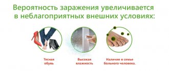
We can definitely say that the predisposition to the formation of corns varies from person to person. For each person, a whole complex of external and internal factors is important here. We have to come to terms with those of them that we are not able to completely change:
- The structure of the foot, abnormalities in the development of the legs (flat feet, bone growths);
- Features of the skin;
- Distribution of subcutaneous adipose tissue;
- Condition of the vascular system of the legs;
- Metabolism, individual characteristics of metabolism;
- Hormonal background;
- Chronic diseases and anamnesis.
If you notice a tendency to form corns, first of all, you need to pay attention to what could cause increased stress on the feet. A number of such reasons are varied and can be corrected:
- Occupation that involves walking or standing during the day;
- Lifestyle, hobbies and activity (running, dancing, ballet);
- Tendency to obesity, excess weight;
- Low-fat diet, vegetarianism;
- Avitaminosis;
- Tight shoes, predominantly wearing high-heeled shoes;
- Fungal diseases of the feet.
Visit our Dermatology page
How to get rid of calluses on your feet yourself
The easiest way to cure a water callus is to wait for it to burst, or to pierce it yourself. However, it is important to take care of disinfection, otherwise there is a risk of infection.
When deciding how to get rid of dry calluses, you need to take into account the area and depth of the lesion. For example, a small thickening on the little finger can be removed with a pedicure file or pumice stone. You may first need to soften the skin in a hot bath with the addition of various components.
Dry calluses on the foot can also be removed by applying salicylic ointment at night or by sticking a special medicated patch.
However, doctors do not recommend removing calluses yourself, as there is always a risk of damaging the skin and causing infection, which will create additional problems and complicate treatment.
4. Treatment of corns
In especially severe cases or when a quick, perfect result is needed, methods of surgical removal of corns are used. Modern aesthetic medicine offers painless and effective procedures for tidying up the feet:
- Laser therapy;
- Cryodestruction;
- Hardware pedicure.
Sometimes corns suddenly begin to form on perfectly healthy feet. In this case, it is necessary to carefully study what has recently changed in your general condition or lifestyle. It often turns out that the cause is new shoes, a change in profession, unusual hobbies, or a change in diet. To solve such problems, it is enough to restore balance in the body, reduce the load on the legs, and adjust the diet. If there is a connection with the identification of any chronic disease, you need to focus on its treatment. Properly selected therapy will have a comprehensive positive effect, and the corns will cease to bother you again as you recover.
How doctors remove calluses
The fastest, safest and most painless method is hardware procedures. Removal of the formation is carried out using a special cosmetology device equipped with an automatic cutter fixation system. This eliminates the risk of soft tissue damage. The procedure itself is painless and does not require special preparation. The session time ranges from 10 to 60 minutes. It depends on the number of dry calluses and the degree of their neglect.
Before starting work and during the procedure, the skin is thoroughly disinfected. In case of increased sensitivity, the patient is given topical anesthesia. When treating calluses and corns, the skin is cooled using a spray.
After removing a callus or corns, the skin is polished, and the edges of the treated area are smoothed so that there is no discomfort when walking later. After completion of the procedure, the treated surface is protected with a bandage.
In this way, you can remove any corns, including dry calluses on the foot.
Features of hardware procedures:
- absolute safety – the surface being treated is thoroughly disinfected, which eliminates the risk of infection;
- speed – unlike home procedures, the removal of corns and calluses in a cosmetology clinic is performed quickly and efficiently. One procedure is enough;
- painlessness – automatic fixation of the cutter on the equipment protects the skin from accidental injury and pain;
- no contraindications – anyone can use hardware callus removal.
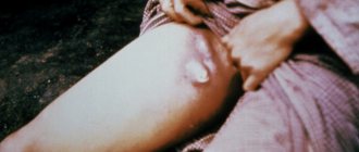
Callus
Calluses can be especially painful. In the center of the callus there is a hole from which the head of the callus protrudes. This head covers the root, which goes deep into the soft tissues and causes pain when walking.
Such a callus can form as a result of a foreign body (for example, a splinter or a pebble) getting under the skin. If the splinter is not removed, when it moves, it begins to irritate the tissues surrounding it, and the body, in response to irritation, forms a corpus callosum, but not from the outside, but from the inside. Another reason for the appearance of calluses is a dermatotropic virus.
Recommendations after removing dry calluses on feet
After hardware removal of calluses and corns, it is recommended to wear loose shoes made from natural materials. It is better to give up high heels for 2 weeks, and also not visit swimming pools and saunas for a month. It is better to treat the skin with a disinfectant composition.
Patients who successfully get rid of dry calluses experience lightness in their legs and no pain while walking.
It is better to prevent the occurrence of corns and regularly remove rough skin on the feet. By periodically undergoing a medical hardware pedicure procedure, you will not be aware of such problems as rough skin on your feet and the occurrence of complications.
If you want to remove dry callus, contact the SM-Cosmetology clinic. At the initial consultation, the podologist will conduct an examination and select the optimal method to solve your problem. You can remove calluses and corns after one visit to a cosmetology clinic.
—>
3.Prevention of corns
For many people, the problem of corns is chronic.
Folk remedies, cosmetic procedures and foot care bring only temporary relief. In this case, it is worth considering the problem as chronic and approaching treatment comprehensively.
The constant formation of corns always indicates a serious imbalance in the body or disturbances in lifestyle. In this case, the problem should be solved in two ways - along with cosmetic methods, it is necessary to focus on identifying the reasons that cause increased keratinization of the skin of the feet.
Seeking medical help often includes consultations with a dermatologist, orthopedist, and endocrinologist. A systematic approach, the study of anamnesis and lifestyle make it possible to identify the main factors of increased death of epidermal cells. If a connection between corns and health problems is discovered, treatment of the underlying disease always helps to solve the problem with the skin of the feet. The effectiveness of the therapy can be accelerated by local procedures aimed at eliminating skin growths and further prevention:
- Anti-callus plasters;
- Keratolytic cream;
- Hot foot baths;
- Natural and synthetic pumice;
- Softening cream with medicinal herbs.
About our clinic Chistye Prudy metro station Medintercom page!
Callus removal methods
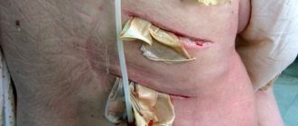
Depending on the size of the callus, hardware targeted removal, radio wave removal, or surgical excision may be recommended. Soft calluses and growths of small diameter, for example, between the fingers, are often removed using a hardware method, using special softeners and cutters, and then local healing therapy is prescribed. In some cases, the procedure is carried out in two stages with a break of 7-14 days. The service includes: consultation with a surgeon, local anesthesia, use of sterile disposable material, application of an unloading bandage. The cost of removing plantar and periungual calluses will depend on the depth of tissue damage.
Stages and symptoms
Bedsores can develop quickly, but the formation of bedsores and ulcers occurs through four stages (each of which has its own symptoms):
- Stage 1
Redness of intact skin. It is slightly inflamed and may be painful, itchy and warm to the touch. You may also notice that the texture of your skin has changed slightly. At this stage, bedsores on the heels resemble a sunburn. This initial stage is difficult to detect in people with dark skin.
- Stage 2
In the second stage, the first signs of skin damage appear. They look like abrasions, blisters or small pits. The outer layer of skin is broken, red and painful. The surrounding tissue may be pale, red, and swollen. The upper layers of tissue, as the most damaged ones, begin to die and peel off. Bruising at this stage indicates suspicion of possible deeper tissue damage, involving fat or muscle tissue.
- Stage 3
Complete loss of tissue. The damage extends to the deeper layers of tissue in the heel. Crater ulcers are present and are susceptible to infection. Subcutaneous fat may be visible, but no bones, tendons or muscles are affected. The depth of a stage 3 ulcer depends on its anatomical location. Most often, bedsores on the heels progress to stage 3, until others notice them and begin to treat them.
- Stage 4
Loss of tissue, opening of bone and tendons. Ulcers with black edges spread to the heel bone and can lead to the development of osteomyelitis (a purulent-necrotic process in the bone). The exposed calcaneus is visible or directly palpable at the bottom of the trophic ulcer. These ulcers are extremely difficult to treat and may require many months and activities to heal. Therefore, preventing the development of ulcers should be taken more seriously.
Diagnosis of calluses
For an experienced podologist, a visual examination of the patient is sufficient to identify the core callus. Correct diagnosis is essential to creating an effective treatment plan. That is why it is important to consult a specialist in a timely manner - a callus can easily be confused with a plantar wart, which forms on the same areas of the foot, but requires different therapy.
As additional studies, the patient may be prescribed blood tests for:
- sugar level;
- glycated hemoglobin;
- human papillomavirus (HPV);
- human immunodeficiency virus (HIV).
How to recognize the symptoms of calluses?
The main sign of a callus is an unpleasant sensation in a local area of the skin. The affected area turns red and may swell, after which a white-yellow blister forms. Any contact with a callus ends in unpleasant sensations.
A characteristic symptom of dry callus is also a dull pain in the area of its formation when pressed. It happens that it hurts on its own.
1.General information
Calluses are a widespread phenomenon, which in the vast majority of cases does not come to the attention of outpatient or, especially, inpatient surgeons. The exception is rare cases of complications that require surgery and subsequent intensive treatment.
This development of events is mainly due to the fact that “ordinary” calluses are opened at home without observing the basic rules of asepsis and the principles of primary medical self-help. Therefore, it will not be amiss to remember what a skin callus is and what measures need to be taken to eliminate and prevent it.
We emphasize that in this case we are talking only about skin calluses; Bone calluses are a different kind of pathology that should be considered separately.
A must read! Help with treatment and hospitalization!
Types of dry calluses
A formation such as dry callus is classified into:
- Tough. It forms on dry and smooth areas of the skin where there is no hair. Most often it can be found on the fingers.
- Soft. As a rule, it appears between the fingers. This type of callus is distinguished by the presence of a hard core, while the skin around it remains in normal condition.
2. Reasons
The immediate cause of the formation of calluses on the skin is a local mechanical effect, stereotypical in nature (repeated friction, vibrating pressure, etc.). If in a given area the combination of intensity and duration of such exposure exceeds a certain threshold of strength, stability, and density of the skin, the cellular layers are damaged, regenerative mechanisms do not have time to restore the tissue, and certain pathological changes begin in it, which, again, depend on the strength , duration and vectors of mechanical irritation.
With strong friction, unusual for any area, a callus can form and spontaneously open in a matter of minutes; on the contrary, with soft, almost imperceptible, but constant pressure and rubbing, years may pass until the skin thickens and hardens adequately to the impact. In general, the adaptive capabilities of skin tissue are very great: it can adapt (hardening, as they say, to stone hardness) even to oars, a shovel or a heavy lumberjack axe.
The most typical origin of calluses is working with hand tools and equipment, playing musical instruments (especially stringed ones), and regular training on sports equipment. An extensive and diverse group is also formed by calluses associated with shoes: improperly selected, tight or too loose shoes that do not correspond to individual orthopedic features (flat feet, high instep, joint inflammation, etc.), rough and simply poorly cut and sewn shoes can become cause serious problems.
Visit our Therapy page
Radio wave removal of calluses
Callus cannot be removed using external preparations or traditional methods. Therefore, modern treatment methods are used. The operation takes no more than 5 minutes. The surgeon uses a so-called “radio wave scalpel” to excise tissue in the area of hyperkeratosis, which is removed after the procedure. Advantages of removing calluses using the Surgitron device:
- absence of pain;
- the risk of infection is minimized;
- no bleeding, scarring or bruising;
- 24-hour surveillance;
- fast healing.
The disadvantage is the higher price compared to conventional surgical removal. When do you need a doctor's help?
- The callus hurts a lot.
- pain syndrome leads to changes in gait, limping
- if you have vascular diseases and diabetes.
- if the temperature in the callus area has increased, cracks, redness, and bleeding have appeared.
The indication for the procedure is the presence of a core dry callus. The doctor may choose another method, depending on the examination performed.
Important! Calluses cannot be removed on your own. You can introduce an infection and also increase the affected area. Also, you should not trust the removal of calluses to pedicurists. Outdated methods can cause significant harm to your health! Be sure to seek the services of a specialist!
Contraindications:
- Pregnancy and lactation period;
- Decompensated diabetes mellitus;
- Oncology;
- Herpes in the intervention area.
Plantar hyperkeratoses: clinical picture, diagnosis, treatment
Leonardo da Vinci has the following statement: “The human foot is a work of art, consisting of 26 bones, 107 ligaments and 19 muscles.” The great artist, writer, scientist and thinker of the High Renaissance was certainly right in treating the foot as a work of art. The interaction of the bone and ligamentous apparatus with the muscular system is brought here to impeccable perfection. And one can only marvel at how, despite the colossal load that our feet experience every day, they combine functional reliability and aesthetic appeal. This is due not only to the muscles, ligaments and bones, but also to the skin, the structure of which on the feet is significantly different due to anatomical and physiological characteristics.
The human foot is a rather complex mechanism designed to hold the entire body in an upright position when standing and walking. This small element of the entire musculoskeletal system in terms of mass and size constantly has to withstand significant static and dynamic loads throughout a person’s life. These features determined the structure of the foot - the lowest part of the limbs. The part of the foot in direct contact with the ground is called the foot or sole, the upper side opposite to it is called the dorsum of the foot. The foot as a whole has an arched structure, thanks to its articulations it has mobility, flexibility and elasticity. Externally, the foot is divided into fore, middle and rear sections. The anterior section includes the toes and from the sole side the ball of the foot, the middle section the arch of the foot, and the rear section from the sole side forms the heel. The arch is that part of the foot that normally does not touch the ground on the side of the sole, but on the back side forms the instep of the foot. According to the bone structure, the foot is divided into tarsus, metatarsus and phalanges. The convex part of the arch is made up of five metatarsal bones located in the body of the foot; the outer extensions of these bones form the fingers and are called phalanges. The ball of the foot is located at the lowest part of the arch in front of the toes and protects the joints from impact [1].
The heel bone is the strongest and heaviest of all 26 bones in the foot. It is she who is a continuation of the axis of the human body, and therefore it bears all his weight. Just like the heel, 6 more bones of the foot (tarsal bones) have a spongy structure, that is, inside they are almost completely filled with strong bone tissue, allowing them to withstand heavy loads. The remaining bones of the foot look like light hollow tubes of varying lengths. Their main task is to ensure mobility and shock-absorbing properties of the foot when walking, jumping and running. All bones of the foot have articular surfaces covered with smooth and slippery cartilage tissue, facilitating their mutual friction. The joints of the hind and midfoot are less mobile than the joints of the toes. Each joint is covered with a capsule, inside which a small amount of fluid is constantly formed, which promotes additional sliding of the articular surfaces of the bones.
Etiology and pathogenesis of plantar hyperkeratoses
The skin of the sole is thick, rough, hairless and rich in sweat glands. The skin on the dorsal surface is elastic and easily shifts, so with any inflammatory processes, swelling appears on the dorsum of the foot. A distinctive feature of the skin of the soles is that in this area there is the thickest epidermis, which, like on the palms, consists of five layers: basal, spinous, granular, shiny and horny. It is worth noting that the stratum lucidum is found only in the epidermis of the palms and soles. The keratinocytes of this layer contain a specific protein, eleidin, an intermediate product of the transformation of keratohyalin into keratin, which gives a characteristic shine upon histological examination. In places that serve to support bones: on the heel, on the heads of the metatarsal bones, on the nail phalanges, between the bones and the outer integument, there is a fairly well-defined third layer of skin - subcutaneous adipose tissue, which protects the bone from external pressure. At the level of the metatarsal heads, the transverse margin is a fat pad, also called the ball of the foot. A deep fold traces it in front of the plantar surface of the fingers, interrupted by individual interdigital spaces. This makes the fingers appear shorter on the side of the sole in relation to their size on the back side.
Under constant stress, with foot deformities, wearing uncomfortable shoes, and active sports, in response to mechanical stress on the skin of the feet, a response occurs in the form of increased proliferation of keratinocyte cells, which ultimately leads to the development of plantar hyperkeratosis. The concept of “hyperkeratosis” comes from two Greek words: ὑπέρ - many and keratosis - formation of keratin. Hyperkeratosis makes the skin rigid, less elastic, and reduces its sensitivity to external influences. There are many reasons that cause plantar hyperkeratosis. The main ones are presented in Table 1.
Hyperkeratoses caused by mechanical causes are among the most common among healthy young people involved in sports, as well as among the elderly and patients with chronic diseases. According to the literature, more than half of people over 65 years of age and more than 65% of patients with rheumatoid arthritis have hyperkeratoses that require treatment [1]. According to Springett, when examining men and women of all age groups at an outpatient appointment, hyperkeratosis is detected in the area of the 1st metatarsophalangeal joint (MTP) in 27% of cases, 2–4 MTP joints in 36% and 5 MTP joints in 17% of cases. In the Grouios studies, men involved in running took part, and approximately comparable results were obtained: hyperkeratosis of the 1st PFJ was detected in 23%, 2–4 PFJs in 32%, and 5 PFJs in 12.5% of cases [2, 3].
Clinical picture
The presence of hyperkeratoses, especially in the heel area, often leads to disruption of the integrity of the skin and the formation of cracks, which are accompanied by severe pain, which reduces the ability to work and limits the possibility of active sports. Based on the above, organizing proper care for the skin of the feet is quite important.
Podological classification of plantar hyperkeratoses from mechanical impact:
- Dry callus;
- core callus;
- soft callus;
- subungual hyperkeratosis;
- fibrous callus;
- vascular callus.
Dry callus (callus, tylosis) is a limited area of thickening of the stratum corneum of the epidermis with clear boundaries, relatively uniform thickness, usually yellowish in color, usually found in areas subject to stress, on the plantar and lateral surfaces of the feet [4]. Most often located on the skin of the heels and in the PFJ area (Fig. 1). Depending on the location and thickness of the underlying tissues, pressure on a dry callus may be subjectively accompanied by pain. Foot pain, or metatarsalgia, is often caused by painful calluses in the PFJ area [1].
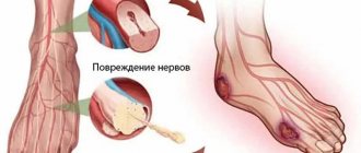
Core callus (tyloma, clavus durus) is a dense and sharply limited area of hyperkeratosis of the epidermis, small in size, round in shape with clear boundaries, smooth edges, located in the area of pressure of bony protrusions and processes on the underlying soft tissue. Most often, dry calluses are located in the area of the dorsal surface of the interphalangeal joints, the lateral surface of the 2–5 toes, as well as in the area of the PFJ with transverse flatfoot [4]. Callus must be differentiated from plantar warts. When a core callus is formed, in addition to the focus of hyperkeratosis, a very hard translucent core is also formed, located in the center of the callus and consisting of very dense horny masses. When pressure is applied to the core callus, sharp pain occurs due to compression of the dermal nerve endings located between the dense core and the bony process (Fig. 2). The same pain occurs when pressure is applied to the plantar wart. However, a plantar wart is painful not only with vertical pressure, but also with lateral compression; a change in the skin pattern is always noted above the wart; there are brownish inclusions, represented by microhemorrhages from the capillaries. In addition, around the “mother” wart we often note numerous “daughter” plantar warts of smaller sizes (Fig. 3).
For a soft callus, the typical location is on the skin between the fingers. Due to the increased humidity in this area, the callus macerates and acquires a soft consistency. Soft calluses are also very painful and are often complicated by a secondary bacterial infection.
Subungual hyperkeratosis is quite common and can be observed with onychomycosis, traumatic onychia and other types of dystrophy [5]. It is characterized by a gradual enlargement of the nail plate from the distal edge, while gray-yellow horny masses accumulate between the free edge of the nail and the hyponychium. Subungual hyperkeratosis is considered one of the pathognomonic signs of onychomycosis, since it is assumed that keratinocytes respond to invasion of fungal infection by hyperproliferation. Therefore, if such a symptom is present, it is imperative to conduct a test for pathogenic fungi.
In recent years, there has been a steady increase in the incidence of diabetes mellitus (DM). About 8–10% of patients with diabetes suffer from diabetic foot syndrome. Diabetic foot syndrome is a complex of anatomical and functional changes that develop against the background of the main manifestations of diabetes mellitus: neuropathy, micro- and macroangiopathy, osteoarthropathy, contributing to increased trauma and infection of the skin and soft tissues of the foot, the development of a necrotic process and, in advanced cases, leading to amputation. Diabetic foot syndrome manifests itself in the form of purulent-necrotic processes, ulcers and osteoarticular lesions that occur against the background of specific changes in peripheral nerves, blood vessels, skin and soft tissues, bones and joints. Prevention of the development of diabetic foot syndrome is of utmost importance in the treatment of patients with diabetes. The skin of patients with diabetes, especially type 2, is prone to excessive dryness, hyperkeratosis and cracking, which is a favorable condition for the development of an infectious process.
Thus, the risk of developing onychomycosis of the feet in patients with diabetes is 2–8 times higher than in the general population [6]. Every third of the 175 million patients with diabetes has mycosis of the feet. In the USA alone there are about 7 million such patients [7]. According to various authors, the frequency of onychomycosis in people with diabetes ranges from 20 to 60% [8]. In patients with diabetes, the skin of the feet is most often affected. Moreover, of all forms of mycosis of the feet, the most common is squamous-hyperkeratotic, but intertriginous and dyshidrotic are also found. In diabetes, the squamous-hyperkeratotic form is manifested by dry flat papules and slightly lichenified nummular plaques of a bluish-reddish color, usually located on the arches of the feet. The surface of the rash, especially in the center, is covered with layers of grayish-white scales of varying thickness; along the periphery there is a “border” of exfoliating epidermis; Upon careful examination, you can notice single bubbles. The rashes, serpiginating and merging, form diffuse foci of large sizes, which can spread to the entire sole, lateral surfaces and back of the feet. Along with such scaly lesions, patients with diabetes often have hyperkeratotic formations of the type of limited or diffuse yellowish calluses with frequent cracks on the surface. In diabetes due to angiopathy, the trophism of the nail bed and matrix is disrupted, the growth rate of the nail plates decreases, the nails change shape and thicken. Trophic disorders lead to the fact that hypertrophic forms of nail damage are most common in patients with diabetes (Fig. 4). In this case, the nails change color and pronounced subungual hyperkeratosis develops; the nail loses its shine, becomes dull, thickens and deforms until onychogryphosis forms; partially collapses, especially from the sides; patients may experience pain when walking. Often, a thickened, deformed nail affects the skin of the lateral ridges, which leads to the formation of paronychia and ingrown nails. In elderly patients, nails changed according to the type of onychogryphosis can lead to the formation of bedsores. These features of the clinical picture and course of onychomycosis of the feet in patients with diabetes increase the risk of ulcerative-necrotic complications, which can lead to the development of gangrene. As a rule, with diabetes there are multiple lesions of the nail plates, which complicates the treatment of onychomycosis in this group of patients.
Treatment methods for plantar hyperkeratoses
Treatment of plantar hyperkeratoses should be comprehensive and include elimination of the causes that caused excess pressure on the skin of the feet, selection and wearing of comfortable shoes, treatment of concomitant pathologies, including mycosis of the feet.
In most foreign countries, patients with plantar hyperkeratosis turn to podiatrists or podiatrists - specialists involved in the diagnosis and treatment of both biomechanical disorders and dermatological diseases of the feet [1]. In Russia, podiatry practice is not properly developed and does not have state certification. There are only a small number of private centers that offer podiatry services on a paid basis. In these centers, using special instruments, medical pedicure devices with rotating burs and cutters, areas of limited hyperkeratosis are removed layer by layer and painlessly. In addition, these centers produce special individual orthopedic insoles, prostheses and correctors that can redistribute the load to other areas of the skin of the foot, which also has a beneficial effect on reducing the severity of plantar hyperkeratosis.
At home, there are many methods to combat plantar hyperkeratosis. As a means for removing horny masses, you can use various pedicure brushes, pumice stones, blades, scrubs, etc., which are presented in abundance on our market, and their arsenal is constantly increasing. In addition, today there is a large selection of cosmetics for foot skin care.
The necessary requirements for an external product used in the presence of plantar hyperkeratoses are as follows: this product must have a pronounced keratolytic effect and at the same time moisturize the skin of the feet with increased dryness, which is often observed with hyperkeratotic lesions (Fig. 5). Of course, such a remedy is urea, which is part of many moisturizing and keratolytic products. For more than 100 years, urea has been successfully used in dermatological practice. Back in 1957, Kligman wrote: “Sometimes in our enthusiastic search for new therapeutic substances we do not pay enough attention to old remedies whose luster has long since worn off, but which nevertheless may at certain times be much more useful than new miracle drugs that fail . In the world of external therapy, such a drug is urea” [9].
Urea can be used, depending on the concentration, when treating the wound surface, treating hyperkeratosis and increased dryness, atopic dermatitis, psoriasis, ichthyosis, eczema, keratosis, keratosis Pilaris, keratoderma, for traumatic and ingrown nails. In low concentrations (2–10%), urea has proven itself well as a basic moisturizing therapy for inflammatory dermatoses; in high concentrations – 40% or more – it can even dissolve the nail plate, therefore it can be used in therapy in combination with antifungal drugs [10– 12].
Foretal Plus cream can occupy a special place, in our opinion, in the treatment of plantar hyperkeratosis and increased dryness of the skin of the feet. This is one of the few drugs on the domestic market that combines a combination of urea and phospholipids. The concentration of urea in it is 25%. This, on the one hand, has a pronounced keratolytic effect, helps to cope with increased dryness, and gets rid of rough skin on the heels. On the other hand, this product also has a pronounced moisturizing effect due to urea and phospholipids, which are known to be necessary for skin cells, since they are the main components of plasma membranes and their main suppliers. The essential polyunsaturated fatty acids contained in phospholipids ensure the mobility of the cell membrane, which is necessary for the normal functioning of cells and the synthesis of lipids in the stratum corneum of the skin, which are responsible for its barrier functions. Phospholipids spontaneously organize into layered structures and create storage reservoirs of moisture for the skin. Having a high affinity for the skin, they bind to its cells (keranocytes) and create a long-lasting moisturizing effect. The moisturizing effect of phospholipids is explained not only by their ability to bind water, but also due to their ability to form bilayer structures in water, they form a thin film on the surface of the skin, which protects it from moisture loss. It is also important to note the pronounced regenerating ability of phospholipids, taking into account the characteristics of the skin of the feet, where the processes of epidermal renewal occur especially intensively due to mechanical stress.
Do not forget that urea in such a concentration (25%), which is used in Foretal Plus cream, can have an auxiliary effect in the treatment of hyperkeratotic forms of mycosis of the feet in combination with antifungal agents and can be used as a prophylaxis for fungal infection, because It eliminates entrance gates well – foci of hyperkeratosis and cracks. In addition, Foretal Plus cream fights well against increased dryness and eliminates excess keratin, which mushrooms use as a food substrate. Therefore, this cream has both a therapeutic and preventive effect in the treatment of mycoses of the feet.
Thus, the constant use of Foretal Plus cream can replace patients with a visit to the podiatry office, since this drug actively helps fight the manifestations of plantar hyperkeratosis.
Podologist's advice
- Treat wet calluses in a timely manner, which over time can transform into dry calluses and then into core calluses.
- Wear shoes that fit comfortably. Avoid shoes with too thin soles. Don't wear stilettos all the time.
- Socks should be made from natural fabrics.
- Use antiperspirant for your feet if you suffer from excessive sweating.
- Maintain personal hygiene and do not wear other people's shoes.
- Watch your weight and use orthopedic insoles if necessary. It is not recommended to select them yourself; to do this, contact a specialist.
Advantages of SM-Clinic
Our medical center employs some of the best specialists in St. Petersburg. The latest equipment is used to conduct research and any interventions. We have our own laboratory. With us you can undergo an examination and take the necessary tests quickly, comfortably, and without queues. Recovery after the procedure takes place within one day. The clinic has a hospital with comfortable rooms where patients are accommodated if necessary. You can make an appointment by phone or online.