Competition "bio/mol/text"-2017
This work was published in the “Free Topic” category of the “bio/mol/text” competition 2017.
The general sponsor of the competition is: the largest supplier of equipment, reagents and consumables for biological research and production.
The sponsor of the audience award and partner of the “Biomedicine Today and Tomorrow” nomination was.
"Book" sponsor of the competition - "Alpina Non-Fiction"
One monk, wandering around the world, met the Plague, which was heading to his city. -Where are you going, Plague? - he asked her. “I’m going to your hometown,” she answered. “I need to take a thousand lives there.” After some time, the monk again met Plague on his way. - Why did you deceive me then? - he asked her reproachfully. “You said you had to take a thousand lives, but you took five thousand.” “I told you the truth then,” Chuma answered. “I really took a thousand lives.” The rest died of fear.
Chastnikova V. Jewish parables. The sage is higher than the prophet [1]
The victims of the plague numbered in the hundreds of thousands and even millions of people, cities died out, entire regions became deserted, and the horror of plague pandemics eclipsed the horrors of all wars known in human history. For thousands of years, people did not understand what was the source of the disease [2].
The Bible is one of the oldest surviving accounts of plague epidemics (1 Kings, chapter 5; 2 Kings, chapter 19, verses 35–36). There have been three pandemics of this disease in world history:
- the first pandemic - the “Justinian plague” - named after the Byzantine emperor Justinian, began in Egypt (542 AD) and spread throughout the entire civilized world;
- the second pandemic - the “Black Death” (1340) - raged furiously from China to Western Europe and was accompanied by the death of about 25 million people (about a quarter of the then population of Europe), and the number of victims worldwide was estimated at 75 million people [3 ];
- The third pandemic, which originated in the Chinese province of Yunnan in 1855, spread to all inhabited continents within a few decades. In China and India alone, the total number of deaths was more than 12 million. According to the World Health Organization, echoes of the pandemic were also observed in 1959 [4].
Large outbreaks of plague are recorded with a certain periodicity (India - 1994; Madagascar - 2011 and 2013). In the USA, from 1965 to the present, up to 40 cases of human plague infection are recorded annually (an average of 10 patients per year) [5]. In Russia, in September 2014 and August 2015, for the first time in the last 35 years, two cases of human infection with plague were recorded [6], [7].
Bubonic plague is the most common form of the disease and, if untreated, leads to the death of 40–60% of those affected. The pulmonary form occurs either as a complication of bubonic or septic forms, or when inhaling air contaminated with the plague pathogen. If treatment is not started within the first 24 hours after the onset of symptoms, death occurs within 48 hours [8].
In nature, the plague microbe is found on almost all continents, with the exception of Australia, Antarctica, and the Arctic, which leads to annually recorded cases of this disease. The rapid evolution of microorganisms leads to the emergence of populations of bacteria (strains) resistant to antibiotics [9], which is especially dangerous in the case of the plague pathogen. In addition, these bacteria can be used as an agent of bioterrorism. All of the above explains the need to study the plague microbe.
The causative agent of plague, Yersinia pestis, is the most dangerous bacterium in the world [10]. What makes it so deadly?
Symptoms of bubonic plague
The name of this form of the disease comes from the Arabic and Greek words "juma" and "bubo", meaning "bean" and "groin". A characteristic symptom is inflammation of the lymph nodes in the groin (more than 50% of cases). Inflamed lymph nodes look like beans. Buboes appear within a few hours after the true onset of the disease. They also develop in the armpits (about 20% of cases), under the jaw and on the neck. The small ones are the size of a hazelnut, the large ones are the size of a chicken egg. The buboes are very painful, so the patient tries to take a position in which the inflamed lymph node is not affected.
- Most often, a person develops one bubo in the groin. The inflammation affects neighboring tissues and skin, so after a day the bubo resembles a dense tumor with clear boundaries, which is characterized by acute pain.
- In the armpit, the tumor may have a jelly-like consistency. The danger is the spread of infection to the lungs.
- Buboes on the neck are the most dangerous, accompanied by massive swelling of the tissues up to the shoulder blades and lower chest. The patient's condition worsens significantly as the swelling spreads throughout the head. Possible development of meningitis.
Buboes on the leg of a plague patient. Photo: PHIL CDC
Virulence factors, or armed and very dangerous
Any pathogenic bacteria must have a number of properties: “abilities” for invasion (invasion), colonization, resistance to the host’s immune reactions and toxicity. Biomolecules that perform these functions are called pathogenicity (virulence) factors.
Since the discovery of the plague causative agent in 1894 by the Frenchman Alexandre Yersin and the Japanese Kitasato Shibasaburo, scientists have been trying to figure out what determines the pathogenicity of Y. pestis. As a result of many years of hard and risky work, which continues to this day, the following pathogenicity factors have been identified:
- outer membrane proteins (Yersinia outer proteins—called Yop proteins, effector proteins, or the Yop-virulon complex) [11];
- pigmentation area complex [12];
- plasminogen activator [13];
- capsular antigen [14];
- pili adhesion or pH6 antigen [15].
Proteins of the outer membrane, or why does the plague pathogen need a syringe?
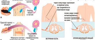
The study of individual Yop proteins, namely V- and W-antigens, began back in 1956 [16]. YopM, YopN, YopH, YopE, YopD and other proteins have also been described [17]. They suppress the development of immune reactions, in particular, phagocytosis, and when microbes are “swallowed” by macrophages (immune cells), they ensure the multiplication of the pathogen inside macrophages [11]. Outer membrane proteins are synthesized only at a temperature of 37 °C and under conditions of calcium ion deficiency (low calcium response) [18]. The mechanism of action of Yop proteins—type III secretion system—was discovered at the end of the last century [19]. According to this mechanism, a plague microbe, approaching a eukaryotic cell, injects effector proteins into the cytoplasm (according to the principle of a syringe with the formation of a special channel - a “needle”) (Fig. 1) [20]. Scientists pay special attention to the V-antigen, since it is used to create a chemical vaccine against plague [21].
Figure 1. Diagram of the action of the type III secretion system.
[19]
Complex area of pigmentation, or can the need for something become a factor of pathogenicity?
During laboratory work, researchers found that the plague microbe can become avirulent (safe) for mice. When such bacteria are sown on nutrient media with hemin (an iron-containing substance formed by the action of hydrochloric acid on hemoglobin), they form uncolored colonies, in contrast to pigmented colonies of virulent strains. This is how one of the simplest ways to determine the virulence of plague pathogen strains without the use of laboratory animals appeared. If a bacterial population on a medium with hemin (Jackson-Burrows medium) “gives” the growth of pigmented colonies, then it is dangerous (virulent), if unpigmented, then it is not dangerous (avirulent) [22].
Subsequently, it turned out that the genes of the hms locus of the pigmentation region on the bacterial chromosome are responsible for the color of virulent colonies. This pigmentation region also contains the Y. pestis pathogenicity island, HPI. What is remarkable about the genes of this island and why was it given such a meaningful name? It turns out that iron ions Fe3+ are necessary for the life of the plague bacterium. The capture and transport of Fe3+ into the cell is carried out by low molecular weight molecules with a high affinity for iron - siderophores [23]. The genes encoding siderophores form the pathogenicity island. At least one siderophore system, namely the yersiniobactin protein synthesis and transport system, provides active iron transport in Yersinia. However, the entire pigmentation region is extremely unstable and can be spontaneously deleted from the chromosome. The loss of the siderophore system from the genome leads to a significant decrease in the virulence of the plague pathogen [24]. Therefore, the need for iron began to be considered as a determinant of the virulence of the plague microbe. And since deletion of the pigmentation region also removes the hms locus, which encodes the pigment sorption trait, they began to separate dangerous and non-dangerous Yersinia populations using this “mark.”
Plasminogen activator, or two-faced Janus
Much attention is paid to omptins, a family of outer membrane proteases that perform many functions in the bacterial cell, including the transport of various substances across the outer membrane, and, in general, contribute to the adaptation of the microorganism to environmental conditions. One of them is plasminogen activator (Pla) [25]. Even before Pla was isolated, experts came to seemingly contradictory conclusions. On the one hand, the plague microbe coagulates blood plasma (plasma-coagulating activity), on the other hand, it prevents the formation of a blood clot (fibrinolytic activity). It is all the more surprising that both of these activities, as later turned out, are possessed by the same protein—plasminogen activator. It turned out that at temperatures below 30 °C Pla exhibits plasmacoagulating activity. In the proventriculus of an infected flea (a carrier of plague microbes), the blood coming from a rodent sick with plague coagulates and a “block” is formed - a reservoir for the reproduction of Yersinia. In this case, a hungry flea begins to actively bite an animal or person without feeling full. When a fresh portion of blood arrives, the pathogen with the flea’s “belching” penetrates the wound and infects it. Once in another environment with a temperature of 36–37 ° C - the temperature of a human body (or slightly higher - a warm-blooded animal), the plasminogen activator begins to act in exactly the opposite direction: it exhibits fibrinolytic activity - it prevents the formation of a blood clot at the site of the bite and thereby ensures the spread pathogen [26].
The article “Black Death. The story of how a harmless bacterium became a merciless killer" [35]. - Ed.
When plague microbes are inhaled (and the development of pneumonic plague), this protein ensures the rapid proliferation of bacteria in the lung tissues and leads to the development of fulminant pneumonia and pulmonary edema, whereas in the absence of Pla the infection does not develop into fatal pneumonia. It has been established that plasminogen activator disrupts the constancy of the internal environment of the host body and blocks immune reactions aimed at destroying the pathogen [27].
Capsular antigen, or slippery type, this plague pathogen
The bacteria are surrounded by a capsule of mucous substance (fraction I, Fra1), which prevents the absorption and neutralization of Y. pestis by the host immune cells during the process of phagocytosis. Many modern methods of laboratory diagnosis of plague are based on the identification of this antigen substance; it is part of many experimental chemical vaccines against plague. However, bacterial populations lacking a capsule were later discovered [28]. In addition, many other microorganisms have a mucous capsule, for example, the causative agent of anthrax and tularemia. The capsular substance of Yersinia is formed at a temperature of 37 °C.
Pili adhesion (pH6 antigen), or "agent 007"
The specific antigen pH6 (adhesion pili - small villi, PsaA protein) is also synthesized only at a temperature of 37 ° C, but there is one more condition - pH below 6.4 (which is reflected in its name). It is believed that this pH value is close to the pH of lysosomes of macrophages or the necrotic contents of abscesses, where this antigen is probably synthesized. The pH6 antigen is responsible for the attachment of plague microbes to the epithelial cells of the respiratory tract and their colonization. This antigen is also interesting because it suppresses phagocytosis: when Yersinia enters the blood, pH6 antigens combine with plasma apolipoprotein B, which makes bacterial cells “invisible” to macrophages [29]. Consequently, in an initially aggressive warm-blooded organism of the host, populations of the plague microbe with the pH6 antigen have a greater chance of survival than populations of the plague pathogen without the pH6 antigen—a phenomenon of selection of PsaA+ bacteria [30]. In natural plague foci, only Y. pestis strains with pH6 antigen are isolated.
Antigens similar to pH6 were found in a number of pathogens that cause less dangerous diseases - intestinal infections (Y. pseudotuberculosis [31], Y. enterocolitica [32], Escherichia coli [8]).
Pathogenesis
The causative agent of plague penetrates the human body through the skin, gastrointestinal tract and respiratory tract. It is the mechanism of penetration of the pathogen that determines the development of a specific clinical form of plague. The human body (its adaptation mechanisms) is not adapted to resist the introduction/development of the plague bacillus, which is due to the rapid reproduction of the pathogen and the massive production of permeability factors ( fibrinolysin , neuraminidase , pesticin ), antiphagins that suppress the process of phagocytosis (V/W-Ar, F1, PH6- Ag, HMWPs), which contributes to their massive/rapid dissemination by the lymphogenous/hematogenous route into the organs of the mononuclear-phagocytic system and its subsequent activation. The release of inflammatory mediators and massive antigenemia contribute to the rapid development of microcirculatory disorders, and in the absence of timely assistance, DIC syndrome with the subsequent development of infectious-toxic shock.
In the cutaneous form, a specific reaction (primary affect) may occur at the site of the entrance gate - in the form of an ulcer/pustule with hemorrhagic contents. Then the pathogen migrates through the lymphatic vessels to the regional lymph nodes (plague bubo), in which the reproduction process occurs, accompanied by an inflammatory reaction. During the process of reproduction, lymph node macrophages (bubonic form) undergo enlargement, fusion, and in some cases, the formation of a conglomerate from lymph nodes. At this stage, the causative agent of the disease, due to the lack of specific antibodies/the protective effect of the capsule, is resistant to phagocytosis by leukocytes. Subsequently, hemorrhagic necrosis , in which microorganisms enter the bloodstream and invade various internal organs, causing generalization of the process. During their breakdown, endotoxins , which causes the manifestations of intoxication.
The septic form of plague is characterized by the rapid formation of multiple secondary foci of infection, bacteremia/toxemia. Developing endotoxemia leads to microcirculation disorders, capillary paresis, development of disseminated intravascular coagulation syndrome and metabolic disorders in the tissues of the body, which is clinically manifested by encephalopathy , acute renal failure , infectious-toxic shock , which determine unfavorable outcomes.
Temperature factor, or what really matters
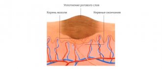
It is necessary to focus on the special role of temperature in the physiology of the plague microbe. It is at a temperature of 37 °C that its nutritional needs increase [33] and almost all known virulence determinants are synthesized (Fig. 2) [34]. In other bacteria, this dependence is less pronounced, which suggests the leading role of the temperature factor in the virulence of the plague pathogen [8].
Figure 2. “Protein portraits” of a plague microbe grown at temperatures of 28 and 37 °C, respectively.
[34]
Who carries the infection
The carriers of the infection are various rodents (rats, field mice, shrews, gerbils and voles) and some animals, mainly living in the steppe zone. These are hares, gophers, marmots, tarbagans, and hedgehogs. They excrete the plague pathogen in their droppings; the bacteria are contained in dead animals - this is how they get into the water and soil, from where they are further spread by other individuals. The carriers are most often fleas, which parasitize on carriers and live in the soil³.
The yellow gopher is a carrier of the infection. Photo: Joint831 / Wikipedia (CC BY-SA 3.0)
Genome or everything important inside
Modern technologies have made it possible to decipher the genome of the plague microbe, which, as it turned out, has more than 98% similarity with the relatively harmless bacterium Y. pseudotuberculosis, the evolutionary predecessor of the plague causative agent (an event 5-7 thousand years ago) [35]. Interestingly, the acquisition of the unique virulence of Y. pestis was accompanied by the loss of some genes. For example, the loss by a plague microbe of the gene encoding adhesin A (YadA), one of the most important virulence factors of the pathogen pseudotuberculosis, leads to blocking the formation of extracellular neutrophil traps, the most effective process of pathogen destruction at present [36]. Neutrophil leukocytes in the absence of adhesin A cannot form a “catching net” from their own DNA and proteolytic enzymes, trap bacteria in it and break them down [37]. The plague causative agent also has a “poorer” set of genes for the Yop-virulon effector proteins than other representatives of the Yersinia genus [8].
Read about sequencing the genome of Yersinia, which caused the Black Death of 1340, in the material “It’s a Plague” [3]. - Ed.
In addition to the chromosome, the plague microbe has plasmids—extrachromosomal sections of DNA [38]. Most protein virulence factors are encoded on plasmids: effector proteins on the pCad plasmid; capsule - pFra; plasminogen activator - pPla (pPst, pPCP). Plasmids pFra and pPla are found only in Y. pestis (species-specific), pCad is common with the causative agent of pseudotuberculosis (genus-specific) [20].
List of sources
- Supotnitsky M.V., Supotnitskaya N.S. Essays on the history of the plague: In 2 books. - Book I: Plague of the prebacteriological period. - M.: University Book, 2006. - 468 p.
- Bryukhanova G.D. Current aspects of the epidemiology and microbiology of plague in modern conditions: Dissertation of the Doctor of Medical Sciences. – 2004.- M.- 250 S.
- Anisimov A.P. Factors of Y. pestis that ensure the circulation and preservation of the plague pathogen in the ecosystems of natural foci // Molecules, genetics. 2002. - No. 3. - P. 3 - 23.
- Innokentyeva T.I. Modern aspects of plague surveillance // Journal. infectious pathol. 1997. - T. 4., No. 1. - P. 8 - 14.
- Nikiforov V.V., Avdeeva M.G., Namitokov H.A. Plague. Educational and methodological manual. 2016. - 121 p.
How does pneumonic plague develop?
When the patient coughs and breathes, the infection releases into the air - this should be taken into account when observing safety measures. An important symptom is a wet cough with sputum. It most often appears towards the end of the first day of illness (in some cases from the very beginning). Sputum contains a huge amount of “plague microbe”. Sputum has a thin consistency (for example, with pneumonia it is thicker). Her character changes as the disease progresses. At first it is transparent, then it comes out with foam, becomes glassy, and then traces of blood appear in it. Eventually the sputum becomes permanently bloody.
On the 1st day the condition quickly and significantly worsens. The patient has difficulty breathing, he is acutely short of air, constantly agitated, trying to get up and get closer to the open window or door. The height of the disease lasts from several hours to 2 days. During this period, the body experiences severe toxic damage.
On the 2nd day, excitement gives way to lethargy due to general depression of the central nervous system. Patients become delirious, their speech becomes slurred, coordination is impaired, and sensitivity to light increases. They still try to stay close to sources of fresh air. Arrhythmia, acute renal failure intensifies, and blood circulation is disrupted. Chest pain becomes more acute and makes it difficult to breathe.
On the 3rd day, pulmonary edema develops and bloody, foamy sputum appears. The patient may fall into a coma, the pulse can barely be felt. All over the body and especially on the face, bluish spots of internal hemorrhages appear. In such cases, death is inevitable.
How does bubonic plague occur?
The peak of clinical manifestations of bubonic plague occurs on days 4-6, when intoxication of the body reaches its maximum. The patient constantly has a high temperature of about 40°C and is febrile. The face is red and swollen, and an expression of horror often appears on it. On physical examination, cardiac dysfunction and liver enlargement are noticeable. Blood pressure decreases. Already at this stage, a patient with low immunity may have a fatal outcome.
If the patient experiences the peak of the disease, then on days 6-8 the bubo noticeably turns red and swells, and after another 2 days it opens. It produces yellow-green pus, which may contain traces of blood. The pus has no obvious odor. During this period, the patient becomes much better. If the body is not attacked by a secondary infection, the prognosis is favorable. Overgrowth of a purulent fistula occurs slowly - within 3-4 weeks. A deep scar forms in its place.
At any time during the disease before the bubo ruptures, the infection may spread to other lymph nodes and the formation of secondary buboes. This is one of the most common complications.
Temperature during bubonic plague
- In some cases, 1-2 days before the appearance of the bubo, the patient is worried about fever with high temperature. After the onset of inflammation, the temperature is consistently high - 39-40°C.
- If it begins to decrease gradually and steadily, this is a positive sign.
- If there is a sharp decrease and a subsequent sharp increase, this, on the contrary, indicates sepsis and a significant deterioration and an unfavorable prognosis.
How does septicemic plague occur?
2-4 hours after the onset of the disease, the patient’s psyche is disturbed - most often he is in an excited state. The temperature is above 40°C, blood is visible in the vomit, and a rash appears on the skin. The face takes on a pained expression.
A doctor’s examination reveals disturbances in the heart’s function, an increase in heart rate to 130 beats/min. In the next 24 hours, signs of tachycardia intensify, and focal traces of internal hemorrhages appear on the body. In some patients, they merge into large spots - the so-called “killing fields”. There are obvious traces of blood in the urine. Patients suffer from diarrhea, and blood is visible in the discharge. Kidney function is disrupted and urine production stops. If medical assistance is not provided in the first day, usually by the end of the second day the disease ends in death.
Preventing the spread of plague
People living in areas of natural outbreaks of the disease are vaccinated. Rodents and fleas are regularly controlled in these areas through disinsection. In private homes, you need to store food in places inaccessible to rodents, remove garbage and waste that attracts them. When traveling outdoors, you should use insect repellent. If there is a need to spend the night in the steppe, you need to carefully choose a place away from rodent burrows, and the area should be inspected for the presence/absence of droppings.
Symptoms of pneumonic plague
The disease is quickly transmitted by airborne droplets, therefore it is extremely dangerous from an epidemiological point of view. Patients fall ill suddenly. Just an hour ago, a person who felt great begins to shake with chills. He develops all the main symptoms of the plague (headache, high fever, nausea, vomiting, aches in muscles and joints) plus cutting pains in the chest that are characteristic of this form.
An important distinguishing feature is that shortness of breath develops in the first hours. Physical examination reveals tachycardia and arrhythmia. However, there are initially no functional abnormalities in the lungs.
Symptoms of septicemic plague
This form is characterized by a rapid course - only 2-4 days. In some cases, the duration of the illness is only a few hours. Requires immediate treatment, as without medical assistance death is inevitable. The incubation period is also characterized by the same rapidity - from the moment of infection to the first symptoms it can take from several hours to 1-2 days.
The onset of the disease is always sudden, violent and acute. The patient immediately develops symptoms of severe intoxication:
- acute headache;
- temperature 39-40oC;
- chills;
- pain in muscles, joints;
- nausea with vomiting.