Aplastic anemia is a serious disorder of the formation, development and maturation of blood cells. It is characterized by inhibition of the hematopoietic function of the bone marrow, which is manifested by a deficiency in the formation of white and red blood cells, as well as platelets. Sometimes there is a lack of formation of only red blood cells. The disease is considered one of the most severe disorders of hematopoiesis and, in the absence of adequate treatment, can cause death within several months. It affects male and female patients equally between the ages of ten and twenty-five or over fifty. According to medical statistics, two cases of pathology per one million people are diagnosed every year.
The Department of Hematology at CELT offers diagnosis and treatment of aplastic anemia in Moscow. Our multidisciplinary clinic was one of the first in the Russian Federation to begin operating in the market of paid medical services and has been successfully continuing it for the third decade. The hematology department receives consultations from leading domestic specialists, who have a modern diagnostic and treatment facility that allows them to accurately diagnose and carry out treatment in accordance with international standards. The cost of services is available in our price list, which we update regularly. To avoid misunderstandings, we ask you to check the numbers with our information line operators.
Aplastic anemia: etiology
According to origin, congenital and acquired anemia are distinguished. The first develops as a result of chromosomal mutations, the second - under the influence of chemicals, radiation, and infections. Experts believe that suppression of bone marrow hematopoiesis can be initiated by the appearance of cytotoxic T-lymphocytes in it and in the blood. They produce TNF (extracellular protein) and interferon “y”, which have a suppressive effect on hematopoietic germs. The reason for starting the mechanism may lie in:
- Exposure to ionizing radiation or chemicals in the form of aromatic compounds, arsenic, pesticides;
- Entering the body of infectious agents (causative agents of hepatitis “D”, “B”, cytomegalovirus, DNA-containing Epstein-Barr virus);
- Taking myelotoxic drugs while undergoing treatment with tranquilizers, anticonvulsants, antithyroid and antitumor drugs;
- Development of a number of autoimmune processes (systemic lupus erythematosus, connective tissue damage - Sjögren's syndrome).
In 50% of cases, the cause of the development of the pathology cannot be determined. This form of aplastic anemia is called idiopathic.
And a few more words about the forecast...
{banner_banstat10}
Although the prognosis of aplastic anemia was briefly touched upon during the description of the disease, I would like to add a few more words.
The main causes of death in patients with AA are infections and bleeding. Some authors believe that very bad prognostic signs are: taking chloramphenicol (even in short courses) and the development of the disease after infectious hepatitis (severe bone marrow aplasia is considered an indication for bone marrow transplantation at the very beginning of the disease).
However, you should not immediately despair and perceive AA as a death sentence.
The use of modern treatment methods, in general, improves the situation and allows many patients to significantly increase their life expectancy, or even get rid of a serious illness altogether.
More than half of AA patients live 10 years or longer after using immunosuppressive drugs. The chances increase markedly in people (up to 75%) who have had a successful donor bone marrow transplant.
In general, the prognosis is based primarily on the form of the disease.
For example, idiopathic AA, if it
does not
occur in an acute, super-severe form, gives more hope for long-term survival or recovery. But only the attending physician can predict something in advance (and even then with great caution); our task is only to provide general information about such a pathological condition as aplastic anemia.
Classification of aplastic anemia
| Form of pathology | What is the difference? |
| By duration | |
| Acute | No more than one month |
| Subacute | From one month to six months |
| Chronic | More than six months |
| According to the severity of selective aplasia | |
| Moderate | Granulocytes less than 0.0x10⁹/l, platelets less than 20.0x10⁹/l. |
| Heavy | Granulocytes less than 0.5x10⁹/l, platelets less than 20.0x10⁹/l. According to diagnostic results, bone marrow cellularity is less than a third of normal. |
| Very heavy | Granulocytes more than 0.5x10⁹/l, platelets more than 20.0x10⁹/l. |
Aplastic anemia: symptoms
The disease begins acutely, it is accompanied by a feeling of severe weakness and fatigue. The patient's skin and visible mucous membranes look pale, and he himself suffers from the following clinical manifestations:
- noise in ears;
- the appearance of shortness of breath even with insignificant efforts;
- unpleasant tingling in the chest area.
When the number of platelets per unit volume of blood decreases, hemorrhagic syndrome appears:
- even after slight compression or shock to the skin, bruises and hemorrhages appear on it;
- a rash in the form of small dots can be seen on the body, arms and legs;
- bleeding gums are observed;
- spontaneous nosebleeds;
- heavy menstruation (in women).
A decrease in the number of leukocytes per unit volume of blood is characterized by the regular frequent development of infectious diseases of the skin and structures of the urinary system, inflammatory processes of the oral mucosa, and pneumonia.
The congenital form of anemia develops in children under ten years of age and is accompanied by a number of other disorders:
- underdevelopment of the skull and brain;
- reduction in the size and weight of the kidneys (hypoplasia);
- intense coloring of certain areas of the skin - hyperpigmentation;
- severe hearing loss and speech impairment due to it.
Symptoms
Suppression of bone marrow hematopoiesis will certainly change the situation in the periphery. A blood test will show pancytopenia (a sharp decrease in all populations of formed elements), which will undoubtedly sooner or later affect the patient’s well-being. Idiopathic aplastic anemia sometimes occurs in an acute form: the disease begins suddenly, progresses rapidly and practically does not respond to treatment. But more often it comes little by little, slowly changing the blood picture. The patient does not notice anything for a long time, since the process proceeds smoothly and adapts to the decrease in blood cells. But for the time being, because sometimes a critical moment comes that forces you to seek help.
When pancytopenia reaches a great depth, the remaining amount of formed elements, which still continues to circulate in the blood, cannot cope with the tasks of a normal community, depression of hematopoiesis begins to manifest itself with symptoms:
- Anemia, which is caused not only by impaired formation of red blood cells, but also by bleeding that occurs due to a decrease in blood platelets - platelets. Anemia is manifested by hypoxia, pale skin, headache, dizziness, shortness of breath, general weakness and malaise, inflammatory reactions of the oral mucosa;
- Thrombocytopenia with clinical signs of hemorrhagic syndrome (skin rashes, bleeding from the gums, nose, uterine cavity);
- A decrease in white blood cells - neutrophil leukocytes, which makes the body unable to resist infections. Neutropenia can be quite severe and cause various inflammatory diseases (otitis media, pneumonia, suppuration of hematomas, sepsis and other infectious complications accompanied by fever);
- Impaired cardiac activity during anemia will manifest itself as a systolic murmur, which the doctor will note during auscultation;
- Concomitant hemolysis - it occurs in some patients (hypoplastic anemia with a hemolytic component) due to the early death of red blood cells, which indicates not only quantitative abnormalities of red cells in the periphery, but also indicates their qualitative defects (the cause of hemolysis is a special syndrome of a combination of AA and paroxysmal nocturnal hemoglobinuria - PNH). Due to an increase in bilirubin in the blood, the patient’s skin becomes jaundiced;
- signs of the initial stage of AA
The change in the size of the spleen and liver is imperceptible in the first stages, but this happens somewhat later, for example, massive transfusions of red blood cells will cause hemosiderosis and splenomegaly, and circulatory failure will affect the liver (it will increase).
Acute anemia is usually an acquired variant of the disease. It should be noted that the acute super-severe form progresses quickly, it is difficult to fight it; within a few weeks, regardless of the intensive measures taken, the patient dies. Super-severe anemia often (10 times more often than in others) develops in people who were treated with chloramphenicol, known as chloramphenicol.
In congenital and hereditary forms, the disease has a predominantly chronic course.
Chronic anemia continues for a long time - sometimes subsiding, sometimes worsening, leaving the patient a chance to live, since sometimes it leads to complete relief from the disease, that is, recovery.
Aplastic anemia: diagnosis
Before starting treatment of the disease, CELT hematologists carry out a comprehensive diagnosis aimed at accurately making a diagnosis and identifying the etiological factor. It includes:
- examination by a hematologist;
- general and biochemical blood tests;
- collection of a bone marrow sample and its examination - sternal puncture.
If the disease is present, the patient is diagnosed with a serious decrease in hemoglobin, up to a critical level of 20-30 g/l, and agranulocytosis is observed - a decrease in granular leukocytes and monocytes. The number of lymphocytes may be normal or reduced, platelets are always reduced, sometimes they are not detected at all. Erythrocyte sedimentation rate – increases to 4-60 mm/h. A study of a bone marrow sample reveals an increased content of adipose tissue - 90%, which includes elements of stroma and lymph, but hematogenous cells are present in very small quantities.
Diagnostic measures
Diagnosing aplastic anemia is not that difficult. Most cases are found during routine examinations. One general blood test is enough to suspect the presence of aplastic anemia.
Then the doctor begins a detailed collection of the patient’s complaints, anamnesis, and notes symptoms characteristic of aplastic anemia.
And then the doctor prescribes more detailed studies to make an accurate diagnosis and determine the degree of complexity.
So, if AA is suspected, the doctor prescribes a bone marrow aspiration. Next, the bone marrow is examined under a microscope, the presence and condition of blood cells are assessed at the stage of their development. In aplastic anemia, the bone marrow is depleted of all cell lineages. The main diagnostic method for AA is bone marrow biopsy, which allows the most accurate assessment of the state of hematopoiesis.
The Doctor also prescribes:
— Serological tests for hepatitis and other viral formations;
— Laboratory tests to study kidney and liver function;
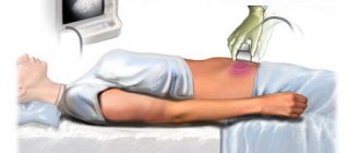
— Ultrasound diagnostics. Enlarged lymph nodes can be specifically examined.
— Test for antibodies to infections;
- number of reticulocytes
— presence of a PNG clone;
Sometimes it is necessary to undergo other tests to clarify the diagnosis and establish the cause of the development of aplastic anemia.
Although, quite often, the cause remains unknown, due to the large number of factors and triggers that can provoke it.
https://www.cinj.org/sites/cinj/files/documents/December%20National%20Aplastic%20Anemia%20Awareness%20Month%202018.pdf
Treatment of aplastic anemia
Treatment of idiopathic and other types of aplastic anemia is a very complex task that requires a comprehensive individual approach. When developing tactics, CELT specialists take into account the diagnostic results and the patient’s indications. The patient is placed in an isolation room with aseptic conditions, which eliminates the risk of developing infections and their complications. Drug therapy consists of taking:
- Glucocorticoids – in identifying autoimmune mechanisms and the formation of antibodies against one’s own blood cells;
- Cytostatics – in the absence of effect from treatment with glucocorticoids for autoimmune anemia;
- Cyclosporine “A” – to suppress the production of TNF and interferon “y”;
- Anabolic steroids – to stimulate hematopoietic function;
- Androgens – to stimulate the formation of red blood cells.
All patients with aplastic anemia receive transfusions of red blood cells and/or platelets, in volumes determined based on the clinical picture and peripheral blood parameters. In addition, the patient may be prescribed a splenectomy, a surgical procedure aimed at removing the spleen. Bone marrow transplantation can provide the most favorable prognosis. It consists of transplanting donor or own hematopoietic stem cells, previously removed from the iliac bones by puncture. Unfortunately, the procedure is not widely available due to the difficulty of selecting a compatible donor. If this is not possible, the patient is prescribed palliative therapy with cyclosporine A.
The hematology department of our clinic welcomes candidates, doctors and professors of medical sciences with over twenty-five years of practical and scientific experience. You can make an appointment with them online or by contacting our operators. Highly qualified specialists also work in the urology department. You can make an appointment with them for cystoscopy of the bladder.
At CELT you can consult a hematologist.
- Initial consultation – 3,500
- Repeated consultation – 2,300
Make an appointment
By making an appointment with a hematologist, you can get a comprehensive consultation. The doctor is competent to treat various blood diseases, most of which can be identified in the early stages and prescribe timely treatment to cope with the disease quickly and easily.
Anemia, pathology of hemostasis, oncohematology
Materials are presented from the RUDN textbook
Anemia. Clinic, diagnosis and treatment / Stuklov N.I., Alpidovsky V.K., Ogurtsov P.P. – M.: Medical Information Agency LLC, 2013. – 264 p.
Copying and reproducing materials without indicating the authors is prohibited and is punishable by law.
Aplastic anemia (AA) is a disease resulting from the disappearance or sharp decrease in pluripotent stem cells in the bone marrow, the number of which decreases to 1% or below, which leads to devastation or aplasia of the bone marrow. Morphologically, this is manifested by pancytopenia in peripheral blood tests, the absence of hematopoietic cells in bone marrow puncture. In the blood, pancytopenia is characterized by leukopenia due to neutropenia with relative lymphocytosis, the absence of reticulocytes. If such changes are detected, it is necessary to exclude all possible causes of aplasia (viral infections, malignant diseases, blood diseases (lymphoproliferative diseases), systemic collagenoses, contact with chemicals, radiation), and in adults, perform a trephine biopsy for histological examination of the bone marrow.
AA was first described by Ehrlich in 1888. The incidence of AA is 2–4 cases per 1 million population in the USA and Europe, while in some East Asian countries AA is much more common. Thus, in Thailand and Japan, the incidence of AA is 11–14 cases per 1 million population.
AA is most often detected in two age groups: 20–25 years and over 60 years. There is no statistically significant difference in incidence between men and women.
Etiology and pathogenesis
AA can be congenital, but more often it is acquired. Congenital, genetically determined AA were first described by Fanconi in 1927.
Congenital AA is clinically manifested by bone marrow failure in the first years of a child’s life and is often combined with congenital dysplasias such as skin dyspigmentation, hypoplasia of the kidneys and spleen, absence or hypoplasia of the radius, microcephaly, congenital heart defects and mental or sexual underdevelopment. Karyological studies in this variant of AA often reveal various chromosomal abnormalities. Among children with Fanconi anemia, there is also a high incidence of acute leukemia and other neoplasias.
In secondary AA, bone marrow failure is caused either by direct toxic effects of radiation or chemical compounds on stem cells, or by an aberrant response caused by viral infections (hemophagocytic syndrome). The most common diseases that provoke the development of AA are malignant diseases, blood diseases (lymphoproliferative diseases), and systemic collagenoses. Enlargement of peripheral lymph nodes, liver and spleen is not typical for AA. Moreover, the discovery of an enlarged spleen at an early stage of the disease casts doubt on the diagnosis of AA and argues in favor of hepatitis-associated aplasia. However, a long course of the disease may be accompanied by an enlargement of the liver and spleen due to post-transfusion hemosiderosis. In cases with an identified cause of AA, this condition should be considered as secondary hematopoietic aplasia.
True (idiopathic acquired) AA is a condition associated with the death of unchanged stem cells. In idiopathic forms, in which the cause of bone marrow aplasia is unclear, T-cell-mediated destruction of pluripotent hematopoietic stem cells of the bone marrow is assumed. In patients with the idiopathic form of AA, an increased number of activated cytotoxic T-lymphocytes, increased production of γ-interferon and tumor necrosis factor, which cause the death of their own bone marrow stem cells, were found in the blood. Moreover, the reason for the autosensitization of T lymphocytes against their own stem cells remains unclear.
Classification of aplastic anemia.
1. Idiopathic aplastic anemia
- congenital (Fanconi anemia)
- acquired
2. Secondary aplastic anemia caused by:
- medications (chloramphinecol, non-steroidal anti-inflammatory drugs, anticonvulsants, cytotoxic drugs)
- due to exposure to ionizing radiation
— chemical influences (benzene and its derivatives, pesticides, paints and varnishes)
— viral infections (Epstein-Barr, hepatitis, parvavirus, cytomegalovirus, HIV)
— autoimmune diseases (SLE, eosinophilic fasciitis, hyperimmunoglobulinemia)
- other reasons (pregnancy, thymoma)
AA Clinic
All clinical manifestations of AA are a consequence of bone marrow failure, their intensity depends on the severity and rate of progression of pancytopenia. The course of AA can be acute, subacute and chronic.
Patients with AA usually have an anemic syndrome in combination with hemorrhages on the skin, mucous membranes and infectious complications. The dominant symptoms of AA differ depending on the timing of the disease.
It is known that red blood cells circulate in the peripheral blood for 3–4 months, so symptoms of anemia can develop only with prolonged suppression of hematopoiesis for more than 1–2 months. As a result of a prolonged lack of red blood cell production, a gradual decrease in hemoglobin concentration occurs by 25–50%, that is, to 90–70 g/l, which may cause symptoms of anemia.
On the contrary, with the rapid death of the overwhelming number of stem cells, within a week hematopoiesis is depleted, the production of platelets and leukocytes stops, and the number of circulating red blood cells remains normal. Then, within a week, the number of platelets decreases, which leads to the appearance of hemorrhagic syndrome, against the background of a complete absence of anemic complaints. Before bleeding occurs, the body compensates for the lack of platelets using existing platelets, which are normally viable for an average of 10–11 days. Moreover, the decrease in platelets in the circulation occurs several days earlier than the appearance of hemorrhages, since the main number of them is represented by the parietal pool, which makes up the bulk of peripheral blood platelets and is consumed more slowly. Hemorrhagic syndrome with thrombocytopenia is associated with inadequate trophism and damage to the endothelium, increased fragility of small vessels and is manifested by bleeding of the mucous membranes (nasal, uterine bleeding), petechial rashes on the skin. Cutaneous hemorrhagic syndrome on the upper half of the body, especially on the face, is considered a life-threatening condition, which sharply increases the risk of bleeding in the brain.
As for leukocytes, especially neutrophils, their number decreases most intensively. The lifespan of granulocytes is no more than a week, and they circulate in the peripheral blood for several days, then enter the surrounding tissues, where they perform the main phagocytic function. Therefore, even against the background of a complete absence of neutrophils in the peripheral blood (agranulocytosis), infectious complications do not occur immediately, but, as a rule, after 5–7 days, and have their own characteristics. These patients often have a high fever in the absence of a source of infection. Thus, with shortness of breath, it is not possible to hear wheezing; with abdominal pain, the symptoms of peritoneal irritation are extremely blurred. To establish the diagnosis of pneumonia and peritonitis in this case, instrumental research methods (x-ray and ultrasound) are extremely important, with the use of which one can see the infiltration of lung tissue, the appearance of fluid levels in the intestines with atony, thickening of the intestinal wall and others. In patients with agranulocytosis, when fever appears, the risk of developing septic shock is extremely high due to the lack of barrier function of leukocytes. Such patients, when fever develops, necessarily need a blood test for sterility. Moreover, the diagnosis of sepsis in agranulocytosis can be established even contrary to the classical definition of sepsis, that is, without the presence of a “primary focus.” The most common cause of sepsis in agranulocytosis is “normal” intestinal flora (saprophytes or opportunistic bacteria).
In the acute course of AA, there is a rapidly progressing hemorrhagic syndrome caused by deep thrombocytopenia, and severe infectious complications due to the almost complete absence of granulocytes. Patients with a similar course of AA require emergency hospitalization in the hematology department and antibacterial, replacement, often intensive therapy, and specialized treatment. Without adequate care, such patients usually die within a few days or weeks of the first signs of the disease appearing.
In the moderate (subacute) course of AA, weakness and increased fatigue remain the patient’s main complaints for a long time; then, due to thrombocytopenia, symptoms of hemorrhagic diathesis may appear.
For secondary AA, which can clinically manifest itself weeks and even months after contact with the etiological factor, a chronic course is more typical. With secondary AA, more often than with the idiopathic form, remission of the disease develops and complete recovery may occur after the action of the etiological factor ceases.
Laboratory data
The picture of peripheral blood in 90% of patients with AA is characterized by pancytopenia: anemia, leukocytopenia and thrombocytopenia.
Anemia is normochromic in nature. The number of reticulocytes is reduced to 0 - 0.3%.
Leukopenia is caused by granulocytopenia, and the content of lymphocytes is usually not changed, which creates the impression of lymphocytosis, which is relative in AA. The severity of leukopenia largely determines the severity of the disease: the number of leukocytes <0.5 x 10⁹/l is observed in patients with severe AA, the number of leukocytes <0.2 x 10⁹/l is observed in patients with super-severe AA.
Thrombocytopenia is detected already in the early stages of the disease and is the most persistent hematological symptom.
The bone marrow aspirate is in the vast majority of cases very poor, i.e. contains a small number of hematopoietic cells, most of which are lymphocytes. However, since the process of bone marrow aplasia occurs unevenly and individual foci of normal hematopoiesis are preserved, it is possible to obtain active bone marrow with an almost normal cellular composition (with pancytopenia in peripheral blood analysis!). All this allows us to conclude that the myelogram in AA does not have a decisive diagnostic value.
The key method for diagnosing AA in adults is histological examination of the bone marrow. To conduct this study, a trepanobiopsy is performed, the results of which in AA indicate a total predominance of adipose tissue over active bone marrow.
Biochemical blood tests in most patients with AA reveal high levels of serum iron, increased levels of LDH and transaminases, and increased levels of erythropoietin.
The diagnosis of AA is based on a combination of peripheral blood pancytopenia, a decrease in bone marrow cellularity during sternal puncture and the detection of fatty bone marrow during histological examination, while other causes of hematopoietic aplasia are excluded.
Criteria for pancytopenia: Hb < 110 g/l, granulocytes < 2.0 x10⁹/l, platelets < 100 x109/l.
The classification of AA according to severity is shown in the table.
Classification of AA by severity
| Form of the disease | Criteria |
| Mild AA | — the patient does not meet the criteria for severe and super-severe forms; — granulocytes > 0.5 x 10⁹/l. |
| Heavy AA | — granulocytes <0.5 x 10⁹/l; — platelets < 20 x 109/l. |
| Super heavy AA | — granulocytes <0.2 x 10⁹/l. |
As can be seen from the table, the severity of AA is not determined by the hemoglobin concentration; it only takes into account severe thrombocytopenia, and the main factor determining the severity of the patient’s condition is the level of neutrophils in the peripheral blood. This is due to the existing ability to replace erythrocyte and platelet function by transfusion of erythrocyte and platelet mass obtained from donors, and the lack of methods for correcting neutropenia.
Differential diagnosis
AA sometimes has to be differentiated from diseases that may be accompanied by pancytopenia:
- acute leukemia;
- megaloblastic anemia;
- idiopathic myelofibrosis;
- hypersplenism;
- myelodysplastic syndrome (MDS).
In acute leukemia with hypoplasia of hematopoiesis, in contrast to AA, accumulations of blast cells are found in the bone marrow against the background of bone marrow hypoplasia, and in megaloblastic anemia - megaloblastic hematopoiesis.
In patients with idiopathic myelofibrosis and hypersplenism, severe splenomegaly is almost always present, which is always absent in AA. In addition, trepanobiopsy reveals myelofibrosis in the first case, and bone marrow hyperplasia in the second.
The combination of pancytopenia and bone marrow hypoplasia can be observed in 10% of patients with myelodysplastic syndrome. However, with MDS, along with hypoplasia of hematopoiesis, its dysplastic features are present in the form of megaloblastoidity, the presence of binuclear erythroblasts, Havell-Jolly bodies, sideroblasts (erythrocytes containing iron granules), sometimes an increased content of blast cells, as well as the presence of Pelger anomaly of granulocytes ( violation of nuclear segmentation) and microforms of megakaryocytes. In addition, in MDS, as a rule, chromosomal abnormalities are found that are absent in AA.
Treatment
The only treatment method for patients with AA that provides high survival rates (78–90%) and even complete recovery of patients is allogeneic bone marrow transplantation (ABMT).
ATKM from an HLA-matched donor is considered the method of choice in patients with AA if they meet the following criteria:
1. presence of an HLA-matched related bone marrow donor;
2. severe or super-severe form of AA;
3. the patient’s age is not older than 40 years;
4. short blood transfusion history.
As a rule, only siblings are used as related donors.
The standard mild regimen for preparing a patient (conditioning) for bone marrow transplantation includes the use of cyclophosphamide at a dose of 50 mg/kg from days 5 to 2, antilymphocyte globulin (ALG) from days 5 to 3, and methylprednisolone from days 5 to 3 . The main purpose of this conditioning is immunosuppression aimed at preventing transplant rejection, which with this regimen is reduced from 30% to 5%.
As a post-transplant immunosuppression aimed at suppressing graft-versus-host disease (GVHD), which is observed both in acute and chronic form (more often), cyclosporine A is used at a dose of 5 mg/kg per day for 12 months in combination with short courses of methotrexate 15 mg/m2 on days +1, +3, +6, +11.
The same regimen of conditioning and GVHD prevention is used when using bone marrow from HLA-matched unrelated donors. However, bone marrow transplantation from unrelated donors produces significantly worse results: only 29% of AA patients were alive 2 years after unrelated donor transplantation.
Previous blood transfusions are important in the effectiveness of bone marrow transplantation: in patients who did not receive blood transfusions, complete remission is achieved in 80% of cases with a five-year survival rate of about 70%, while in patients with sensitizing blood transfusion therapy, complete remission is achieved only in 50% of cases.
Experience in the treatment of AA with the introduction of hematopoietic cells has revealed the advantage of bone marrow transplantation over the use of mobilized stem cells obtained from the peripheral blood of a donor.
The number of mononuclear cells during bone marrow infusion should be at least 3.0x10⁹/kg with a minimum number of CD 34+ stem cells of 2.6x10⁶/kg.
Based on the assumption of the role of the suppressive effect of one’s own sensitized T-lymphocytes on bone marrow stem cells (with their subsequent apoptosis), the use of immunosuppressants with anti-T-lymphocyte action in the treatment of AA in patients was proposed. Like this:
- do not have HLA-compatible donors,
- patients with mild AA who depend on blood transfusions,
— patients with severe and super-severe forms of AA over 40–45 years of age.
The main therapy for AA is a regimen with a mandatory combination of pathogenetic treatment (immunosuppressive drugs and splenectomy), transfusion therapy and antimicrobial drugs.
Pathogenetic treatment
Most often, treatment begins with antilymphocyte (antithymocyte) globulin (ATG: atgam, thymoglobulin). The drugs are administered intravenously slowly over 12 hours, after a sensitivity test, the dose of the drug is 20 mg/kg/day. The total number per course is 4 introductions. To prevent serum sickness, prednisolone is used at a dose of 1 mg/kg or methylprednisolone 125–250 mg/day (before and after ATG) in combination with antihistamines (before and after ATG). From the 14th day of the course (day 1 of the course is counted from the first day of ATG administration), prednisolone must be discontinued. Cancel it a week in advance, gradually reducing the dose (daily by 1/3 - 1/2). The effect of treatment for ALH is observed in half of the patients, with complete remissions achieved in 15%.
The second immunosuppressant that is widely used in the treatment of AA is cyclosporine A (CyA), which blocks the production of interleukin-2 (IL-2), responsible for the proliferation of cytotoxic T-lymphocytes. CyA is used in a dose of 5 - 10 mg/kg per day for a duration course for at least 12 months. While taking SuA, magnesium preparations (Magne B6, Magnerot) should be prescribed, and liver (bilirubin, ALT, AST) and kidney parameters (creatinine) must be monitored. As with the use of ALG, its effectiveness is 50–60%, however, apparently, this drug does not provide sufficient immunosuppression and its withdrawal leads to relapse of the disease.
Currently, the most commonly used combination treatment regimen includes the administration of ALG for 4 days, and from the 14th day - the use of CyA for 12 months. The response rate to combination therapy is quite high and reaches 80% in severe AA with a five-year survival rate of 75%. If the combined use of ATG + CyA is ineffective, a second (after 3–6 months) and third (after 6–12 months) administration of ALG or replacement of one course of ATG with splenectomy is possible.
Treatment regimen for aplastic anemia (* - instead of ATG, splenectomy is possible)
When using ATG, serum sickness often develops. It manifests itself in 50% of patients 5–14 days after the end of drug administration with a rash, arthralgia, fever, laboratory signs of hepatitis, and increased blood pressure. In such cases, it is advisable to prescribe prednisolone at a dose of 0.5 - 1 mg/kg until the symptoms are relieved; in the absence of effect or severe symptoms of serum sickness, plasmapheresis is performed.
Transfusion therapy
In recent years, splenectomy has been performed in patients who have responded to ALG+SyA therapy, which has demonstrated its positive effect even before the introduction of immunosuppressive drugs into practice.
Maintenance (replacement) therapy, ensuring a satisfactory quality of life for patients, is of great importance in the treatment of aplastic anemia:
1. Red blood cells should be transfused when hemoglobin decreases to 80 g/l, when patients usually develop symptoms of tissue hypoxia.
2. The indication for platelet transfusion is a decrease in platelet count less than 20 x 109/l or hemorrhagic syndrome in the form of bleeding of the mucous membranes and/or in combination with rashes on the skin of the upper half of the body (during treatment with ATG, transfusions should be carried out when the platelet level is 40 - 60 109 /l).
Transfusions of blood components should be avoided in cases where the patient has HLA-compatible relatives and a bone marrow transplant is proposed. This prevents sensitization of patients and reduces the risk of transplant rejection. Long-term blood transfusion therapy leads to the development of hemosiderosis, since the erythromass packet contains 200–250 mg of iron.
Chelation therapy is used to combat post-transfusion hemochromatosis, which aggravates bone marrow failure and contributes to the development of cirrhosis of the liver and pancreas. It is started when serum ferritin increases > 1000 μg/L. The main drugs that form a complex with iron in the blood and remove it in the urine, which prevents the development of hemosiderosis, are desferal (deferoxamine) 500 mg per day intravenously (usually used against the background of blood transfusions or once a week), as well as a new one that removes excess iron is the drug exjade (deferasirox), which is conveniently administered - orally at 20 mg/kg/day.
Antimicrobial therapy
In patients with severe granulocytopenia (<0.5x10⁹/l), CSF is prescribed, which stimulates granulocytopoiesis, but does not affect other hematopoiesis.
Patients with AA with profound neutropenia and fever are recommended to be treated with broad-spectrum antibiotics under the control of bacteriological examination of blood and urine; isolation of patients in sterile rooms is mandatory. Antibiotics must be prescribed when neutrophils drop below 0.5 x 10⁹/L to all patients, and at normal temperatures, use prophylactic doses; when the temperature rises above 37.5 0C, use 3rd and 4th generation cephalosporins, only maximum therapeutic doses.
If there is no effect of antibiotic therapy in febrile patients, it is necessary to undergo examination for aspergillosis (galactomonan in blood serum), Pneumocystis infection (bronchoscopy with bronchoalveolar lavage - BAL), herpetic infection (PCR - in blood serum). Even with negative culture results, antifungal drugs are used: ketoconazole, fluconazole, intraconazole.
Under the influence of modern therapy, the period of extensive clinical manifestations can be replaced by a state of clinical and hematological compensation and even complete remission, in which all signs of the disease disappear. In the future, the state of clinical and hematological remission may be replaced by a relapse of the disease. Complete recovery is observed in 15–50% of patients with AA after allogeneic bone marrow transplantation.
The effectiveness of therapy is assessed after 3, 6, 9, 12, 18, 24 months from the start of therapy:
Remission (complete, partial) – complete normalization, partial normalization of blood parameters (Hb> 100 g/l, granulocytes > 1.5 x 10⁹/l, platelets > 100 x 109/l) and absence of dependence on blood transfusions.
Clinical and hematological improvement - improvement in blood counts (Hb> 80 g/l, granulocytes > 1.0 x 10⁹/l, platelets > 20 x 109/l) and absence or reduction of dependence on blood transfusions.
Course and prognosis
Before the use of bone marrow transplantation and immunosuppressive therapy, 25% of AA patients died within 4 months of diagnosis, and less than half lived for more than a year. Bone marrow transplantation leads to a significant improvement in the prognosis of AA and cures 80% of patients who did not receive transfusions of blood components and 60% of patients who received transfusions. Immunosuppressive therapy causes complete or partial remission in half of the patients, but some of them (15%) develop a relapse of the disease. Moreover, against the background of almost ten years of complete remission of the disease, in 40% of patients the existing stem cell defect can manifest itself in the development of paroxysmal nocturnal hemoglobinuria, myelodysplastic syndrome or acute myeloid leukemia.
When diagnosing the disease, the prognosis largely depends on the absolute number of neutrophils and platelets. An extremely poor prognosis for patients with posthepatitis aplasia.