Differences in the acute form of pancreatitis
There are two forms of pancreatitis - acute and chronic. Acute pancreatitis, the symptoms of which are not always strong, is indicated by:
- pain in the upper abdomen or around the navel;
- shooting pains in the area of the scapula or sternum;
- vomiting, nausea, fever, bowel dysfunction.
The pain feels dull or cutting, intense, and does not stop.
Symptoms of acute pancreatitis are elevated body temperature, low or too high blood pressure. Symptoms appear from the progress of inflammatory processes in the body.
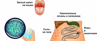
In addition to pain, with pancreatitis the complexion also changes. It becomes yellowish and pale.
The appearance of white plaque and dry mouth is also a manifestation of pancreatitis . If after an attack of vomiting it does not become easier, you need to fast. Otherwise, food will make the situation worse.
Diarrhea is characterized by a foul odor and a foamy mass containing food particles that have not been digested. The opposite situation is bloating or constipation. The abdomen becomes hard, this is the first signal that an acute attack of pancreatitis .
Signs of acute pancreatitis appear as a result of drinking alcohol or fatty foods and may occur suddenly. Pain in acute pancreatitis lasts up to several days.
Publications in the media
Regarding the treatment of this disease, you can contact the Department of X-ray surgical methods of diagnosis and treatment of the Clinic of Faculty Surgery named after. N.N. Burdenko
Acute pancreatitis is an inflammatory-necrotic lesion of the pancreas caused by enzymatic autolysis caused by various reasons. Etiology • Diseases of the biliary tract (cholelithiasis, choledocholithiasis, stenosis of the papilla of Vater) • Alcohol excess and rich fatty foods • Surgical interventions on the pancreas and adjacent organs, abdominal trauma with damage to the pancreas • Acute circulatory disorders in the gland (vessel ligation, thrombosis, embolism) • Severe allergic reactions • Diseases of the stomach and duodenum (peptic ulcer, parapapillary diverticulum, duodenostasis) • Endoscopic retrograde cholangiopancreatography • Viral infections (mumps) • Drugs (azathioprine, estrogens, thiazides, furosemide, sulfonamides, GC and valproic acid) • Hypercalcemia, hyperparathyroiditis, uremia • Kidney transplantation.
Pathogenesis • Enzymatic autolysis of gland tissue with the development of a demarcation inflammatory reaction and the formation of microthrombi • The progressive course of the disease is characterized by pancreatogenic toxemia, hemodynamic disturbances, inhibition of the activity of parenchymal organs and post-necrotic complications. Pathomorphology. Autolysis, interstitial edema, hemorrhages, cellular and fat necrosis are noted in the pancreas.
Clinical and morphological classification • Edematous form of pancreatitis • Fatty pancreatic necrosis • Hemorrhagic pancreatic necrosis. Clinical picture • Constant severe girdling pain and pain in the epigastric region, accompanied by nausea and vomiting • The abdomen on palpation is painful, tense and moderately swollen • Positive symptoms of Shchetkin-Blumberg, Voskresensky, Mayo-Robson, Razdolsky. The severity of symptoms depends on the form of the disease, the degree of intoxication and complications • The skin and mucous membranes are often pale, sometimes cyanotic or icteric. Mondor's syndrome, Gray Turner's symptom, and Cullen's symptom appear. The body temperature with edematous pancreatitis is normal. • With pancreatic necrosis, pain is most pronounced in the epigastric region. With the progressive course of pancreatic necrosis, on the 7th–10th day of the disease, abdominal pain decreases due to the death of sensory nerve endings in the pancreas. Also characteristic is a serious condition, vomiting, increased body temperature (37.7–38.3 °C), cyanosis of the skin, tachycardia, arterial hypotension, oliguria, and symptoms of peritonitis. Symptoms of Grunwald and Davis are characteristic. Often, symptoms of intoxication prevail over local manifestations of the disease • With parapancreatic phlegmon and abscess of the pancreas, deterioration of the condition is noted: increased body temperature, chills, inflammatory infiltrate in the upper floor of the abdominal cavity, leukocytosis with a shift in the leukocyte formula to the left • Severe inflammation and necrosis of the pancreas can cause bleeding into the retroperitoneal space, which can lead to hypovolemia (arterial hypotension, tachycardia) and accumulation of blood in soft tissues. Laboratory tests • CBC - leukocytosis (10–20109/l) with a shift of the leukocyte formula to the left • Biochemical blood test •• Increased content of α-amylase - 95% of cases (decreases with pancreatic necrosis) •• Increased ratio of amylase to creatinine clearance (higher 5%), which is normally 1–4% •• Increase in Ht to 50–55% •• Moderate increase in ALT and/or AST with concomitant alcoholic hepatitis or choledocholithiasis •• Moderate increase in ALP concentration with concomitant alcoholic hepatitis or choledocholithiasis •• Hyperbilirubinemia - in 15–25% of patients •• Increased levels of serum lipase •• Hyperglycemia in severe cases •• Hypocalcemia on the first day of the disease.
Special studies • Plain radiography of the abdominal organs - signs of dynamic intestinal obstruction, accumulation of gas in the area of the lesser omentum (abscess inside or near the pancreas); displacement of the abdominal organs (exudation and swelling of the lesser omentum and organs adjacent to the pancreas); blurred shadows of the iliopsoas muscles with retroperitoneal necrosis of the pancreas • X-ray contrast study with a barium suspension is used to diagnose pathology of the upper gastrointestinal tract: an increase in the radius of the horseshoe of the duodenum due to edema of the pancreas is possible; with relaxation duodenography, a pillow symptom can be detected • X-ray examination of the chest organs - pleural effusion (rare) • Ultrasound of the pancreas - decreased echogenicity, edema, thickening in the anteroposterior direction, virtual absence of tissue between the pancreas and the splenic vein • CT scan of the pancreas (high resolution) • Selective celiacography: with edematous pancreatitis - increased vascular pattern; with pancreatic necrosis - narrowing of the lumen of the celiac trunk, deterioration of the blood supply to the gland with areas of vascular bed exclusion • Radioisotope study with pancreatic necrosis - lack of fixation of the isotope in the pancreas, decreased excretory function of the liver • Endoscopic retrograde cholangiopancreatography • Laparoscopy - foci of fat necrosis, hemorrhage and edema of the gastrocolic ligaments, exudate in the abdominal cavity (serous or hemorrhagic), assess the condition of the gallbladder.
Differential diagnosis • Penetrating or perforating gastric and/or duodenal ulcer • Acute cholecystitis • Choledocholithiasis • Mesenteric vascular obstruction and/or infarction • Perforation of internal organs • Obstructive intestinal obstruction • Aortic aneurysm • Pancreatic cancer • Acute appendicitis • Ectopic pregnancy • Posterior MI • Hematoma of the muscles of the anterior abdominal wall • Blunt trauma or penetrating injury to the spleen.
TREATMENT Diet. Fasting is prescribed for up to 7 days; after reducing the severity of pain, you should eat small meals with a high carbohydrate content, limiting fats and proteins (to reduce the secretion of pancreatic enzymes). Expansion of the diet in accordance with the patient's condition. Management tactics • For edematous form of pancreatitis •• Nasogastric tube and gastric drainage - for vomiting, nausea •• IV solutions of glucose, Ringer-Locke (1.5–2 l), rheopolyglucin (lowers blood viscosity, prevents the aggregation of formed blood elements, leading to improved microcirculation and reduced swelling of the pancreas), hemodesis (binds toxins and quickly removes them in the urine) •• Lytic mixture: trimeperidine, atropine, diphenhydramine, procaine •• Protease inhibitors: aprotinin •• Moderate forced diuresis •• To relieve spasm of the sphincter of Oddi and blood vessels - papaverine hydrochloride, atropine, platyphylline, drotaverine, aminophylline •• Antihistamines (promethazine, chloropyramine, diphenhydramine) - to reduce vascular permeability, analgesic and sedative effects •• Perinephric procaine blockade and splanchnic nerve blockade for relief inflammatory process and pain reaction, reducing the external secretion of the pancreas, normalizing the tone of the sphincter of Oddi, improving the outflow of bile and pancreatic juice. These manipulations can be replaced by the administration of 0.5% procaine solution intravenously •• On 3–5 days, patients are discharged, as a rule, in satisfactory condition.
• For pancreatic necrosis (treatment of fatty and hemorrhagic pancreatic necrosis is carried out in the intensive care unit) •• For rapid restoration of blood volume and normalization of water and electrolyte metabolism - intravenous administration of solutions of glucose, Ringer-Locke, sodium bicarbonate, as well as rheopolyglucin, hemodez, and then plasma, albumin with simultaneous stimulation of diuresis •• Lytic mixture, protease inhibitors, cytostatics •• Cytostatics (for example, fluorouracil) have an anti-inflammatory, desensitizing effect and (most importantly!) inhibit the synthesis of proteolytic enzymes •• Protease inhibitors (aprotinin) suppress the activity of trypsin, kallikrein, plasmin, forming inactive complexes with them. They are administered IV every 3–4 hours in loading doses (for example, aprotinin up to 80–320 thousand units/day) •• To force diuresis - IV mannitol 15% solution 1–2 g/kg or furosemide 40 mg •• For an infectious process (for example, in the lungs, bile ducts or urinary tract) - broad-spectrum antibiotics. Data on the effectiveness of antibiotics for the prevention of purulent complications in the pancreas are contradictory. In case of an infectious lesion of the pancreas, antibiotic therapy is necessary •• To reduce the external secretion of the pancreas, cold applied to the epigastric region, aspiration of gastric contents, intragastric hypothermia are indicated •• UV laser irradiation of blood (15 min each, 2-10 sessions) relieves pain and inflammation , improves the rheological properties of blood and microcirculation •• Extracorporeal detoxification methods (plasmapheresis, lymphosorption) are aimed at removing pancreatic enzymes, kallikrein, toxins, and cellular decay products from the body •• Close-focus radiation therapy has an anti-inflammatory effect. 3–5 sessions are performed •• In case of progression of signs of peritonitis, surgical drainage of the cavity of the lesser omentum and abdominal cavity is indicated.
Surgical treatment is carried out in case of • Ineffectiveness of conservative therapy - drainage of the omental bursa and abdominal cavity, necrectomy • Inability to exclude acute surgical disease of the abdominal organs • Symptoms of diffuse peritonitis • Combination of acute pancreatitis with destructive cholecystitis • Pancreatitis caused by an impacted gallstone - endoscopic retrograde cholangiopancreatography , sphincterotomy and stone removal • Severe blunt trauma or penetrating injury to the abdomen.
Complications • Toxic •• Pancreatic shock •• Delirium •• Hepatic-renal and cardiovascular failure • Post-necrotic •• Pancreatic abscess •• Phlegmon of the retroperitoneal tissue •• Peritonitis •• Erosive bleeding •• Cysts and pancreatic fistulas. Prognosis • Signs detected upon admission •• Age over 55 years •• Leukocyte count in peripheral blood more than 16109/l •• Fasting blood glucose concentration above 11 mmol/l •• LDH activity in the blood above 350 IU/l •• AST content more than 25 IU/l • Signs detected 48 hours after admission •• Drop in Ht by more than 10% •• Increase in blood urea nitrogen content to 1.8 mmol/l •• Serum calcium concentration below 2 mmol /l •• paO2 below 60 mm Hg •• Base deficiency more than 4 mEq/l •• Loss of fluids into the third space - more than 6 l • If less than 3 of the above signs are present, mortality is 1%, 3–4 - 16% , 7 - 90%, more than 7 - 99% • 85–90% of cases of edematous form of acute pancreatitis resolve spontaneously, mortality 3–5%.
ICD-10 • K85 Acute pancreatitis • K90.3 Pancreatic steatorrhea
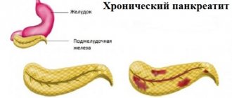
Differences in the chronic form
The chronic form of pancreatitis is characterized by prolonged inflammation, pain occurs periodically. Crises alternate with remissions. If pancreatitis on time, periods of exacerbation will become more frequent, and the patient’s weight will decrease due to intestinal or digestive system disorders. Symptoms of chronic pancreatitis are:
- pain in the upper abdomen radiates to the left hypochondrium, back and lower abdominal cavity;
- loss of appetite;
- nausea;
- vomit;
- bloating;
- stool is disturbed.
Signs of the disease may appear after consuming fatty foods that are difficult to digest, or alcohol. Chronic pancreatitis often appears under constant stress.
In chronic pancreatitis, the pain is girdling, clearly localized and radiates to the back. Appears immediately after eating.
If appetite and blood pressure decrease, temperature rises, tachycardia appears and weakness is felt throughout the body - this indicates drug-induced pancreatitis .
Chronic pancreatitis appears as bright red spots on the abdomen, chest, and back that do not disappear with pressure. Hypoglycemia and diabetes mellitus also appear.
If pancreatitis is diagnosed, treatment in women may be aggravated by additional symptoms :
- hair fragility increases;
- the skin becomes dry;
- nails break;
- the patient gets tired faster;
- loses weight sharply;
- anemia is pronounced.
It is impossible to ignore the signs of the onset of the disease; this is fraught with complications such as abscesses, cysts, enterocolitis, gastric bleeding, diabetes and even cancer.
Causes of pancreatitis
Pancreatitis , like any disease, is preceded by certain causes. If a person develops pancreatitis, treatment is prescribed after taking a history and determining the cause. Pancreatic tissues are affected:
- through excessive and constant consumption of cigarettes and alcohol;
- genetic predisposition;
- abdominal injuries;
- after surgical operations;
- poisoning with chemicals and poor-quality food;
- unbalanced diet - spicy, fatty, fast food, lack of fruits and vegetables in the diet, long time intervals between meals;
- prolonged and uncontrolled use of medications.
These are the main sources of pain in pancreatitis. But the main causes of the disease are frequent and uncontrolled consumption of alcoholic beverages and the appearance of gallstones. Stones are a problem that occurs in up to 50% of people with pancreatitis . They act as blockers of the pancreatic duct. Stones prevent the flow of enzymes into the duodenum. Proteins, instead of breaking down food, direct their action to healthy tissues, thereby destroying them.
Treatment of pancreatic necrosis
The best treatment tactic is urgent hospitalization of the patient in the surgical department for a comprehensive examination and emergency measures. Conservative therapy is effective when there is a chance of recovery. The patient is given drugs that block the production of secretions. A complete refusal to eat for several days is required. Cell nutrition is carried out intravenously. If the bile ducts are blocked and the required amount of bile is not produced, intravenous feeding is indicated for a long time.
Dehydration is eliminated, intoxication is relieved, and if necessary, hemosorption is carried out when the blood is cleansed of toxic components.
Treatment requires rest of the inflamed organ. Analgesia using analgesics provides adequate pain relief. Novocaine blockades are practiced. After taking diuretics, the swelling of the gland subsides, the tension of the pancreatic capsule is weakened. For purulent complications, antibiotics are prescribed.
In a hospital setting, if necessary, anti-shock measures are carried out aimed at relieving pain and improving the outflow of enzymes produced by the gland. Complex therapy also includes drugs that restore the functions of other digestive organs and the cardiovascular system.
How to help yourself with pancreatitis
A crisis of pancreatitis is accompanied by severe pain . To reduce them, you need to apply a heating pad with cold water to your stomach in the place where the pain is the strongest. As a result, the frequency and severity of pain will decrease, and the inflammatory process will decrease. Lying down in a horizontal position is prohibited; the best option is the fetal position.
A prerequisite is an examination by a doctor. treat yourself on your own ; this is fraught with exacerbations. Pancreatitis does not go away on its own, and the symptoms mentioned above may also indicate other diseases. If pancreatitis starts, treatment in adults can take a lot of time and money. Children are also diagnosed with this disease.
Surgeons, therapists, and gastroenterologists treat pancreatitis To correctly diagnose a person and establish the form of the disease, a number of examinations are carried out:
- clinical blood tests;
- feces and urine are examined;
- MRI, radiography and ultrasound of the abdominal cavity are performed;
- CT scan (if indicated).
These studies are identical for acute and chronic pancreatitis . The difference is that for the accuracy of the chronic picture, tests are taken during the period of exacerbation .
When should you make an appointment with a doctor?
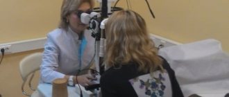
If nausea does not go away for a long time, it is accompanied by vomiting, diarrhea, or an increase in body temperature, you need to consult a doctor. Often these symptoms are confused with food poisoning and mild stomach upsets. This may be an exacerbation of pancreatitis. A gastroenterologist treats pancreatitis; he will diagnose, prescribe treatment, and monitor the healing process. The examination is carried out using new equipment, doctors use modern treatment methods. JSC "Medicine" (clinic of academician Roitberg) is located in the center of Moscow, at 2nd Tverskoy-Yamskaya lane 10, not far from the metro stations Chekhovskaya, Mayakovskaya, Belorusskaya, Novoslobodskaya, Tverskaya.
Preventive measures to prevent pancreatitis
To prevent exacerbation of pancreatitis or to prevent its development, you need to follow several recommendations from doctors.
- Do not drink alcohol more than twice a month. Alcohol provokes inflammation of pancreatic tissue.
- Stop smoking. Carcinogenic elements entering the body through tobacco smoke have a detrimental effect on all organs, especially the pancreas, and lead to pancreatitis .
- Refusal of harmful products. If a person does not want to “get” pancreatitis , you need to exclude fried, salty and fatty foods from the diet. You should not eat foods with dyes and artificial additives; they destroy the structure of pancreatic cells and block their restoration.
- Maintaining an active lifestyle. Elimination of stress, daily walks in any weather, sufficient time for sleep and physical activity are the basis for the prevention of any disease, including pancreatitis
Even following all the rules does not guarantee that the disease will not make itself known. Genetics are often the cause of pancreatitis .
To completely eliminate pancreatitis, symptoms and treatment in adults should be prescribed by a doctor, and not by the patient himself. Initial treatment may include medications, diet, and lifestyle changes. Self-medication, as well as delay, lead to complications and even death.