Shereshevsky-Turner syndrome
(Turner syndrome, Ullrich syndrome) is a typical form of dysgenesis (underdevelopment) or primary agenesis (complete underdevelopment) of the female gonads - the ovaries, caused by genetic deformation and accompanied by short stature and a number of developmental anomalies of the body and some internal organs. The clinical picture of this syndrome was described in the 20s of the twentieth century by endocrinologist N.A. Shereshevsky. This syndrome develops only in females.
Shereshevsky-Turner syndrome is a chromosomal disorder
, which is characterized by either the complete absence of one chromosome or the presence of a defect in one of the X chromosomes. The karyotype of such women is 45 X0, or the mosaic type is 45 X/ 46 XX, 45 X/ 46 XY. According to medical statistics, Shereshevsky-Turner syndrome occurs in one case per 4000 live births of girls, despite the fact that 98-99% of pregnancies with fetal karyotype 45 X0 result in spontaneous abortions in the early stages, therefore this pathology is classified as one of the most common chromosomal anomalies among women.
Causes of development of Shereshevsky-Turner syndrome
In half of the patients with Shereshevsky-Turner syndrome, the complete absence of one of the chromosomes is detected, and karyotyping determines the corresponding 45 X karyotype. Moreover, in 70-80% of cases the father's X chromosome is absent. In the other half of the patients, a mosaic type karyotype is detected, in which one X chromosome is normal and one is ring (consisting of preserved sections of the short and long chromosome arms). In a number of patients, one regular X chromosome is found in the karyotype, and the other is an isochromosome, which loses the short arm and consists of two long arms of the X chromosome. Depending on what karyotype the patient has, the external manifestations of the syndrome vary from typical signs, according to the clinical picture of the disease, to their complete absence.
As a result of the absence or deformation of the sex chromosome in girls, the process of ovarian maturation is disrupted, there is a delay or complete absence of puberty and, as a consequence, the development of infertility. The chromosomal imbalance observed in Shereshevsky-Turner syndrome also leads to various anomalies of somatic development (body development).
Bonnevie-Ulrich syndrome
Bonnevie-Ulrich syndrome is defined as a variant of Turner syndrome. It manifests itself as a consequence of numerous intrauterine developmental defects. There are two types of syndrome. The symmetrical form of bilateral endocrine origin is a combination of Bonnevie-Ulrich syndrome and Shereshevsky-Turner syndrome. The unilateral asymmetric form is defined as simple Bonnevie-Ulrich syndrome, which is also called “multiple abortion” syndrome.
When this syndrome manifests itself, its obligatory sign is the presence of a pterygoid fold on the neck. In addition, the patient has dysmorphia of the maxillofacial type, paresis and paralysis of the motor nerves, deformation of the auricles, lymphatic edema of the feet and hands, and congenital dislocation of the hip. Visual defects are observed. As with Turner syndrome, a number of malformations of the heart, blood vessels, etc. may be observed. Symptomatic treatment is carried out. As a rule, surgical intervention is necessary to correct congenital developmental pathologies.
Shereshevsky-Turner syndrome - signs of the disease
Patients with Shereshevsky-Turner syndrome complain of lack of menstrual flow, short stature and some cosmetic defects in appearance.
Most newborn girls with this disease have only mild clinical manifestations, but a certain group of infants have lymphedema of the hands and feet, as well as skin folds on the back of the neck. Some of the most common anomalies found in Shereshevsky-Turner disease are a short neck with low hair growth and wing-shaped cervical folds, an expanded chest, poorly developed genitals - the labia minora, clitoris, uterus, as well as widely spaced inverted nipples. Some patients have abnormalities in the shape and location of the ears (protruding ears), which are combined in half of the cases with hearing loss.
Compared to their relatives, girls are shorter - no more than 150 cm, but at the same time their body weight is quite impressive. Less commonly, signs such as multiple pigmented nevi or vitiligo, anomalies of the metacarpal and metatarsal bones of the hands and feet, deformation of the elbow and shoulder joints, and hypoplastic narrow nails are detected. The facial part of the patients is also modified - the jaw bones are reduced in size, the palate is high, and drooping eyelids are also observed.
From the internal organs, it is possible to detect cardiac anomalies in the form of coarctation of the aorta, its dissection, disruption of the integrity of the interventricular septum and bicuspid aortic valve. Congenital anomalies and malformations of the kidneys (horseshoe kidney, duplication of the pelvis and ureters), and blood vessels of a tumor nature (hemangiomas, telangiectasia) are also common.
The female gonads are replaced by strands of connective tissue that are unable to produce germ cells, as a result of which the vast majority (90%) of sick girls do not go through puberty, the mammary glands do not enlarge, and primary amenorrhea develops. In the remaining 10%, the spontaneous onset of menstruation is possible, but even in this case, the woman’s fertility remains in question.
The mental sphere of patients with Shereshevsky-Turner syndrome practically does not suffer. True, some authors note a decrease in their attention and some processes of perception, in particular spatial, which affects the quality of processing of nonverbal (nonverbal) information.
There are three forms of gonadal dysgenesis - erased, pure and mixed, which differ from each other in clinical manifestations. When the form is erased
In patients, a mosaic karyotype of 45 X0/46 XX is detected, and the severity of the clinical picture is associated with the ratio of cells with a normal and deformed karyotype. If the percentage of cells with the absence of one of the X chromosomes predominates, then the external signs of the patient will be similar to the classic appearance of patients with Shereshevsky-Turner disease. Otherwise, spontaneous development of external sexual characteristics will be noted, but patients will have primary amenorrhea and signs of underdevelopment of the genital organs.
Pure form of ovarian dysgenesis (Swyer syndrome)
characterized by karyotype 46 XX or 46 XY. It is difficult to recognize the presence of chromosomal defects by appearance, since the growth of patients is normal, somatic defects and anomalies are not detected. However, external sexual characteristics are poorly developed. In addition, upon examination, genital infantilism is revealed, i.e. the genitals are also underdeveloped.
Mixed form of ovarian dysgenesis
manifested by primary amenorrhea, virilization - enlargement of the clitoris, male-type hair growth, which is due to the presence of an inferior Y-chromosome in the karyotype. With this form of the disease in the postpubertal period, combined type ovarian tumors are possible - gonadoblastomas, embryonal carcinomas.
Clinical picture and diagnosis
Skin folds in the neck area are a characteristic sign of the disease.
In the photo: a girl before and after plastic surgery. The lag in physical development of patients with Shereshevsky-Turner syndrome is noticeable already from birth. In approximately 15% of patients, the delay occurs during puberty. Full-term newborns are characterized by small length (42-48 cm) and body weight (2500-2800 g or less). Characteristic signs of Shereshevsky-Turner syndrome at birth are excess skin on the neck and other malformations, especially of the osteoarticular and cardiovascular systems, “sphinx face”, lymphostasis (lymph stagnation, clinically manifested by large edema). The newborn is characterized by general anxiety, impaired sucking reflex, regurgitation, and vomiting. At an early age, some patients experience delayed mental and speech development, which indicates a pathology in the development of the nervous system. The most characteristic feature is short stature. The height of patients does not exceed 135-145 cm, and their body weight is often excessive.
Lymphedema of the feet.
In Shereshevsky-Turner syndrome, the pathological signs are distributed according to the frequency of occurrence as follows: short stature (98%), general dysplasticity (irregular physique) (92%), barrel-shaped chest (75%), shortened neck (63%), low hair growth on the neck (57%), high “Gothic” palate (56%), wing-shaped folds of skin in the neck (46%), deformation of the auricles (46%), shortening of the metacarpal and metatarsal bones and aplasia of the phalanges (46%), deformation of the ulnas joints (36%), multiple pigmented moles (35%), lymphostasis (24%), heart defects and large vessels (22%), high blood pressure (17%).
Sexual underdevelopment in Shereshevsky-Turner syndrome is distinguished by a certain originality. Frequent signs are geroderma (pathological atrophy of the skin, reminiscent of senile skin) and a scrotal appearance of the labia majora, high perineum, underdevelopment of the labia minora, hymen and clitoris, and a funnel-shaped entrance to the vagina. The mammary glands in most patients are not developed, the nipples are low located. Secondary hair growth appears spontaneously and is scanty. The uterus is underdeveloped. The gonads are not developed and are usually represented by connective tissue. With Turner syndrome, there is a tendency to increase blood pressure in young people and to obesity with tissue malnutrition.
The intelligence of most patients with Shereshevsky-Turner syndrome is practically preserved, but the frequency of oligophrenia is still higher. Mental retardation in patients with this syndrome is more common than in the general population. In the mental status of patients with Shereshevsky-Turner syndrome, the main role is played by a kind of mental infantilism with euphoria with good practical adaptability and social adaptation. Many patients have decreased cognitive interests, insufficient spatial concepts, underdevelopment of the emotional-volitional sphere and a lack of creative needs.
The diagnosis of Shereshevsky-Turner syndrome is based on characteristic clinical features, determination of sex chromatin (the substance of the cell nucleus) and the study of karyotype (chromosomal set). Differential diagnosis is carried out with:
- Dwarfism (dwarfism), to exclude which the content of pituitary hormones in the blood, especially gonadotropins, is determined
- Perrault syndrome
Diagnosis of Shereshevsky–Turner syndrome
General diagnostic criteria for all forms of Shereshevsky-Turner syndrome are height less than 150 cm, primary amenorrhea, underdevelopment of the genital organs, absence or poor development of secondary sexual characteristics, according to ultrasound examination - a reduced uterus with a linear endometrium, and ovaries without follicles. Blood tests reveal elevated levels of gonadotropins, particularly follicle-stimulating hormone, and decreased levels of estrogen. Karyotyping allows you to identify a defective set of chromosomes, with the absence or significant reduction of sex chromatin.
Differential diagnosis of Shereshevsky-Turner syndrome is carried out with primary amenorrhea of hypothalamic origin. Unlike the latter, patients with Shereshevsky-Turner syndrome do not have neuropsychiatric symptoms.
Diagnostics
Shereshevsky-Turner syndrome in newborn girls allows one to suspect the presence of lymphedema or a pterygoid fold in the neck. If such signs are absent, then suspicion of the presence of this pathology may appear later due to short stature, amenorrhea, and also in the absence of puberty. A karyotype study allows you to confirm or refute such a diagnosis. In general, the diagnosis can be established by the presence of a clinical picture characteristic of this disease.
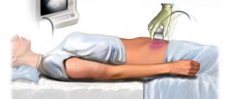
MRI or echocardiography is performed to identify congenital heart defects. Some specific examinations are also carried out, in particular, cytogenetic research . A blood test during the development of this disease demonstrates a decrease in the amount of estrogens and at the same time a high content of pituitary . X-ray examination often reveals osteoporosis , as well as pathologies of the development of the bone skeleton. In addition, during the diagnostic process, an examination is carried out aimed at identifying concomitant diseases.
Also in modern medicine, screening tests , which make it possible to find out about the risk of a fetus having Shereshevsky-Turner syndrome.
Treatment of Shereshevsky–Turner syndrome
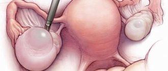
The choice of treatment tactics for patients with Shereshevsky-Turner syndrome depends on the presence or absence of the Y chromosome in the karyotype. If elements of the Y chromosome are detected in the karyotype, the ovaries are surgically removed at a young age (up to 20 years), in order to prevent the degeneration of the gland tissue into a malignant tumor.
In cases where there is no Y chromosome in the karyotype of patients, or after surgical removal of the ovaries, hormone replacement therapy with estrogen is prescribed at the age of 16-18 years, the purpose of which is the development of secondary sexual characteristics (female hair growth, enlarged mammary glands), reducing the level of gonadotropins, restoring menstrual cycle, preventing the development of estrogen deficiency conditions - osteoporosis, metabolic disorders, diseases of the cardiovascular system, improving the quality of life and adaptability to society. If necessary, patients are provided with psychological assistance.
Short stature is corrected by administering growth hormone until the growth plates are closed. The prognosis of Shereshevsky-Turner syndrome is favorable in terms of life expectancy and intellectual development. With regard to restoration of reproductive function, the prognosis is unfavorable; most patients remain infertile.
Treatment
At the first stage, therapy consists of stimulating body growth with anabolic steroids and other anabolic drugs. Treatment should be carried out with minimal effective doses of anabolic steroids intermittently with regular gynecological monitoring. The main type of therapy for patients is estrogenization (prescription of female sex hormones), which should be carried out from 14-16 years of age. Treatment leads to feminization of the physique, the development of female secondary sexual characteristics, improves trophism (nutrition) of the genital tract, and reduces the increased activity of the hypothalamic-pituitary system. Treatment should be carried out throughout the entire childbearing age of patients.
If, with the help of hormonal therapy, it is possible to grow the uterus to a normal size, then pregnancy in such patients is possible using IVF with a donor egg. Cases where eggs have been preserved are rare.
Recently, growth hormone therapy has been used to increase final growth rates.
Turner syndrome: primary ovarian insufficiency
The article discusses current issues related to the pathogenesis, epidemiology, and clinical manifestations of Turner syndrome - a chromosomal pathology caused by partial or complete monosomy. An algorithm for verifying the diagnosis is given. The choice of treatment at the stages of growth and development of women with Turner syndrome is described in detail, with special attention paid to hormonal therapy with sex steroids. An atypical clinical case of Turner syndrome is analyzed.
Introduction
In the recent past, medicine had very limited opportunities to improve the quality of life of patients with Turner syndrome, although the existence of such a disease has been known for a long time. For the first time, the case of a patient with such a disease was examined in 1925 by N.A. Shereshevsky. However, the classic description (infantilism syndrome in combination with webbed neck and valgus deformity of the elbow joints) was made in 1938 by N. Turner. In 1942, F. Albright, PH Smith and R. Frazer found that this syndrome is combined with elevated levels of gonadotropin titers in the urine [1, 2]. Already in 1944, L. Wilkins and W. Fleischmann discovered in such patients the absence of gonads, but the presence of correctly formed, but immature, Müllerian ducts [1]. The gonads were mainly represented by primitive cords of white or yellow color, and microscopically contained only stroma. In 1954, a number of researchers independently established that the majority of these patients had negative sex chromatin [1, 2]. Finally, in 1959, C.E. Ford et al. published data that at least some patients with negative sex chromatin had not the XY chromosome set, but the XO configuration of sex chromosomes [1]. This discovery became the cornerstone for the scientific explanation of the syndrome, first described by N. Turner.
Pathogenesis
Turner syndrome, or a typical form of gonadal dysgenesis, is the most common cause of ovarian dysfunction due to loss of genetic material [1–4]. For the proper development of the gonads, the presence of two sex chromosomes is necessary, but during the first divisions one of the sex chromosomes may be “lost,” which occurs sporadically. An embryo containing one Y chromosome is not viable, but if it has one X chromosome, it survives and develops as a girl with Turner syndrome. This is the first reason for the development of the syndrome [2, 4].
If the second sex chromosome is lost early in embryonic development, then not all, but only some, cells of the growing organism contain one X chromosome. This condition is called “mosaicism”, and this is the second mechanism for the formation of Turner syndrome. The clinical manifestations of the syndrome directly depend on the percentage of cells with an abnormal 45,X karyotype [4]. If the proportion of the abnormal cell clone is small, then the phenotype of a patient with Turner syndrome differs little from the norm, and women can have regular menstrual cycles and even become pregnant. Genetic diagnosis in such cases often requires the study of a very large number of blood cells and/or the study of cells from other tissues, in particular the skin. An example of such a genotype is 45,X(10)/46,XX(90) which means that only 10% of cells have monosomy, and 90% have a normal set of chromosomes [2–4].
The third reason for the development of Turner syndrome is not the loss, but defects of one of the X chromosomes that prevent normal gene expression (for example, fragmentation of the X chromosome, loss of part of it, or formation of a ring shape) [4, 5]. The clinical consequences of such structural rearrangements vary widely. Small deletions may result in isolated problems such as ovarian dysfunction and short stature in the absence of other symptoms. More severe deletions, or deletions that affect critical regions and regulate the entire X chromosome, may be responsible for the full range of problems in Turner syndrome.
Sometimes monosomy appears to be preferable to defects in the retained X chromosome. Thus, the presence of a small ring-shaped X chromosome leads to severe consequences, firstly, due to the absence of several important genes, and secondly, due to the expression of those genes that are normally latent or inactivated [5]. Diagnosis of an abnormal X chromosome may require specialized molecular cytogenetic testing to identify small deletions or inversions of chromosomal material [5].
Epidemiology
Turner syndrome affects all races, nationalities, and regions of the world with equal frequency. In the population, this syndrome occurs with a frequency of 1 in 2000–2500 live births of girls [2–4]. Karyotype changes are observed much more often and reach 1–2% of all conceptions. However, only 1% of such pregnancies persist and result in a live birth [6]. Monosomy 45,X occurs in approximately 40–60% of karyotypes of patients with Turner syndrome. Among the mosaic options, 45,X/46,XX, 45,X/46,XiXq, 45,X/46,XY, 45,X/46,XrX are often defined [6]. It has been established that the risk of developing Turner syndrome does not depend on the age of the pregnant woman. Parents who have several healthy children are not insured against the birth of a child with this disease [6]. No toxins or environmental factors have been identified to increase the risk of having children with Turner syndrome [6].
Clinical manifestations and diagnosis
In some countries, Turner syndrome is included in prenatal screening [7]. The disease can be suspected in the fetus by ultrasound examination [7, 8], and karyotyping using amniocentesis or chorionic villus biopsy is performed to confirm the diagnosis [7].
The social and medical significance of diagnosing Turner syndrome is determined by the variety of disorders in various body systems characteristic of the disease. To identify and correct the manifestations of Turner syndrome, patients need interdisciplinary care from birth [6].
In the first days after birth, Turner syndrome can be suspected by characteristic lymphatic swelling of the hands and feet, low weight that does not correspond to the gestational age, and body length close to the third centile in the growth charts. At older ages, indicators and growth rates may be below the third centile [6]. In addition, during the newborn period there may be difficulties with feeding and weight gain and problems sleeping. In the classic version, the syndrome is characterized by the presence of skin folds on the neck. Cardiovascular anomalies, the most serious of which include aortic coarctation, aortic stenosis, and aortic valve disease, can be detected both prenatally and in the early neonatal period [6, 9].
At an early age and in subsequent years, children with Turner syndrome often suffer from ENT diseases, especially repeated middle ear infections, exudative otitis media, and they may develop conductive hearing loss and sensorineural deafness.
Abnormalities of the urinary system are described in the following variants: duplication of the urinary system, absence of one kidney, incomplete rotation, horseshoe kidney, and asymptomatic hydronephrosis.
Intelligence in Turner syndrome is not impaired, but children are distinguished by behavioral difficulties and psychological problems with exaggerated fears [6, 9]. Naturally, such problems, coupled with short stature and the absence of spontaneous timely puberty, lead to learning difficulties, social problems, long-term psychological immaturity and less social activity [6, 9].
Immune disorders become a serious problem in adolescence - autoimmune thyroiditis, inflammatory bowel disease and celiac disease, which occur along with the early development of hypertension, osteoporosis, obesity, aortic dilatation and infertility [6, 9].
The gynecologist’s area of responsibility is a significant delay in sexual development or even its absence in cases of untimely prescription of hormone replacement therapy [1–3, 6, 9].
Levels of gonadotropins, especially follicle-stimulating hormone (FSH), may be elevated at birth, but this fact does not warrant neonatal screening in the same way as congenital hypothyroidism [6]. By the fourth year of life, FSH levels gradually decrease, but then increase again, reaching menopausal values by puberty [6, 9].
In adolescents and adults, the basis for diagnosis is usually delayed puberty, impaired fertility, and short stature. The onset of pubertal hair growth occurs at normal times, but this does not mean that sexual development will progress [3, 6]. The mammary glands and even the menstrual cycle form spontaneously in no more than 13% of girls. Such cases are usually characterized by a mosaic karyotype of 45.X/46.XX and a normal gonadotropin response to stimulation with gonadotropin-releasing hormone [6]. The presence of Turner syndrome can be suspected in primary or secondary amenorrhea, as well as in adult women with unexplained infertility, especially if they are short [6].
To make a diagnosis of Turner syndrome (a typical form of gonadal dysgenesis), the presence of characteristic phenotypic features and the complete or partial absence of the second sex chromosome is necessary. If there is only one X chromosome, then in 2/3 of cases it is maternal in origin [1, 7]. Most features of Turner syndrome, including short stature, are associated with a deficiency of the short stature gene containing the homeobox, the second X chromosome [5]. Women with Turner syndrome may also have other pathological conditions of the musculoskeletal system: congenital hip dislocation, arthritis, scoliosis, mandibular malformation (retrognathia), lymphoid edema [6].
Most of the listed symptoms are a consequence of impaired growth regulation, which manifests itself already in the prenatal period and, starting from early childhood, continuously progresses [9, 10]. Pubertal growth acceleration does not occur. The final height of girls with Turner syndrome without treatment is on average approximately 20 cm lower than that of healthy peers [6, 9]. In contrast to Turner syndrome, short stature is not characteristic of primary hypergonadotropic hypogonadism without karyotype abnormalities [1–3]. Since growth hormone levels are usually normal in Turner syndrome, short stature is most likely a consequence of an underlying form of skeletal dysplasia [3, 10]. This may be due to disturbances in gene expression due to the absence of one X chromosome. The administration of growth hormone often accelerates growth and improves final growth indicators, but not as significantly as in case of growth hormone deficiency [3, 10]. Higher final height can be achieved with early and long-term use of growth hormone until induced or spontaneous puberty [6, 9, 10]. With this treatment, the height in approximately 50% of cases can be 150 cm or more, while in untreated adults it does not exceed 142 cm [6, 9].
Turner syndrome is not an inherited disorder and its recurrence risk is considered low [6]. Special genetic counseling is required in situations where Turner syndrome is diagnosed in utero or a possible diagnosis is suspected in a patient with a normal karyotype [6, 7, 9]. In all cases of gonadal dysgenesis in the presence of high FSH levels, patients should be tested for the presence of Y chromosome material using Y-centromere assays [6].
When prescribing any type of therapy, it is necessary to take into account the high probability of diseases concomitant with Turner syndrome: Crohn's disease, Hashimoto's thyroiditis, hypertension, type 1 diabetes mellitus [9]. A particular problem is the higher incidence of malignant neoplasms than in the population: cases of gonadoblastoma, meningioma, childhood brain tumors, bladder cancer, Williams tumor, melanoma and uterine cancer have been described [6].
Diagnosis verification
The clinical manifestations of Turner syndrome are so characteristic (short stature, “wing-shaped” folds on the neck, low hairline, high palate, barrel-shaped chest and widely spaced nipples, many nevi, lack of mammary gland growth and scant sexual hair growth) that a specialist can assume the presence of this disease at the first glance at the patient [1–3]. However, even the most typical combination of external manifestations of the syndrome and delayed sexual development requires mandatory research to make a diagnosis.
1. Karyotyping with mandatory clarification of monosomy or mosaicism, and in the case of mosaicism, mandatory exclusion of the presence of the Y chromosome.
2. Before starting hormone therapy, study the levels of gonadotropins: luteinizing hormone and FSH, which can be elevated before the age of four years. Later, gonadotropin levels decrease to normal age levels, and then, upon reaching the age of 10 years, increase to menopausal values [6, 9, 11].
3. Assess bone age before initiating growth hormone therapy and/or estrogen administration. Bone age in children may be within normal limits, but later, due to estrogen deficiency, it is delayed. When growth plates are closed (bone age above 14 years), the use of growth hormone is contraindicated [10]. In adult patients, regular testing of bone mineral density is recommended due to the early development of osteoporosis [6].
4. Electro- and echocardiography with consultation with a cardiologist before starting hormonal therapy [11].
5. Assessment of thyroid function - analysis of hormones (thyroid-stimulating hormone, free thyroxine and thyroid antibodies) [2, 12].
6. Screening for diabetes mellitus (determining the level of glycosylated hemoglobin or fasting glucose level) [6, 9, 12].
7. Determination of electrolyte levels in the blood, general urine analysis, exclusion of asymptomatic bacteriuria, as well as ultrasound of the urinary system to test the consistency of the urinary system [6, 9, 12].
Patients should be jointly observed by an endocrinologist and gynecologist, consult a psychologist, ophthalmologist, ENT doctor, urologist and cardiologist. The need for an interdisciplinary approach to the examination and treatment of patients with Turner syndrome is recognized throughout the world [12].
Observation and choice of therapy at the stages of the patient’s growth and development
Elimination of cosmetic defects (pterygoid folds of the neck) is carried out by pediatric surgeons at an early age.
Before the age of 10–11 years, short stature becomes one of the main problems for the patient and her family. It is recommended to begin growth hormone therapy from an early age, when the child’s lag behind his peers becomes noticeable and on this basis attempts are made for the first time to clarify the diagnosis [9]. Recombinant growth hormone (rGH) is recommended for the treatment of short stature associated with Turner syndrome. When given 0.3 to 0.378 mg/kg rGH per week, girls were able to gain approximately 3 cm in height during the first year and 2 cm during the second year of treatment [9, 10]. Recent studies have shown that the combination of rGH and ultra-low dose estrogen during the prepubertal period improves final growth rates [13]. The use of growth hormone is recommended until bone age is 14 years. Some endocrinologists recommend the use of rGH in combination with anabolic steroids [9]. The effectiveness of rGH therapy is monitored using growth dynamics and insulin-like growth factor 1 levels, which should be maintained below the upper limit of normal [10]. If you start therapy at age eight and use growth hormone daily for five years, you can achieve a height gain of +7 cm compared to those who did not receive therapy [9, 10]. Failure to respond adequately to growth hormone therapy is usually associated with hypothyroidism, celiac disease, or noncompliance with the drug [6, 9, 10]. Side effects of therapy include increased intracranial pressure, femoral head dislocation, scoliosis, pancreatitis, and a possible increased risk of developing type 1 diabetes [3, 10].
Hormone replacement therapy with sex steroids
Approximately 95% of patients with Turner syndrome are characterized by short stature and delayed sexual development [2, 3, 6, 9]. In a small proportion of patients, spontaneous development of some signs of puberty and even the appearance of menstruation is possible [1, 2].
The use of hormonal therapy to initiate puberty and ensure sufficient estrogen saturation during adolescence and the conditional reproductive period is a complex task. This process must be carefully monitored while providing psychological support to the patient and her family [6, 9].
The question of when to start using estrogen and choosing the right dose remains controversial. For a long time, the prevailing opinion was that the use of estrogens accelerates the closure of growth plates and should be started no earlier than the completion of growth hormone therapy and/or the achievement of bone age of 12 years, which for patients with Turner syndrome corresponds to the passport age of 14–16 years [10, 14].
According to another point of view, it is necessary to begin the use of estrogens at a time corresponding to the average age of the appearance of the first signs of puberty. The first physiological symptom of puberty is the onset of mammary gland growth – thelarche, which occurs on average at 10–11 years of age. It is at this age that there is a significant increase in gonadotropin levels in patients with Turner syndrome, reaching menopausal levels. Starting estrogen therapy at this age is “layered” on ongoing growth hormone therapy and is associated with a number of difficulties: what dose of estrogen drug should be offered to the patient? Which dosage regimen is preferable - intermittent or continuous? And finally, which drug is better to choose? There are no clear answers to these questions yet; each country creates its own recommendations. For example, British clinical guidelines recommend starting estrogen therapy at age 13 years, although growth hormone therapy is ongoing and families have expressed their wishes for earlier or delayed initiation of estrogen therapy [6].
Hormonal therapy should begin with natural analogues of estradiol (17-beta-estradiol, estradiol valerate) or conjugated estrogens in initial doses of 0.5-1 mg. According to some authors, conjugated estrogens are contraindicated in childhood [15]. Tablet preparations of estradiol valerate present on our market contain 2 mg of estradiol, a lower dosage is not provided. Therefore, the only possible choice is a transdermal dosage form of 17-beta-estradiol, which can be dosed depending on the current need. Estradiol is prescribed to initiate the development of sexual characteristics and growth of the genital organs, initially as monotherapy with a gradual increase in dose (usually the dose is recalculated every six months) under ultrasound control and annual determination of bone age. When bleeding from the genital tract appears or the thickness of the endometrium increases to 1 cm, analogues of natural progesterone are added to estradiol, and therapy becomes cyclical. It is believed that the duration of estrogen monotherapy should not exceed one and a half years.
Subsequently, it is recommended to switch to a cyclic regimen or use combined drugs for hormone replacement therapy (HRT). Dosage regimens adopted for menopausal hormone therapy should not be extrapolated to the population of patients with Turner syndrome. The BSPED study is currently ongoing in the UK, which is designed to assess the effectiveness and acceptability of different types of estrogens and compare different regimens of HRT [6, 9].
The Institute of Endocrinology of Ukraine has developed its own method of hormonal therapy for Turner syndrome [14]. The guideline for starting estrogen therapy is a decrease in growth rates at a bone age of 11 years, which for patients with Turner syndrome corresponds to the age of 14–16 years. For the first two to two and a half years, estrogen monotherapy is offered in minimal doses, which are not able to block growth zones, but, on the contrary, stimulate the growth of tubular bones. This approach, according to the authors, allows patients to gain 4–5 cm per year. Switching to cyclic hormone therapy is suggested when growth slows to 1 cm per year or when growth stops completely.
A pilot study conducted in 2009 by N. Zuckerman-Levin et al. showed the effectiveness of adding small doses of androgens during HRT both at the time of puberty induction and in the subsequent period [16]. Such comprehensive replenishment of missing ovarian function allows one to obtain better results in terms of sexual development, quality of life and cognitive functions. The authors themselves emphasize the need for further study of this issue.
Timely adequate hormone therapy allows you to achieve the formation of secondary sexual characteristics, correct growth, ensure full function of the uterus before a planned pregnancy, prevent the progression of osteoporosis and lipid metabolism disorders, improve cognitive functions, and psychosocial adaptation.
Implementation of reproductive function
In patients with the mosaic form of Turner syndrome, spontaneous pregnancy may occur, for which medical genetic counseling and invasive prenatal screening are recommended.
In other cases, women with Turner syndrome are infertile [2, 3], and only assisted reproductive technologies using a donor egg can achieve pregnancy. At the same time, the reproductive outcomes of resulting pregnancies should be carefully assessed. It should be remembered that the risk of death from aortic dissection or rupture during pregnancy can be as high as 2% [17]. This risk continues after childbirth due to changes in the aorta that occur during pregnancy.
Turner syndrome is recognized as a relative contraindication to pregnancy, and in the case of cardiovascular system defects - an absolute contraindication. This should be taken into account when including a patient with Turner syndrome in an assisted reproductive technology program.
Forecast
In general, the prognosis for life is assessed as satisfactory in the absence of gross defects of the cardiovascular system [6, 9]. The height of patients with Turner syndrome, even after growth hormone therapy, is usually less than the average in the population. Life expectancy in patients with Turner syndrome is somewhat shorter than in the general population, but with careful correction of problems such as obesity and hypertension, life expectancy can be increased.
Clinical example
Let's consider an atypical situation that expands our views on Turner syndrome and poses many questions to us.
Patient N, 33 years old, is planning a pregnancy. Objectively: correct physique, petite, height 145 cm, weight 37 kg. Secondary sexual characteristics are well developed: P3Ax2Ma3 (the hair on the entire triangle of the pubis is long, curly, thick, the hair on the central part of the armpit is sparse, the body of the mammary gland takes on a rounded shape, the nipples rise above the areola), menstruation from the age of 13.
The patient was born from her third birth with a weight of 2500.0 g. From birth, there was a lag in height and weight compared to her peers. Mother's height is 157 cm, father's height is 160 cm. Other children in the family: the first sister is 160 cm tall, married, three healthy children; second sister, height 160 cm, married, one child; brother, not married, height 179 cm.
At the age of nine, the patient was examined due to her short stature; no pathology was detected. The use of growth hormone was not considered. At 11 years old and with short stature, the bone age corresponded to the passport age. Pubertal development began spontaneously at the same time as in female peers with normal growth. Menarche at 13 years old, heavy menstruation for seven days, irregular cycle. Sexual life since 22 years old. For six years, she used combined oral contraceptives irregularly (with interruptions) for contraceptive purposes.
From common diseases in the anamnesis of ARVI.
During the examination, a doubling of the left kidney was revealed.
Gynecological diseases: ectopic endocervix of the cervix. At the age of 24, due to heavy uterine bleeding, she was taken to the emergency gynecology department, where separate diagnostic curettage of the cervical canal and uterine cavity was performed for the purpose of hemostasis. Histological conclusion: glandular cystic endometrial hyperplasia. Fibrous endometrial polyp. Endometritis.
At the same age (24 years), a hormonal examination was carried out: FSH 5.1 IU/l, luteinizing hormone 4.3 IU/l, prolactin 199.0 IU/l, testosterone 0.43 ng/ml. Thyroid function is within normal limits. However, according to ultrasound data, the size of the thyroid gland is smaller than standard: the volume of the right lobe is 4.29 cm3, the volume of the left lobe is 3.33 cm3.
The husband's spermogram is without pathology. Subsequently, the patency of the fallopian tubes was assessed - they are passable.
At the age of 27 years, due to short stature, karyotyping was performed for the first time, the result was 45.X. Soon the karyotype was re-examined, the result was 45.X/46.XXdelX in a ratio of 90/10%. Geneticist's conclusion: Turner syndrome. In vitro fertilization with a donor egg is recommended.
At the age of 28 years, she received micronized progesterone 200 mg in the second phase of the cycle for six months; one cycle of ovulation stimulation with clomiphene citrate 50 mg was performed from the fifth to the ninth day of the cycle. Against the background of ovulation stimulation, dominant (preovulatory) follicles with a diameter of 1.4 and 2.0 cm were recorded, but ovulation did not occur.
At the age of 30, uterine bleeding began at the expected time of menstruation. The woman was hospitalized on the eighth day from the onset of bleeding, hemoglobin was 93 g/l, and ultrasound showed the thickness of the endometrium to be 1.2 cm. For the purpose of hemostasis, a diagnostic curettage of the uterine cavity was performed. Histological conclusion: simple typical endometrial hyperplasia. The patient is currently receiving progestogen therapy.
This clinical example is indicative in that of the variety of clinical manifestations of Turner syndrome, patient N does not have stigmata of disembryogenesis, only short stature and an altered karyotype occur. According to the karyotype data, there should have been pronounced manifestations of Turner syndrome, but this did not correspond to the clinic.
Folliculogenesis in the ovaries was not impaired, since there was timely puberty, independent menstrual function, although without ovulation, and levels of gonadotropic hormones within the reference values. Accordingly, the patient did not need HRT. In this case, in the absence of ovulation, endometrial hyperplasia developed again. The considered example, exceptional, but probably not an isolated one, violates the established understanding of the phenomenon of Turner syndrome, allowing for a broad interpretation of clinical manifestations. In our opinion, in this case the genetic material responsible for sexual development and menstrual function was not lost. How should the karyotype result be interpreted? It can be assumed that a certain zone of the second X chromosome could take over the lost genetic function, or with the loss of the second X chromosome, an important part of the genetic material could be transferred to other chromosomes [18]. Such reasoning, especially coming from gynecologists rather than geneticists, can cause a flurry of objections. In the recent past, patients who met the clinical requirements of Turner syndrome were sent for karyotype research: short stature, delayed sexual development, amenorrhea, the uterus and ovaries practically indistinguishable by ultrasound with a female structure of the external genitalia, and, finally, significantly high levels of gonadotropic hormones [3] , and the result of karyotyping confirmed or excluded the final diagnosis. Currently, the karyotype is being studied more widely, and clinicians are forced to assume the opposite, making a diagnosis of “Turner syndrome” without clinical manifestations, only on the basis of the karyotype. Unfortunately, in modern conditions it is not possible to conduct a targeted search for female genetic material using fluorescent in situ
, as is available to the Y chromosome.
Conclusion
Despite the long and successful history of studying the problem of a typical form of gonadal dysgenesis, new problems arise that require solutions. Today, it is necessary to discuss the timing of induction into puberty, adequate doses of estrogens, the choice of optimal drugs for long-term therapy, as well as the features of the use of progestogens in patients with low body weight (type of gestagen, dosage, regimen, etc.).
In connection with the given clinical example, the following must be taken into account:
- introducing clarifications into the formulation of the diagnosis in cases similar to the one considered, since the combination of such diagnoses as recurrent uterine bleeding, endometrial hyperplasia, Turner syndrome, contains an internal contradiction. Turner syndrome is one of the main causes of primary amenorrhea;
- indications for biopsy and karyotyping of cells from other tissues, including ovaries, as a basis for recommending in vitro fertilization and the use of a donor egg.