Terminology
The concept of “tubulointerstitial nephropathies” in the broad sense of the word includes inflammatory, metabolic or toxic diseases of the kidneys, which occur in contrast to glomerular (glomerular) diseases with primary and predominant damage to the tubules and interstitial tissue of the kidneys. In a narrower sense, the term “tubulointerstitial nephropathies” is more often used to refer to metabolic and toxic lesions without a clear inflammatory component, applying the term “tubulointerstitial nephritis” or, equivalently, “interstitial nephritis” to inflammatory, immunoinflammatory lesions of the tubules and interstitium. In addition to tubulointerstitial nephropathies as independent nosological forms, a tubulointerstitial component can be isolated in a number of glomerular kidney lesions, including chronic glomerulonephritis.
The term “interstitial nephritis” (IN) has existed since the mid-nineteenth century, but over the course of a century and a half it included various concepts. The term “chronic interstitial nephritis” for a long time corresponded to the modern understanding of kidney damage in hypertension - nephroangiosclerosis, primary wrinkled kidney, i.e. designated those types of kidney pathology, which are based on vascular damage; this group then began to include secondarily wrinkled kidney, chronic nephritis of the hypertensive type. IN was contrasted with tubular, parenchymal nephritis with edematous syndrome (corresponding to modern nephritis with nephrotic syndrome). In parallel, in the first half of the twentieth century, the term “tubulointerstitial nephritis” existed to refer to acute renal failure (ARF).
IN, corresponding to the modern interpretation of this concept, was first described in 1898 by Councilman in patients who had diphtheria and scarlet fever. However, for a very long time, IN was identified exclusively with pyelonephritis and was the prerogative of study by urologists. And at present, IN remains poorly known to a wide range of doctors, which is due, firstly, to the relative rarity of the disease, and secondly, to the nonspecificity of most symptoms. Poor knowledge of this pathology by doctors is evidenced by the fact that among the 22 patients with acute IN we observed, 20 were admitted with other diagnoses: 12 with a diagnosis of chronic glomerulonephritis, 3 with acute glomerulonephritis, 2 with pyelonephritis, 3 with systemic lupus erythematosus. At the same time, IN is in most cases a disease with an established etiology, promising in terms of treatment and prevention.
The current wave of interest in tubulointerstitial lesions is associated both with the possibilities of treatment and prevention (obviously greater than with glomerular diseases), and with the increasing frequency of iatrogenic pathology in general, as well as with the assumption of the leading role of the interstitium and tubules in the progression of kidney diseases.
What is jade?
Inflammation of the vascular glomeruli of the kidney is called glomerulonephritis
, tubular system ー
tubulointerstitial nephritis
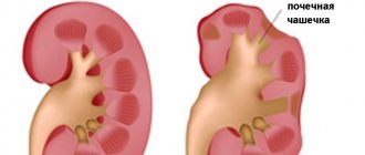
, pyelocaliceal system ー
pyelonephritis
. All of them can occur in acute or chronic forms.
Nephritis can be primary, with inflammation localized only in the kidney, or secondary, in which case the infection is brought into the kidneys from another organ.
Clinical picture
In the clinical picture of tubulointerstitial nephropathies, an important place is occupied by tubular disorders with predominant damage to one or another part of the nephron. Let us recall that the main function of the tubular apparatus, which includes the proximal tubule, loop of Henle, distal tubule and collecting ducts, is to maintain homeostasis. In the tubules, the process of concentration of urine (absorption of water and sodium), reabsorption of part of the organic and inorganic substances filtered in the glomerulus, as well as secretion into the lumen of the tubule of substances from the blood or formed in the cells of the tubules occur. The following symptoms are characteristic of tubular damage: polyuria, decreased relative density of urine, renal glycosuria, renal diabetes insipidus, renal tubular acidosis, hypo- or hyperkalemia, hypouricemia, tubular proteinuria.
According to the course, acute and chronic IN are distinguished.
Acute IN is characterized by an acute onset with fever, hematuria, polyuria, often with acute renal failure, sometimes with lower back pain. Depending on the etiology, acute IN can be drug-induced, viral, bacterial, parasitic, or immune.
Many drugs can lead to the development of drug-induced acute IN, most often antibiotics from the penicillin group (penicillin, ampicillin, oxacillin, methicillin), cephalosporins, tetracyclines, anti-tuberculosis drugs (rifampicin, ethambutol), etc., as well as non-steroidal anti-inflammatory drugs; Acute IN develops especially often after treatment with methicillin. However, cases of acute IN have been described after treatment with many other drugs, including sulfonamides (the first descriptions of acute drug IN), acyclovir, diuretics, allopurinol, cimetidine, phenyline, and even Chinese herbs.
The most recognized immune concept of the pathogenesis of drug-induced acute IN, which is supported by the detection of mononuclear cell infiltrates with non-caseating granulomas in the interstitium in 1/3 of renal biopsies, the occurrence of a delayed-type hypersensitivity reaction after an intradermal test with a damaging drug, and the predominance of T-lymphocytes among the infiltrate cells. Immunofluorescence studies in some cases of acute IN reveal deposits of immunoglobulins and complement in the interstitium and on the tubular basement membrane, less often - linear deposition of IgG.
The clinical signs of acute drug-induced ID are varied and nonspecific; sometimes symptoms of a common allergy can lead one to think about this disease. In some cases, the first clinical sign of drug-induced acute IN is a repeated wave of fever after successful treatment of the infection with antibiotics, often in combination with eosinophilia and skin rashes. Hematuria is characteristic, proteinuria is usually moderate, rarely exceeds 2 g/day, there is a decrease in glomerular filtration rate, an increase in creatinine levels; Oliguria is rare, polyuria is more common. ARF, one of the main and most constant signs of acute IN, is detected simultaneously with urinary syndrome. In our observations, acute renal failure of varying severity was diagnosed in 18 out of 19 patients with acute drug-induced IN; the disease in all of them was non-oliguric in nature. An essential diagnostic sign is a decrease in tubular functions. First of all, you should pay attention to a pronounced decrease in the relative density of urine. Renal diabetes insipidus, renal tubular acidosis, hyponatremia due to sodium loss, and hyperkalemia due to impaired potassium excretion with methicillin IN have been described. Rifampicin is associated with tubular disorders such as increased excretion of potassium and uric acid with a corresponding decrease in the level of these substances in the blood, and glycosuria. Expired tetracyclines and gentamicin can cause Fanconi syndrome, a complex tubular dysfunction.
TUBULOINTERSTITIAL NEPHROPATHIES
The concept of “tubulointerstitial nephropathies” (in the broad sense of the word includes inflammatory, metabolic or toxic diseases of the kidneys, occurring in contrast to glomerular (glomerular) diseases with primary and primary damage to the tubules and interstitial tissue of the kidneys. In a narrower sense of the word, the term “tubulointerstitial nephropathy "is more often used to refer to metabolic and toxic lesions without a clear inflammatory component, applying the term “tubulointerstitial nephritis” or, equivalently, “interstitial nephritis” to inflammatory, immunoinflammatory lesions of the tubules and interstitium. In addition to tubulointerstitial nephropathies as independent nosological forms, the tubulointerstitial component can be identified with a number of glomerular kidney lesions, including chronic glomerulonephritis. The term “interstitial nephritis” (IN) has existed since the mid-nineteenth century, but over the course of a century and a half included various concepts. The term “chronic interstitial nephritis” has long corresponded to the modern understanding kidney damage due to hypertension - nephroangiosclerosis, primary wrinkled kidney, i.e. designated those types of kidney pathology, which are based on vascular damage; this group then began to include secondarily wrinkled kidney, chronic nephritis of the hypertensive type. IN was contrasted with tubular, parenchymal nephritis with edematous syndrome (corresponding to modern nephritis with nephrotic syndrome). In parallel, in the first half of the twentieth century, the term “tubulointerstitial nephritis” existed to refer to acute renal failure (ARF). IN, corresponding to the modern interpretation of this concept, was first described in 1898 by Councilman in patients who had diphtheria and scarlet fever. However, for a very long time, IN was identified exclusively with pyelonephritis and was the prerogative of study by urologists. And at present, IN remains poorly known to a wide range of doctors, which is due, firstly, to the relative rarity of the disease, and secondly, to the nonspecificity of most symptoms. Poor knowledge of this pathology by doctors is evidenced by the fact that among the 22 patients with acute IN we observed, 20 were admitted with other diagnoses: 12 with a diagnosis of chronic glomerulonephritis, 3 with acute glomerulonephritis, 2 with pyelonephritis, 3 with systemic lupus erythematosus. At the same time, IN is in most cases a disease with an established etiology, promising in terms of treatment and prevention. The current wave of interest in tubulointerstitial lesions is associated both with the possibilities of treatment and prevention (obviously greater than with glomerular diseases), and with the increasing frequency of iatrogenic pathology in general, as well as with the assumption of the leading role of the interstitium and tubules in the progression of kidney diseases.
Clinical picture
In the clinical picture of tubulointerstitial nephropathies, an important place is occupied by tubular disorders with predominant damage to one or another part of the nephron. Let us recall that the main function of the tubular apparatus, which includes the proximal tubule, loop of Henle, distal tubule and collecting ducts, is to maintain homeostasis. In the tubules, the process of concentration of urine (absorption of water and sodium), reabsorption of part of the organic and inorganic substances filtered in the glomerulus, as well as secretion into the lumen of the tubule of substances from the blood or formed in the cells of the tubules occur. The following symptoms are characteristic of tubular damage: polyuria, decreased relative density of urine, renal glycosuria, renal diabetes insipidus, renal tubular acidosis, hypo- or hyperkalemia, hypouricemia, tubular proteinuria.
According to the course, acute and chronic IN are distinguished.
Acute IN
is characterized by an acute onset with fever, hematuria, polyuria, often with acute renal failure, sometimes with lower back pain.
Depending on the etiology, acute IN can be drug-induced, viral, bacterial, parasitic, or immune. to the development of drug-induced acute IN
, most often antibiotics from the penicillin group (penicillin, ampicillin, oxacillin, methicillin), cephalosporins, tetracyclines, anti-tuberculosis drugs (rifampicin, ethambutol), etc., as well as non-steroidal anti-inflammatory drugs;
Acute IN develops especially often after treatment with methicillin. However, cases of acute IN have been described after treatment with many other drugs, including sulfonamides (the first descriptions of acute drug IN), acyclovir, diuretics, allopurinol, cimetidine, phenyline, and even Chinese herbs. The most recognized immune concept of the pathogenesis of drug-induced acute IN, which is supported by the detection of mononuclear cell infiltrates with non-caseating granulomas in the interstitium in 1/3 of renal biopsies, the occurrence of a delayed-type hypersensitivity reaction after an intradermal test with a damaging drug, and the predominance of T-lymphocytes among the infiltrate cells. Immunofluorescence studies in some cases of acute IN reveal deposits of immunoglobulins and complement in the interstitium and on the tubular basement membrane, less often - linear deposition of IgG. The clinical signs
of acute drug-induced ID are varied and nonspecific; sometimes symptoms of a common allergy can lead one to think about this disease.
In some cases, the first clinical sign of drug-induced acute IN is a repeated wave of fever after successful treatment of the infection with antibiotics, often in combination with eosinophilia and skin rashes. Hematuria is characteristic, proteinuria is usually moderate, rarely exceeds 2 g/day, there is a decrease in glomerular filtration rate, an increase in creatinine levels; Oliguria is rare, polyuria is more common. ARF, one of the main and most constant signs of acute IN, is detected simultaneously with urinary syndrome. In our observations, acute renal failure of varying severity was diagnosed in 18 out of 19 patients with acute drug-induced IN; the disease in all of them was non-oliguric in nature. An essential diagnostic sign is a decrease in tubular functions. First of all, you should pay attention to a pronounced decrease in the relative density of urine. Renal diabetes insipidus, renal tubular acidosis, hyponatremia due to sodium loss, and hyperkalemia due to impaired potassium excretion with methicillin IN have been described. Rifampicin is associated with tubular disorders such as increased excretion of potassium and uric acid with a corresponding decrease in the level of these substances in the blood, and glycosuria. Expired tetracyclines and gentamicin can cause Fanconi syndrome, a complex tubular dysfunction. Laboratory indicators
by anemia, increased ESR, hyperproteinemia, hypergammaglobulinemia.
In diagnostic terms, other (extrarenal) signs of an allergic reaction are important: fever, skin rashes, arthralgia, drug-induced hepatitis, etc. However, the classic triad - fever, skin rashes and arthralgia - occurs only in 15 - 20% of cases. All of our 19 patients with acute drug-induced IN had one or another extrarenal manifestations of the disease, the most common of which was fever, observed in 2/3 of the patients, then skin syndrome, and arthralgia was much more rare. All patients had a sharp increase in ESR - up to 40 - 60 mm/h, more than half had anemia, a third had hyperproteinemia, and the vast majority had hypergammaglobulinemia. Criteria for diagnosing acute drug-induced ID:
-
temporary connection with taking medications; - moderate urinary syndrome with proteinuria not exceeding 2 g/day, predominance of red blood cells in urine sediment; - non-oliguric acute renal failure of varying severity, not accompanied by hyperkalemia and arterial hypertension; - a high frequency of various tubular disorders, among which a concentration defect occurs in 100% of cases; - protein shifts in the form of an increase in ESR, hyperproteinemia and hypergammaglobulinemia; - anemia; - extrarenal manifestations in the form of fever, skin syndrome, and liver damage. Treatment
consists primarily of immediately stopping the drug causing the kidney damage.
If there is no effect, glucocorticoids are prescribed after 2 - 3 days (60 - 80 mg of prednisolone per day); for acute renal failure, hemodialysis is performed. Acute drug-induced IN usually has a cyclical course
.
If the diagnosis is made quickly enough after discontinuation of the allergenic drug, recovery usually occurs; cases of chronicity are rare, although the period of reverse development can be very long. Only in advanced cases with long-term indiscriminate treatment (usually antibiotics prescribed because of persistent fever) can acute drug-induced IN take a chronic course. Drug-induced acute IN with acute renal failure can develop in chronic glomerulonephritis, and not only after the use of antibiotics, but sometimes also after the use of furosemide, phenylin, allopurinol, etc. Among viral acute IN
, hemorrhagic (Far Eastern) fever with renal syndrome (HFRS) is of greatest importance. .
The disease was first described in the Far East in the 30s of our century. Infection occurs from infected rodents. In 1978, in South Korea, a specific antigen was isolated in cryostat sections of the lung tissue of field mice, and then the virus itself, named Hantaan after the river in the valley of which rodents infected with the virus were caught. Currently, the HFRS virus has been isolated from 55 species of animals that are its carriers. The most infected with the virus are several species of field mice, gray and black rats. However, antibodies to the virus have also been detected in animals such as guinea pigs, dogs, chickens and pigs. In Russia, natural foci have been noted in the Far East, Bashkiria, and the Urals. Outbreaks of HFRS have been described in Northern China, South Korea, Japan, Hungary, and Scandinavian countries. Some cases are diagnosed in Central European countries. We recently observed a patient who became infected in the Moscow region. The disease most often occurs in spring and autumn. The disease begins with fever, malaise, then hemorrhages in the mucous and subcutaneous tissues, thrombocytopenia appear, from the 5-7th day urinary syndrome and oliguric acute renal failure develop with pronounced electrolyte shifts and hypervolemia. Damage to the gastrointestinal tract, respiratory organs, cardiovascular system, and severe intoxication may occur. However, as a rule, it is the severity of renal pathology that determines the prognosis of the disease. Morphologically, IN and multiple hemorrhages in the kidneys, especially in the pelvis, are characteristic. Hemorrhages are also found under the pleura, epicardium, splenic capsule, etc. The role of the direct nephrotoxic effect of the virus, microcirculatory disorders, and disseminated intravascular coagulation syndrome in the pathogenesis is discussed. Treatment is aimed at combating intoxication and manifestations of acute renal failure. Currently, antiviral therapy (ribovirin, specific immunoglobulin) is being used. Bacterial acute IN
is fully consistent with acute pyelonephritis.
Among parasitic acute IN,
kidney damage in leptospirosis is of primary importance. The clinical picture of the disease is characterized by severe intoxication, headache, fever, and tachycardia. On the 3-4th day of illness, often simultaneously with jaundice and hepatolienal syndrome, signs of kidney damage appear: proteinuria, erythrocyte and leukocyturia, oliguric acute renal failure. Death from uremia may occur.
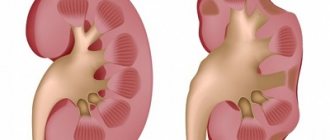
Chronic IN
The development of chronic IN is caused by certain medications, metabolic disorders (hyperuricemia, oxaluria, hypercalcemia), intoxication with heavy metals (lead, cadmium, zinc), malignant neoplasms, as well as some diseases with a clear immune pathogenesis. In some cases, the cause of chronic IN cannot be determined (idiopathic chronic IN). Leading morphological characters
are diffuse lymphohistiocytic infiltration of the interstitium, stromal sclerosis, severe degeneration or atrophy of the tubular epithelium.
The clinical picture
is determined by gradually progressive tubular disorders.
Water and electrolyte disturbances and disturbances in the concentration function of the kidneys develop relatively quickly, i.e. those functional changes that, with primary glomerular lesions, are usually a sign of advanced renal failure. Sometimes one or another part of the nephron is predominantly affected, which leads to certain functional disorders characteristic of this particular part: proximal tubular insufficiency with the development of proximal tubular acidosis, glycosuria, rarely - complete Fanconi syndrome or distal tubular insufficiency with the development of distal tubular acidosis, sometimes with hyperkalemia or loss of sodium (salt-wasting kidney). Most often, chronic IN develops with long-term regular use of non-narcotic analgesics (analgesic mixtures). This is the so-called analgesic nephropathy (AN)
- chronic IN, occurring with necrosis of the renal papillae.
The mechanism of the damaging effect of analgesics is associated with disruption of oxidation processes in the tubular epithelium and interstitial tissue, suppression of the synthesis of prostaglandins - the main regulators of medullary blood flow in the kidneys, as well as with the direct toxic effect of drugs on the medulla of the kidney. Morphological examination, in addition to signs of chronic IN, reveals necrosis of the renal papillae, specific brown pigmentation of the renal papillae and urinary tract, and microangiopathy of the ureteral vessels (a diagnostic sign of analgesic abuse). The incidence of AN worldwide (according to pathological studies) ranges from 0 to 4%; As a cause of end-stage renal failure among patients undergoing hemodialysis or undergoing kidney transplantation, AN has been reported in 3% of patients in Europe and 20% in Australia. In the early years of studying AN, the main damaging role was assigned to phenacetin, which is why the term “phenacetin nephritis” existed. Then it was found that it was not pure phenacetin that was harmful, but analgesic mixtures that included phenacetin. Finally, over the past decade, researchers have shown that AN can also develop in individuals who have never taken phenacetin but have abused other analgesic mixtures. That is, phenacetin is no longer considered the only nephrotoxic drug; the possible damaging role of other analgesic mixtures is emphasized, including two analgesics (combinations of aspirin or analgin with paracetamol or pyrazolones are especially dangerous) and caffeine or codeine; caffeine and codeine affect mood, and it is well established that the addition of these substances can cause psychological dependence on the drug. In a large study conducted in 15 European countries, among 226 patients with AN diagnosed using computed tomography criteria (see below), 219 patients took 2 or more analgesics (in 173 patients one of the analgesics was phenacetin) in combination with caffeine and/or codeine; in 46 patients who did not take phenacetin, nephrotoxicity was documented for combinations of aspirin with paracetamol, aspirin with pyrazolones, paracetamol with pyrazolones and two pyrazolones (Elseviers, De Broe, 1996). AN is observed more often in women over 40 years of age (although the abuse of analgesics begins much earlier in them), suffering from migraines or lumbodynia, clinically manifested by moderate urinary syndrome (hematuria, slight proteinuria), polyuria, nocturia, night cramps. An early sign (even in the preclinical stage) is a decrease in the relative density of urine. In 70% of patients, abacterial leukocyturia is detected, in 1/3 - a urinary infection (usually asymptomatic). The development of severe proteinuria is a poor prognostic sign and indicates the possibility of rapid (in 1 - 2 years) development of end-stage renal failure. Chronic renal failure (CRF) gradually develops, which progresses slowly, accompanied by severe osteodystrophy and severe metabolic acidosis. Half of the patients develop necrosis of the renal papillae with gross hematuria, sometimes with repeated episodes of obstructive acute renal failure with rapid spontaneous recovery of renal function, relapses of the disease with renal colic, and attacks of acute pyelonephritis. In most patients, papillary necrosis is asymptomatic (the so-called incomplete rejection of the necrotic renal papilla) with minimal proteinuria and microhematuria; persistent microhematuria may indicate the development of urinary tract carcinoma. Significantly more often than with other types of chronic IN, hypertension develops, which can sometimes acquire a malignant course. Hypertension may be associated with salt depletion (due to loss of sodium chloride in the urine). Complications of AN:
obstructive acute renal failure (with bilateral papillary necrosis), addition of pyelonephritis, nephrocalcinosis, nephrolithiasis.
Patients with AN have a high risk of developing malignant tumors of the bladder, ureters, and renal pelvis, which are observed much more often than in the population. The disease is diagnosed in 80% of patients in the stage of renal failure. In terms of diagnostics, the combination of persistent aseptic leukocyturia with episodes of renal colic with gross hematuria in the absence of nephrolithiasis, polyuria, decreased kidney size, and anemia that does not correspond to the severity of chronic renal failure are important. Elseviers et al. recently presented diagnostic criteria for AN based on computed tomography without contrast. A decrease in the mass of both kidneys in combination with uneven contours or calcification of the papillae is highly likely to indicate AN, even in patients with slowly progressive (incipient) renal failure. Computed tomography is the most suitable diagnostic method to determine the most pathognomonic sign, renal papillary calcification, with a sensitivity of 87% and a specificity of 97%. When making a diagnosis
, you should pay attention to extrarenal signs of analgesic abuse, among which are hypochromic anemia with a peculiar pale yellow coloration of the skin, damage to the stomach (peptic ulcer), liver (drug-induced hepatitis), early atherosclerosis with progressive coronary heart disease, psychasthenia, premature aging, early graying (patients look older than their years).
The effect on gonadal function (infertility, teratogenic effects) is known. Gastric ulcer sometimes precedes nephropathy for many years. Treatment consists of completely stopping the use of analgesics, correcting water and electrolyte disturbances, hypertension, and fighting urinary tract infections. Reducing calorie intake (without changing protein intake) or a low-protein diet can slow progression. AN is one of the few kidney diseases that can be prevented.
The most rational approach to preventing AN is to prohibit the over-the-counter sale of any analgesic mixtures containing two analgesics in combination with caffeine and/or codeine. In Australia, restrictions on the over-the-counter availability of analgesic mixtures have led to a sharp decrease in the incidence of AN.
to the development of chronic toxic IN
, for example, antitumor drugs (cisplatin), lithium used in psychiatric practice, gold preparations, etc. Speaking about iatrogenic lesions, we should also mention radiation toxic IN, which sometimes develops during radiation therapy patients with tumors of the kidney (Wilms tumor), ovary, testicle, retroperitoneal space.
Among metabolic toxic INs,
the most common are hyperuricemic and oxalate nephropathies.
In the development of IN in hyperuricemia, the role of several pathogenetic mechanisms is assumed - blockage of the ureters, intratubular deposition of urate crystals, and the nephrotoxic effect of hyperuricemia as such. Oxalate nephropathy (kidney damage due to oxaluria) usually develops in childhood as a hereditary IN or urolithiasis. Less common and less well known are metabolic kidney lesions that develop with hypercalcemia and hypokalemia. Hypercalcemia
is observed in primary and secondary hyperparathyroidism, sarcoidosis, malignant neoplasms, etc.
The distinctive morphological feature is nephrocalcinosis, small calcium deposits are often found in the kidneys at biopsy or autopsy, the formation of stones can lead to obstructive nephropathy. Functional changes are especially characteristic, primarily a sharp decrease in the concentration function of the kidneys, associated not only with structural disorders (intraluminal obstruction with aseptic interstitial inflammation), but also with the influence of calcium on the regulation of CP and tubular reabsorption, disruption of intracellular calcium transport, decreased sensitivity of the tubules to vasopressin; the possible role of increased prostaglandin secretion is discussed. Clinically, nephropathy is characterized by polyuria, azotemia, hypertension is noted in half of the cases, and changes in urine may be observed. Structural and functional changes in the kidneys develop with hypokalemia
(or rather, not so much with a decrease in the level of potassium in the extracellular fluid, but with a decrease in the content of total potassium in the body).
Morphologically, the most characteristic lesion is the epithelium of the proximal tubules. Interstitial lesions usually do not develop, i.e. This is more likely a tubulopathy than a toxic IN. Functional disorders are expressed primarily by a decrease in concentration function, changes in the acid-excreting function of the kidneys and a tendency to sodium retention, up to the formation of edema. Among the factors leading to the development of chronic toxic IN, heavy metals should be mentioned: cadmium and lead. Cadmium, widely used in modern industry, leads to nephropathy, characterized by proximal dysfunction, up to full Fanconi syndrome. Finally, toxic interstitial lesions can be caused by tumors (extrarenal) that secrete pathological proteins, vasoactive amines, and hormones. The most studied toxic IN
in myeloma, which forms the basis of the myeloma kidney, is associated with the excessive formation of light chains of immunoglobulins, which are normally catabolized in the kidney - a small amount of light chains, normally filtered in the glomeruli, is absorbed in the proximal tubules and destroyed there by lysosomal enzymes. In myeloma, overproduction of light chains leads to the fact that they cannot be completely absorbed in the proximal tubules, overload them and accumulate without being completely catabolized, with the development of proximal dysfunction, up to Fanconi syndrome (myeloma is the most common cause of Fanconi syndrome in adults). Some of the light chains that are not reabsorbed in the proximal tubules pass into the distal ones, where they are deposited with the formation of cylinders, blockage of the tubules, and the development of inflammatory infiltrates with giant multinucleated cells. The possibility of a direct toxic effect of light chains on the tubules is also discussed. Hypercalcemia and hyperuricemia can also lead to tubulointerstitial kidney damage in myeloma. The clinical picture is determined by progressive nephropathy, leading quite quickly to the development of chronic renal failure. Kidney damage similar to a myeloma kidney sometimes occurs with other tumors in which overproduction of monoclonal immunoglobulins is observed (malignant lymphomas, lymphocytic leukemia, pancreatic tumors).
Literature:
1. Sirotin B.Z. Hemorrhagic fever with renal syndrome. Khabarovsk, 1994. 2. Tareeva I.E., Androsova S.O. Tubulointerstitial nephropathies. In the book: Nephrology, ed. I.E. Tareeva, 1995, vol. 2, p. 101-9. 3. Tareeva I.E., Nikolaev A.Yu., Androsova S.O. Drug-induced kidney damage. Ibid., p. 299-312. 4. Elseviers MM, De Broe ME/ Amer J Kid Dis 1996; 28;48-55. 5. Tubulo-interstitial nephropathies. Churchill, Livingstone, 1983. Ed B Brenner, J Stein.
Diagnostics
Laboratory indicators are characterized by anemia, increased ESR, hyperproteinemia, hypergammaglobulinemia.
In diagnostic terms, other (extrarenal) signs of an allergic reaction are important: fever, skin rashes, arthralgia, drug-induced hepatitis, etc. However, the classic triad - fever, skin rashes and arthralgia - occurs only in 15 - 20% of cases. All of our 19 patients with acute drug-induced IN had one or another extrarenal manifestations of the disease, the most common of which was fever, observed in 2/3 of the patients, then skin syndrome, and arthralgia was much more rare. All patients had a sharp increase in ESR - up to 40 - 60 mm/h, more than half had anemia, a third had hyperproteinemia, and the vast majority had hypergammaglobulinemia.
Criteria for diagnosing acute drug-induced ID:
- temporary connection with taking medications;
- moderate urinary syndrome with proteinuria not exceeding 2 g/day, predominance of red blood cells in urine sediment;
- non-oliguric acute renal failure of varying severity, not accompanied by hyperkalemia and arterial hypertension;
- a high frequency of various tubular disorders, among which a concentration defect occurs in 100% of cases;
- protein shifts in the form of an increase in ESR, hyperproteinemia and hypergammaglobulinemia;
- anemia;
- extrarenal manifestations in the form of fever, skin syndrome, and liver damage.
Symptoms of nephritis
The main signs of acute nephritis:
- pain in the lumbar region;
- swelling that appears in the morning and disappears during the day;
- change in the color and amount of urine (cloudy or red);
- increased body temperature;
- increased blood pressure (more typical is an increase in diastolic - lower);
- constant thirst;
- general weakness and malaise.
Chronic nephritis
has the same symptoms, but less pronounced. Body temperature remains normal. Often the patient only has morning swelling on the face and high blood pressure. Chronic nephritis is cyclical: periods of exacerbation alternate with remissions.
Factors that provoke exacerbation may be hypothermia, illness or severe stress.
Treatment
Treatment consists primarily of immediately stopping the drug causing the kidney damage. If there is no effect, glucocorticoids are prescribed after 2 - 3 days (60 - 80 mg of prednisolone per day); for acute renal failure, hemodialysis is performed.
Acute drug-induced IN usually has a cyclical course. If the diagnosis is made quickly enough after discontinuation of the allergenic drug, recovery usually occurs; cases of chronicity are rare, although the period of reverse development can be very long. Only in advanced cases with long-term indiscriminate treatment (usually antibiotics prescribed because of persistent fever) can acute drug-induced IN take a chronic course.
Drug-induced acute IN with acute renal failure can also develop in chronic glomerulonephritis, and not only after the use of antibiotics, but sometimes also after the use of furosemide, phenyline, allopurinol, etc.
Among viral acute IN, hemorrhagic (Far Eastern) fever with renal syndrome (HFRS) is of greatest importance. The disease was first described in the Far East in the 30s of our century. Infection occurs from infected rodents. In 1978, in South Korea, a specific antigen was isolated in cryostat sections of the lung tissue of field mice, and then the virus itself, named Hantaan after the river in the valley of which rodents infected with the virus were caught. Currently, the HFRS virus has been isolated from 55 species of animals that are its carriers. The most infected with the virus are several species of field mice, gray and black rats. However, antibodies to the virus have also been detected in animals such as guinea pigs, dogs, chickens and pigs.
The disease most often occurs in spring and autumn. The disease begins with fever, malaise, then hemorrhages in the mucous and subcutaneous tissues, thrombocytopenia appear, from the 5-7th day urinary syndrome and oliguric acute renal failure develop with pronounced electrolyte shifts and hypervolemia. Damage to the gastrointestinal tract, respiratory organs, cardiovascular system, and severe intoxication may occur. However, as a rule, it is the severity of renal pathology that determines the prognosis of the disease.
Morphologically, IN and multiple hemorrhages in the kidneys, especially in the pelvis, are characteristic. Hemorrhages are also found under the pleura, epicardium, splenic capsule, etc. The role of the direct nephrotoxic effect of the virus, microcirculatory disorders, and disseminated intravascular coagulation syndrome in the pathogenesis is discussed.
Treatment is aimed at combating intoxication and manifestations of acute renal failure. Currently, antiviral therapy (ribovirin, specific immunoglobulin) is being used.
Bacterial acute IN is fully consistent with acute pyelonephritis.
Among parasitic acute IN, kidney damage in leptospirosis is of primary importance. The clinical picture of the disease is characterized by severe intoxication, headache, fever, and tachycardia. On the 3-4th day of illness, often simultaneously with jaundice and hepatolienal syndrome, signs of kidney damage appear: proteinuria, erythrocyte and leukocyturia, oliguric acute renal failure. Death from uremia may occur.
Causes of nephritis
Nephritis can develop due to exposure to an infectious factor, immune or toxic. Sometimes it becomes a complication of urolithiasis.
Bacteria most often infect the renal pyelocaliceal system. The main causative agents of pyelonephritis:
- staphylococci;
- coli;
- enterococci;
- streptococci.
The infection enters the kidney tissue along with the blood or through the ascending route (from the skin of the perineum through the urethra and bladder).
Immune inflammation of the glomeruli (glomerulonephritis) develops when bacteria enter the body and cause a reactive response of the immune system. A large number of immune complexes (antigen-antibody) are formed in the blood, which damage the thin membranes of the vascular glomeruli (autoimmune disease).
Such glomerulonephritis can develop after a streptococcal infection - tonsillitis, scarlet fever, influenza, pneumonia.
Possible complications of nephritis
Nephritis disrupts kidney function - normal blood filtration and urine formation. With improper treatment, or lack thereof, complications develop such as:
- renal failure;
- pulmonary edema;
- cerebral edema;
- heart failure;
- hypertension;
- hydropericardium (fluid in the pericardium).
All these complications are life-threatening; it is important to identify nephritis at the first stage and begin its treatment.
Recommendations for the prevention of nephritis
To prevent the development of nephritis, follow these recommendations:
- avoid hypothermia;
- provide a balanced diet rich in vitamins;
- normalize the drinking regime, 30 ml per kg of clean water per day - this is the minimum;
- treat colds correctly; if the doctor prescribed antibiotics, you need to drink them completely until the end of the course.
If symptoms of nephritis appear, contact a nephrologist, urologist, or internist for examination and treatment.