Solution for intravitreal injection Eylea from the German company Bayer is one of the newest means of treating complex eye diseases, including the “wet” form of age-related macular degeneration, diabetic macular edema, macular edema due to retinal vein occlusion.
The action of the drug suppresses the growth of abnormal newly formed vessels, prevents the accumulation of fluid in the retina and increases visual acuity. At the same time, Eylea is well tolerated by patients, and the achieved effect lasts quite a long time.
In terms of price, Eylea is comparable to other drugs of similar action (Lucentis, Avastin).
What is Eylea?
Eylea is a solution for intravitreal administration from Bayer (Germany). The product is developed for the conservative treatment of ophthalmological diseases, including the wet form of macular degeneration and diabetic macular edema. The main active ingredient is aflibercept. It is a transparent liquid with a yellow tint. The principle of action involves the fusion of active substance molecules with VEGF molecules. Eylea is used to suppress the growth of abnormal new blood vessels. The drug is characterized by high efficiency, safety, long-lasting effects and is well tolerated by patients. Hospitalization is not required during therapy. The clinical effectiveness of the drug has been confirmed by various international studies. Eylea helps to significantly slow down the progression of the disease and maintain visual acuity. Release form: colorless bottles of 0.1 ml (100 µl).
Therapy with Eylea
The active ingredient of Eylea solution is the recombinant fusion protein aflibercept. It acts as a decoy by binding to vascular endothelial growth factor (VEGF) and placenta growth factor (PIGF). The result of such a reaction is inhibition of the process of neovascularization and reduction of macular edema.
The drug is intended only for intraocular (intravitreal) injections into the vitreous body. Typically, therapy with Eylea solution, like other drugs in this series, lasts three or more months with an interval between injections of at least 30 days. However, the main difference between the drug Eylea is the ability to achieve a therapeutic effect with intravitreal injections in a standard dosage not monthly, but once every two months.
The solution is administered until the maximum level of visual acuity is achieved, which is determined through monitoring at three consecutive visits to the attending physician.
After 12 months of drug therapy, it can be continued according to objective indications.
Eylea in macular degeneration
Macular degeneration (AMD) is characterized by damage to the retina and damage to central vision. The “wet” form of macular degeneration is especially dangerous.
The main symptoms of the disease are:
- decreased visual acuity,
- appearance of spots before the eyes,
- distortion of the contours of objects.
When these symptoms appear, special therapy is necessary, including taking anti-VEGF drugs. These drugs include Eylea solution. The product is used exclusively for intravitreal administration. When therapy is carried out correctly, the process of neovascularization is inhibited. The recommended dosage is 2 mg of aflibercept. The duration of treatment is determined individually by the ophthalmologist. The course of treatment is 3 months, 1 injection per month. In the future, the interval between injections may be more than 2 months. If effectiveness is low, drug therapy is discontinued. Treatment is carried out until visual acuity improves. If visual acuity decreases, the intervals between injections may be reduced.
Self-medication for AMD is strictly prohibited.
Eylea®
Aflibercept is a recombinant fusion protein consisting of fragments of the extracellular domains of human VEGF receptors 1 (VEGFR-1) and 2 (VEGFR-2) combined with the Fc fragment of human immunoglobulin G (IgGl).
Aflibercept is produced by Chinese hamster ovary (CHO) K1 cells using recombinant DNA technology.
Aflibercept acts as a soluble decoy receptor that binds VEGF-A (vascular endothelial growth factor A) and PIGF (placental growth factor) with higher affinity than their natural receptors and thus may inhibit the binding and activation of these related VEGFs receptors.
Mechanism of action
Vascular endothelial growth factor A (VEGF-A) and placental growth factor (PIGF) are members of the VEGF family of angiogenic factors that have potent mitogenic, chemotactic effects on endothelial cells and increase vascular permeability. VEGF acts through two types of tyrosine kinase receptors, VEGFR-1 and VEGFR-2, present on the surface of endothelial cells. PIGF binds only to VEGFR-1, which is also present on the surface of leukocytes. Excessive VEGF-A activation of these receptors can lead to pathological neovascularization and excessive vascular permeability. In these processes, PIGF may synergize with VEGF-A and also stimulate leukocyte infiltration and vascular inflammation.
Pharmacodynamic effects
Neovascular (“wet” form) age-related macular degeneration (“wet” form of AMD)
The “wet” form of AMD is characterized by pathological neovascularization of the choroid. Leakage of blood and fluid from the abnormally neovascularized choroid may cause central retinal thickening (CRT) or swelling and/or hemorrhage into the retina/subretinal space, resulting in decreased visual acuity.
The safety and effectiveness of Eylea® in wet AMD was assessed in two randomized, multicenter, double-blind, active-controlled studies VIEW1 and VIEW2. In these studies, 2412 patients were randomized in a 1:1:1:1 ratio to receive Eylea® (1817 patients) or ranibizumab in the following regimens:
1) Eylea® 2 mg every 8 weeks after 3 initial monthly injections (Eylea® 2q8);
2) Eylea® 2 mg every 4 weeks (Eylea® 2q4);
3) Eylea® 0.5 mg every 4 weeks (Eylea® 0.5q4);
4) Ranibizumab 0.5 mg every 4 weeks (ranibizumab 0.5q4).
The studies included patients aged 49 to 99 years, with a mean age of 76 years. About 89% of patients (1616 of 1817) randomized to Eylea® were 65 years of age or older, and about 63% (1139 of 1817) were 75 years of age or older.
In the second year of the study, patients continued to receive the drugs at the same dose that was prescribed to them initially, but in a modified dosing regimen, which, according to the protocol, was based on the results of changes in visual acuity and anatomical parameters, while the maximum interval between injections was no more than 12 weeks
In both studies, the primary efficacy endpoint was the proportion of eligible patients who maintained visual acuity, defined as a loss of <15 ETDRS (Early Treatment Diabetic Retinopathy Study) letters at week 52. compared to the initial state.
In the VIEW1 study, at week 52, visual acuity improvement was maintained in 95.1% of patients receiving Eylea 2 mg every 8 weeks after the initial 3 monthly injections, compared with 94.4% of patients receiving ranibizumab 0.5 mg. every 4 weeks.
In the VIEW2 study, at week 52, visual acuity improvement was maintained in 95.6% of patients receiving Eylea 2 mg every 8 weeks after the initial 3 monthly injections, compared with 94.4% of patients receiving ranibizumab 0.5 mg. every 4 weeks.
Both studies showed that Eylea® and ranibizumab, administered at a dose of 0.5 mg every 4 weeks, were comparable in clinical efficacy.
A detailed description of the results of the analysis of the pooled data from both studies is provided in Table 1.
Table 1: Efficacy assessment at 52 and 96 weeks; combined data from VIEW1 and VIEW2 studies
| Efficiency mark | Eylea® 2 mg every 8 weeks after 3 initial monthly injections (N=607) | Ranibizumab 0.5 mg every 4 weeks (N=595) | |||||
| 52 week | Week 96 | 52 week | Week 96 | ||||
| Average number of injections | 7,6 | 11,2 | 12,3 | 16,5 | |||
| Average number of injections (weeks 52-96) | 4,2 | 4,7 | |||||
| Proportion of patients with ≤15 letter loss with best-corrected visual acuity (BCVA) compared with baseline | 95,33 % | 92,42 % | 94,42 % | 91,60% | |||
| Mean change from baseline in best-corrected visual acuity (BCVA) measured by ETDRS | 8,40 | 7,62 | 8,74 | 7,89 | |||
| Proportion of patients who gained ≥15 letters on the ETDRS compared to baseline | 30,97 % | 33,44 % | 32,44 % | 31,60% | |||
In patients treated with Eylea® (one injection per month for three consecutive months, then one injection every 2 months), central retinal thickness (CRT) and the average size of the area of pathological neovascularization decreased soon after the start of treatment, which is consistent with the results, obtained with the use of ranibizumab at a dose of 0.5 mg every month. The achieved reduction in the size of pathological neovascularization and TCZD remained stable in the second year of the study until the last assessment at week 96, with 2-4% of patients requiring all injections on a monthly basis, and a third of patients requiring at least one injection with a treatment interval of one month.
In both studies, a decrease in the area of pathological neovascularization was noted in all groups with different dosing regimens.
Macular edema that developed as a result of occlusion of the central retinal vein (CRVO) or its branches (BRVO)
In BRVO and BRVO, retinal ischemia develops, which is a signal for the release of VEGF, which in turn leads to destabilization of tight junctions and stimulates the proliferation of endothelial cells. Increased expression of VEGF is associated with complications such as disruption of the blood-ophthalmic barrier, retinal edema due to increased vascular permeability, and neovascularization. The safety and efficacy of Eylea® were assessed in two randomized, multicenter, double-blind, controlled studies, COPERNICUS and GALILEO, which included 358 patients with macular edema secondary to CRVO. In both studies, patients were randomized 3:2 to receive 2 mg Eylea® every 4 weeks (217 patients) (Eylea® 2q4 group), or a control group to receive sham injections every 4 weeks.
The studies included patients aged from 22 to 89 years, with a mean age of 64 years. In trials for the indication of CRVO, approximately 52% of patients (112 of 217) randomized to the Eylea® group were aged 65 years or older, and approximately 18% (38 of 217) were aged 75 years or older.
After 6 consecutive monthly injections, patients continued to receive treatment only if they met predefined criteria for continuation of therapy, with the exception of control patients in the GALILEO study, who continued to receive sham injections until week 52. From this point on, all patients were treated if they met predefined criteria.
In both studies, the primary efficacy endpoint was the proportion of patients whose best-corrected visual acuity (BCVA) improved by at least 15 letters at 24 weeks compared with baseline. The secondary endpoint was change from baseline in visual acuity at week 24.
The differences between the groups were statistically significant in favor of Eylea in both studies. Improvement in BCVA was achieved after 3 months, followed by stabilization of visual acuity and TCVA until the 6th month. Statistically significant differences persisted until week 52.
A detailed description of the analysis results from both studies is provided in Table 2 and Figure 1.
Table 2: Efficacy assessments in the COPERNICUS and GALILEO studies at 24, 52 and 76/100 weeks.
| Efficiency mark | Proportion of patients who gained ≥15 letters compared to baseline | Mean change from baseline in ETDRS BCVA (SD1) | ||
| COPERNICUS | 24 weeks | Control (N=73) | 12% | -4,0(18,0) |
| Eylea® 2 mg every 4 weeks (N=114) | 56% | 17,3(12,8) | ||
| 52 weeks | Control2 (N=73) | 30% | 3,8(17,1) | |
| Eylea® 2 mg (N=114) | 55 % | 16,2(17,4) | ||
| 100 weeks | Control2,3 (N=73) | 23,3 % | 1,5(17,7) | |
| Eylea® 32 mg (N=114) | 49,1 % | 13,0(17,7) | ||
| GALILEO | 24 weeks | Control (N=68) | 22% | 3,3(14,1) |
| Eylea® 2 mg every 4 weeks (N=103) | 60% | 18,0(12,2) | ||
| 52 weeks | Control (N=68) | 32% | 3,8(18,1) | |
| Eylea® 2 mg (N=103) | 60% | 16,9(14,8) | ||
| 76 weeks | Control4 (N=68) | 29,4 % | 6,2(17,7) | |
| Eylea® 4 2 mg (N=103) | 57,3 % | 13,7(17,8) | ||
1) SD: standard deviation
2) In the COPERNICUS study, patients in the control group could receive Eylea® as needed (PRN) every 4 weeks during the period from 24 to 52 weeks, patients visited the doctor every 4 weeks.
3) In the COPERNICUS study, both the control group and the Eylea® 2 mg group received Eylea® 2 mg as needed (PRN) every 4 weeks from weeks 52 to 96: patients were required to visit the doctor every quarter, but could come to the appointment if necessary every 4 weeks.
4) In the GALILEO study, from weeks 52 to 68, both the control group and the Eylea® 2 mg group received Eylea® 2 mg as needed (PRN) every 8 weeks, with patients attending mandatory appointments every 8 weeks.
The safety and efficacy of Eylea® were evaluated in the randomized, multicenter, double-blind, controlled trial VIBRANT, which included 181 patients with macular edema secondary to BRVO, including hemiretinal central retinal vein occlusion. In this study, patients were randomized 1:1 to receive Eylea 2 mg every 8 weeks after 6 initial monthly injections (91 patients) or to receive laser photocoagulation initially (active control group).
The study included patients aged from 42 to 94 years, with an average age of 65 years. In the BRVO trial, approximately 58% of patients (53 of 91) randomized to Eylea were 65 years of age or older, and approximately 23% (21 of 91) were 75 years of age or older.
Beginning at week 12, patients in the active control group could receive additional laser photocoagulation, called salvage therapy, at a minimum interval of 12 weeks. Beginning at week 24, patients in the active control group who met prespecified criteria could receive salvage therapy with Eylea 2 mg every 4 weeks for 3 months, then every 8 weeks.
In the VIBRANT study, the primary efficacy endpoint was the proportion of patients whose BCVA increased by at least 15 letters at 24 weeks compared with baseline, which was superior to the active control group in the Eylea* group.
In the VIBRANT study, the secondary endpoint was improvement in visual acuity at week 24 compared with baseline, which was statistically significant in favor of Eylea®. The improvement in vision occurred quickly and reached its maximum value in the 3rd month, with subsequent maintenance of the achieved values until the 12th month.
Beginning at week 24, 67 patients in the active control group received rescue therapy with Eylea® (Active Control group/Eylea* 2 mg), which resulted in an average improvement of 5 letters in visual acuity from week 24 to 52 weeks. th week.
A detailed description of the VIBRANT study analysis results is provided in Table 3 and Figure 2.
| Efficiency mark | Proportion of patients who gained ≥15 letters compared to baseline | Mean change from baseline in ETDRS BCVA (SD1) | |
| 24 weeks | Eylea® 2 mg every 4 weeks (N=91) | 52,7 % | 17,0(11,9) |
| Active control (N=90) | 26,7 % | 6,9(12,9) | |
| 52 weeks | Eylea® 2 mg every 8 weeks (N=91)2 | 57,1 % | 17,1(13,1) |
| Active control/Eylea® 2 mg (N=90)3 | 41,1 % | 12,2(11,9) | |
1) SD: standard deviation
2) The treatment interval for all patients in the Eylea® group was increased to 8 weeks during the period from the 24th to the 48th week.
3) Starting at week 24, patients in the active control group who met at least one of the predefined criteria could receive salvage therapy with Eylea® (67 patients in total). Fixed regimen of “rescue therapy” with Eylea® - Eylea® at a dose of 2 mg every 4 weeks for 3 months, then intravitreal injections every 8 weeks.
Patients who received 6 consecutive monthly injections of 2 mg of Eylea® showed a durable, rapid and pronounced morphological response (measured by an improvement in mean TCZP scores). At week 24, the reduction in TCPV was statistically significantly superior to that in the control group in all three studies (COPERNICUS (CRVO): -457 microns versus -145 microns,
GALILEO (CBVS): -449 microns relative to -169 microns, VIBRANT (CBVS): -280 microns relative to -128 microns). The achieved reduction in CVD was maintained until the end of each study: up to 100 weeks in the COPERNICUS study, up to 76 weeks in the GALILEO study and up to 52 weeks in the VIBRANT study.
Diabetic macular edema (DME)
Diabetic macular edema is a consequence of diabetic retinopathy and is characterized by increased vascular permeability and damage to the retinal capillaries, which can lead to loss of visual acuity.
The safety and efficacy of Eylea® in patients with diabetic macular edema were assessed in two randomized, multicenter, double-blind, active-controlled studies. A total of 862 patients were randomized. Of these, 576 patients were randomized to Eylea® in two studies (VIVID-DME and VISTA-DME). In each study, patients were randomly assigned to three groups in a 1:1:1 ratio:
1) Eylea® 2 mg every 8 weeks for the first 5 months;
2) Eylea® 2 mg every 4 weeks;
3) laser coagulation in the macula (active control).
The studies included patients aged from 23 to 87 years, with a mean age of 63 years. In phase III trials for diabetic macular edema, approximately 47% of patients (268 of 576) randomized to Eylea were 65 years of age or older, and approximately 9% (52 of 576) were 75 years of age or older. The majority of patients included in both studies had type II diabetes.
Beginning at week 24, patients meeting predefined vision loss thresholds could receive additional therapy: patients in the Eylea® groups could receive laser photocoagulation and patients in the control group could receive Eylea® therapy.
In both studies, the primary efficacy endpoint was the mean change from baseline in best-corrected visual acuity (BCVA) at week 52, which was Eylea 2 mg every 8 weeks for the first 5 months and Eylea 2 mg every 4 weeks was statistically significant and superior to the control group. This advantage continued until the 100th week.
A detailed description of the results from the VIVID-DME and VISTA-DME studies is provided in Table 4 and Figure 3.
Table 4: Efficacy assessments at weeks 52 and 100 in the VIVID-DME and VISTA-DME studies
In the VIVID-DME and VISTA-DME studies, 36 (9%) and 197 (43%) patients, respectively, had received prior anti-VEGF therapy, with a washout period of 3 months or more. Treatment effects in subgroups of patients pretreated with VEGF inhibitors were similar to those observed in patients newly treated with VEGF inhibitors.
Patients with bilateral disease could receive anti-VEGF therapy in the other eye if deemed necessary by the treating physician. In the VISTA-DME study, 217 (70.7%) patients receiving Eylea® received Eylea® injections in both eyes before week 100; in the VIVID-DME study, 97 (35.8%) patients receiving Eylea® received injections of various anti-VEGF drugs into the other eye.
Efficacy and safety parameters were comparable to those for the general population.
Soon after the start of therapy, patients treated with Eylea® showed a rapid and pronounced response in terms of morphological parameters (TCRS, level on the Diabetic Retinopathy Severity Scale (DRSS)). In the VIVID-DME and VISTA-DME studies, the mean reduction in TCPV compared with baseline values at week 52 was statistically significantly greater in the Eylea® group compared to the laser group: -192.4 microns and -183.1 microns in in the Eylea® group 2 mg every 8 weeks for the first 5 months and -66.2 microns and -73.3 microns in the laser group, respectively.
In the VIVID-DME and V1STA-DME studies, improvement of 2 or more levels of diabetic retinopathy on the DRSS was assessed in a prespecified manner and was found in 73.7% of patients in the VIVID-DME study and in 98.3% of patients in the VISTA-DME study.
An independent comparative study (DRCR.net Protocol T) used a dosing regimen based on strict OCT criteria and visual changes with re-treatment. In the aflibercept treatment group (224 patients), this treatment regimen resulted in patients receiving an average of 9.2 injections, which is similar to the number of doses received in the Eylea® 2 mg every 8 weeks groups for the first 5 months in the VIVID-DME and VISTA- studies. DME, while overall efficacy in the aflibercept treatment group in the Protocol T study was comparable to that in the Eylea® 2 mg every 8 weeks for the first 5 months groups in the VIVID-DME and VISTA-DME studies. In the Protocol T study, there was an average improvement of 13.3 letters, with 42% seeing at least 15 letters of improvement from baseline. Ophthalmic and systemic safety profiles (including arterial thromboembolic events) were comparable to those in the VIVID-DME and VISTA-DME studies.
Myopic choroidal neovascularization (myopic CNV)
Myopic choroidal neovascularization (myopic CNV) is a common cause of vision loss in adults with pathological myopia. It is manifested by the appearance of “varnish cracks”, which are a consequence of ruptures of Bruch’s membrane, and is the most vision-threatening phenomenon in pathological myopia. The safety and efficacy of Eylea® in treatment-naïve patients with myopic choroidal neovascularization was assessed in the randomized, multicenter, double-blind, controlled trial MYRROR. Patients were randomized 3:1 to receive 2 mg of Eylea intravitreal or to receive a sham injection once at baseline, with additional injections if disease persisted or relapsed before week 24. Beginning at week 24, patients receiving sham injections could receive their first dose of Eylea®. Patients in both groups could then receive additional injections if the disease persisted or relapsed. A total of 121 patients received treatment and were evaluated for effectiveness, of which 90 patients received Eylea®. The study included patients aged from 27 to 83 years, with an average age of 58 years. In the study for myopic CNV, approximately 36% (33 of 91) of patients randomized to Eylea® were age 65 years or older, and approximately 10% (9 of 91) were age 75 years or older. Differences between groups were statistically significant in favor of Eylea® for the primary endpoint (change in BCVA) and supportive for the secondary endpoint (proportion of patients whose BCVA increased by at least 15 letters) at week 24 compared with baseline . Differences for both endpoints persisted until week 48.
A detailed description of the MYRROR study results is provided in Table 5 and Figure 4.
Table 5: Efficacy Assessments at Week 24 (Primary Analysis) and Week 48 in the MYRROR Study
| Efficiency mark | MYRROR | |||
| Week 24 | 48 week | |||
| Eylea® 2 mg (N=90) | Sham injection (N=31) | Eylea® 2 mg (N=90) | Sham injection / Eylea® 2 mg (N=31) | |
| Mean change from baseline in ETDRS BCVA (SD1) | 12,1 (8,3) | -2,0 (9,7) | 13,5 (8,8) | 3,9(14,3) |
| Proportion of patients who gained >15 letters on the ETDRS compared to baseline | 38,9 % | 9,7 % | 50,0 % | 29,0 % |
1) SD: standard deviation
In the MYRROR study, in patients treated with Eylea® (one injection at the beginning of therapy, with additional injections if the disease persists or relapses), TCPV decreased soon after the start of treatment, at week 24 it was statistically significantly superior to the Eylea® group (-79 and -4 microns for the Eylea* 2 mg group and the control group, respectively) and persisted until the 48th week. In addition, the average size of CNV lesions decreased.
Preparation for the procedure
Before administering the drug, a preoperative examination is carried out, during which indications and contraindications are assessed. The patient tells the doctor about any medications he is taking.
For rent:
- general blood analysis
- sugar test
- Analysis of urine
You need to consult:
- therapist
- ENT doctor
- dentist
- gynecologist (for women)
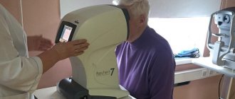
Mandatory instrumental examinations include ECG and fluorography of the lungs. Eye drops are instilled for 3 days before the medication is administered. Eating is allowed no later than 8 hours before the start of the operation. For the operation, the patient must take a passport, eye drops, medications, a hearing aid (if necessary), and a change of clothes.
Preparation and carrying out the procedure
- A few days before IVI, on the recommendation of the attending physician, it is necessary to adjust the schedule for taking certain medications and instill antibacterial drops if necessary.
- The procedure itself is carried out under local anesthesia (drops). After the procedure, the patient’s intraocular pressure readings must be taken. In case of increased IOP, the attending physician prescribes drops to reduce it. The patient is sent home on the same day.
- The next day you need to come to the clinic for an examination and instill drops for a week according to the regimen prescribed by the doctor.
The recovery period after the procedure lasts for a month, and after that you must come for a follow-up examination.
Preparations for intravitreal injections and their action
- Lucentis
is a drug used to treat the wet form of age-related macular degeneration (AMD), as well as diabetic macular edema and other circulatory disorders of the fundus. The active components of the drug localize the affected areas, relieve swelling, stop uncontrolled vascular growth and hemorrhages. The course of treatment usually includes 3 injections with intervals of 1 month. - "Eylea"
is an analogue of "Lucentis", which is also used to prevent pathological vascular growth and reduce retinal edema. - Ozurdex
is a drug for the treatment of thrombosis (blockage) of large retinal veins. It reduces inflammation and swelling, normalizes capillary permeability and blocks abnormal vascular growth. Usually the effect is achieved after one injection of Ozurdex. A repeat procedure may be scheduled after approximately 3-6 months.
Intravitreal injections require mandatory monitoring of dynamics after the procedure, so we immediately set dates for follow-up examinations for our patients.
Doctors at Dr. Belikova's Eye Clinic always approach each patient carefully and select the treatment that is most suitable for you.
Remember that any problem is easier to prevent than to eliminate, so examinations by an ophthalmologist should be regular.
But for almost every eye disease today there is an effective treatment method. We keep up with the times and are always ready to offer you all available ways to restore your vision. Related articles:
- What is IVV and why is it needed?
Preparation for the procedure
Treatment with Eylea is performed by a highly qualified ophthalmologist-surgeon in a sterile room. Before the operation, the patient's skin is treated with a special disinfectant solution. Drip anesthesia is also performed. The administration of Eylea solution 2 mg is carried out using a thin needle. To do this, it is necessary to puncture the ciliary body with a depth of 3 mm. No stitches are required when administering the drug. The duration of the operation is no more than 15 minutes. After completing the procedure, the doctor checks the patient’s IOP volume. If IOP is high, paracentesis is performed.
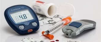
Intravitreal injections
In terms of complexity and technical details, intravitreal administration of Eylea solution is very close to microsurgical operation. In this regard, it is performed by an experienced surgeon in a sterile operating room.
In preparation for the injection, nearby skin is treated with a disinfectant solution. Epibulbar (drip) anesthesia is administered, which completely blocks the sensitivity of the scleral surface.
The injection is performed with a special syringe, which is used to partially puncture a certain area of the ciliary body to a depth of 3 mm. The drug is injected by slowly pressing the piston, after which the needle is removed. No puncture fixation or sutures are required.
At the end of the procedure, monitoring of intraocular pressure is mandatory. If it increases significantly, paracentesis can be performed - a puncture of the cornea in the limbal region.
At the final stage, special drops with antimicrobial and anti-inflammatory effects are instilled into the eye, which must continue to be dripped according to a certain pattern for the next three days at home.
Pregnancy
Throughout therapy, women of reproductive age must use contraception.
The use of Eylea solution is not recommended for women during pregnancy and breastfeeding. It is advisable to interrupt breastfeeding or refuse treatment. The drug Eylea is recommended for use in the treatment of ophthalmic diseases. Regular injections of Eylea help prevent a decrease in visual acuity and ensure its improvement. After the first injection of the solution, positive results are observed in 95% of patients. Throughout the course of treatment, you must consult an ophthalmologist. You can make an appointment by calling +7 (499) 141-13-75.
What diseases are treated with IVV?
The intravitreal injection method is widely used in the treatment of the following diseases:
- Wet form of age-related macular degeneration (AMD)
- Diabetic retinopathy
- Postthrombotic retinopathy
- Macular edema with complicated myopia (myopia).
The listed diseases pose a serious danger to the patient's vision. In advanced cases, they lead to partial or complete blindness. However, timely examination and treatment will protect you from an unfavorable outcome.
Intravitreal injection is a modern method of treating the retina, which has already established itself as one of the most effective methods of treatment.