What is pityriasis versicolor
Pityriasis versicolor, or pityriasis versicolor, is a chronic skin disease of a fungal nature. It appears in the form of specifically pigmented spots of various sizes and shades, without signs of inflammation. Most often, the areas affected by pityriasis versicolor are located on the body (back, abdomen, neck, shoulders) and scalp. Representatives of both sexes are most susceptible to the disease at a young age, and children under 7 years of age are the least susceptible.
External manifestations of the disease usually do not cause concern, so they are often perceived as harmless cosmetic defects. For this reason, the pathology becomes chronic: spots periodically appear, then disappear, and after some time, under the influence of provoking factors, the disease worsens again.
Symptoms of the disease
When infected with pityriasis versicolor, the pathogens multiply in the superficial layers of the skin, disrupting the normal functioning of cells. First of all, melanocytes, which are responsible for the production of the pigment melanin, are affected, thanks to which the skin has one color or another. This means that under the influence of pathogenic microorganisms, the affected areas acquire an uncharacteristic, painful shade. Hence the second name for pityriasis versicolor - versicolor.
The main symptoms of pityriasis versicolor include:
- Spots of yellow, coffee, scarlet or dark brown shades, located mainly on the back, head and neck, shoulders and stomach. In children, they also appear on the limbs, armpits and scalp. The affected areas can be of different sizes, but usually their size does not exceed 1 cm. Subsequently, the spots merge with each other, forming significant areas of the affected skin.
- No signs of inflammation in the form of redness, swelling, hyperemia and pain to the touch.
- Severe peeling of the skin in infected areas, aggravated by touch.
- No tanning on the affected areas.
Pityriasis versicolor is not characterized by itching and discomfort, so patients often mistake the manifestations of the disease for minor cosmetic defects, postponing a visit to the doctor and full treatment.
Such spots begin to itch and scratch only if a secondary infection occurs.
Reasons for development
Versicolor versicolor is a type of fungal skin infection that affects the stratum corneum of the epidermis and hair follicles. Its causative agents are two types of fungi, and infection is possible only through prolonged and close contact with the patient. And in this case, provoking factors play a big role. These include:
- weakened immunity;
- hyperhidrosis;
- disruption of the sebaceous glands;
- diseases of the endocrine system (obesity, diabetes, Itsenko-Cushing syndrome, etc.);
- hormonal imbalance due to pregnancy, menopause or taking hormone-containing medications;
- vegetative-vascular dystonia;
- abuse of antibacterial personal hygiene products;
- excessive exposure to ultraviolet rays (intense tanning, frequent visits to the solarium) and regular overheating of the body.
It is noteworthy that patients with pityriasis versicolor over 60 years of age are extremely rare. This is due to natural age-related changes in the skin, which make it less susceptible to pathogens.
In children under 10 years of age, the main causes of pityriasis versicolor infection are neglect of personal hygiene rules or improper skin care. At this age, with the protective functions of the skin intact, the body independently copes with pathogenic microorganisms attacking it, so the development of the disease does not occur. But closer to adolescence, when hormonal changes begin, the body’s susceptibility to bacteria, viruses and fungi increases, so children over 10 years old become infected with pityriasis versicolor just like adults.
Prevention of erythrasma
First of all, the prevention of erythrasma consists of following the rules of skin hygiene, thoroughly wiping large folds of the skin after taking water treatments, and avoiding tight clothing and synthetic clothing. Secondary prevention involves preventing the development of relapses of the disease. Prevention is carried out within a month after the last symptom of the disease has disappeared. It consists of washing the skin folds with camphor or salicylic alcohol and applying a layer of talc.
Treatment of pityriasis versicolor
The diagnosis of pityriasis versicolor is made to the patient after examination by a dermatologist and dermatoscopy. Additionally, an iodine test and laboratory testing of scrapings can be used.
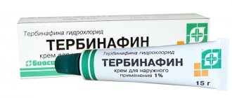
Treatment of pityriasis versicolor is carried out on an outpatient basis until the symptoms of the disease completely disappear. If measures were taken on time, the patient is prescribed local therapy using antifungal ointments and special agents for exfoliating dead cells.
Additionally, immunomodulators, vitamin complexes, antifungal shampoos and antihistamines are used if the patient is bothered by itching. In the most advanced cases, antimycotic agents are prescribed for oral administration. In addition, in order to avoid relapse of the disease and infection of others, the patient’s clothing and bedding are treated with disinfectant compounds.
Disease prevention also plays an important role. The following will help prevent the development of the disease:
- timely solution to the problem of hyperhidrosis (use of medicinal deodorants, creams, powders, compliance with personal hygiene rules, frequent changes of underwear, etc.);
- use of high-quality soaps and skin care products;
- regular water procedures;
- wearing clothes made from natural, hypoallergenic materials;
- avoiding stress;
- balanced diet rich in vitamins and microelements.
Experts also recommend avoiding overheating of the body and promptly seeking advice from cosmetologists and dermatologists in order to identify the problem and begin proper treatment.
Treatment
Treatment of erythrasma involves the use of external agents for exfoliation and disinfection of the skin - erythromycin and sulfur-tar ointment. The ointment is rubbed into problem areas twice a day for a week. In addition, 5% salicylic alcohol, 3% salicylic-resorcinol alcohol, and 2% alcohol solution of iodine are used to treat infectious foci. In case of extensive damage to the skin, a dermatologist prescribes antibiotics for oral administration. To eliminate redness and swelling, aqueous solutions of aniline dyes are used. Physiotherapeutic procedures with ultraviolet light are very useful, as well as daily tanning for 30 - 40 minutes under the morning and evening sun. To successfully cure erythrasma, bedding, clothing, shoes and other household items should be disinfected. Clothes and underwear must be washed and ironed daily. In order to prevent relapses of the disease, it is recommended to regularly wipe the skin folds with a 2% solution of salicylic alcohol or a 1-2% solution of camphor alcohol for one month, and then apply talc or boric acid to them.
What is erythrasma
Erythrasma is a chronic bacterial disease affecting the epidermis layer in the deep folds of the skin. It is characterized by a long course - in some cases, symptoms develop for at least 10 years, without causing significant discomfort to the patient. The clinical picture of erythrasma is similar to a fungal infection of the skin, but modern dermatology classifies it as a group of pseudomycoses.
The following main stages are distinguished in the development of the disease:
- Progression. The first characteristic spots appear on the skin, their size slowly increases, and additional symptoms develop. In some cases, secondary infections occur. The spots gradually merge with each other, forming large areas of damage.
- Stabilization. New spots do not appear, and existing ones stop growing. Peeling of the skin begins. This stage is usually associated with a change in external conditions, for example, cold weather, during which the intensity of sweating decreases and the skin condition stabilizes.
- Exacerbation or relapse. Usually associated with the beginning of the warm season. But in the case of prolonged erythrasma, the disease constantly develops in waves, and after a slight decline its symptoms again actively appear.
- Remission. Occurs with a favorable microclimate, compliance with preventive measures and proper skin care. The color of the affected areas gradually returns to normal, itching and flaking disappear, and the skin is restored.
Without timely, well-chosen treatment, erythrasma can lead to the development of serious complications.
For example, it can provoke eczema and secondary infection in patients with diabetes or obesity. Also, the course of the disease is aggravated by increased humidity and contamination of the affected areas. As a result, its typical symptoms are complicated by burning, itching and pain.
What is erythrasma
Erythrasma is so called due to the fact that when using fluorescent diagnostics, the lesions glow with a reddish tint. This disease occurs most often in adults, mainly among male patients, and lasts a very long time. In this case, the patient does not experience any negative or painful sensations.
For a long period of time, erythrasma, together with actinomycosis, trichophytosis, pityriasis versicolor, microsporia, favus and epidermophytosis, was considered a skin disease of fungal etiology for the reason that when conducting a microscopic analysis of the scales that were removed from the affected skin lesions, it was possible to detect tortuous thin threads that Outwardly they resemble fungal mycelia. In the current classification, dermatologists believe that erythrasma belongs to the group of pseudomycoses. These diseases, in their clinical picture, are very similar to fungal skin lesions, but they have a completely different etiology.
Signs of the disease
Externally, erythrasma manifests itself in the form of light brown, brick-red, brown or yellow-brown spots on the skin, most often round in shape and without signs of inflammation. The diameter of the lesions can reach several centimeters, and they tend to merge, forming large affected areas. First of all, erythrasma spots appear in the folds of the skin, where there is a favorable environment for the proliferation of bacteria.
In addition to spots, erythrasma is characterized by:
- Peeling of the skin on the affected areas, aggravated by touch. Usually this is where the development of the disease begins.
- Mild, irregular itching. It intensifies and begins to cause significant discomfort only if a secondary infection is added to the primary disease.
- Absence of fever, wounds, ulcers and ulcers with copious discharge. This distinguishes erythrasma from most bacterial skin pathologies.
- Getting wet. An optional symptom, the manifestation of which depends on the amount of sweating and the quality of skin care.
It is noteworthy that in children, symptoms of erythrasma appear extremely rarely. The risk group includes adults, primarily men, who are predominantly overweight and prone to excessive sweating. In this case, in men, the skin in the groin, navel and inner thighs is usually affected, and in women, the entire abdomen, armpits and areas under the breasts are affected.
Types of erythrasma
The following types of erythrasma are found.
- Generalized – due to the pathogen entering the blood, all tissues of the body are affected to varying degrees. In this case, a thorough comprehensive examination and complex, multi-stage treatment are required. Externally, generalized erythrasma manifests itself in the form of many characteristic spots throughout the body and limbs (not only in the folds!). They merge into plaques of a large area. This form is most often found in patients with immunodeficiency.
- Disc-shaped - the spots grow, unite into large round formations up to 10 cm in diameter, become denser, change color to a more saturated one, and can become covered with a light flaky coating. They are found not in folds, but on the surface of the body and limbs. Discoid erythrasma is common in tropical countries, especially in women.
- Interdigital. It manifests itself as peeling of the interdigital space of the feet, less often of the hands. The color of the spots on the limbs is less intense than in the folds of the body - from pale to moderate pink.
- Intertriginous form. This is the name given to corynebacteria affecting large folds of the skin. In men, inguinal erythrasma is most often diagnosed; both sexes are characterized by the intergluteal and axillary form; exclusively in women, it is localized in the inframammary folds, around the navel, and on the abdomen. Seasonal spring-summer exacerbation is typical, when sweating increases as the air temperature increases.
- Vesicular - bullous form - it is characterized by the appearance of vesicles: bullae and vesicles. This type of erythrasma is a consequence of the addition of a secondary infection, an allergy to corynebacteria, and excessive mechanical friction of the tissues affected by it.
Each type of erythrasma has its own treatment, the plan and composition of which also depends on the general condition, the presence of allergies and a history of chronic diseases.
Causes of pathology
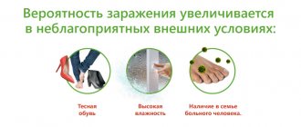
Corynebacteria, which are the causative agents of the disease, are normally always present on human skin. Moreover, they provoke the development of pathology only under certain, favorable conditions. Corynebacteria do not penetrate deeper than the epidermis, and also do not affect nails and hair. Since the appearance of erythrasma is directly related to increased sweating, the disease most often manifests itself in the hot summer season.
Among the main reasons for the development of erythrasma are:
- hyperhidrosis;
- deviation of the normal pH of the skin to the alkaline side;
- diaper rash, constant friction and mechanical damage to the skin;
- dermatitis and other skin diseases;
- neglect of personal hygiene rules;
- wearing synthetic, overly warm clothing;
- the use of low-quality care products or the abuse of soap with an antibacterial effect, which destroys the natural protection of the skin.
Erythrasma is transmitted by contact, most often after the use of clothing, bedding and personal hygiene products of the patient. You can also become infected during sexual intercourse, when visiting a pool or bathhouse, and when walking barefoot on the ground or beach. At the same time, it is not always possible to accurately determine the source of infection, because the carrier may not have obvious external manifestations of the disease in the form of characteristic spots and peeling.
Causes of erythrasma
In the recent past, erythrasma was considered a fungal disease and treatment typical for ailments of this group was prescribed. However, it did not give a guaranteed effect and relapses often occurred. Researchers came to this erroneous opinion because they were confused by the thin whitish threads on the affected areas, which were very reminiscent of the structure of mushroom mycelium. Today, treatment of erythrasma is successfully carried out by dermatologists. In accordance with ICD-10, it belongs to other local skin diseases and is coded as L08.1.
The real cause of unpleasant skin spots is Corynebacterium minutissimum. It constantly lives on the skin of a healthy person, multiplying vigorously and beginning its pathogenic activity only when factors are favorable to it. In their absence, corynebacterium simply lives on the surface of the skin in moderate quantities, without causing inflammation and spots until a certain point. Its environment is only the skin and exclusively the epidermis layer; it does not penetrate deeper. Erythrasma does not appear on nails and hair. The following factors contribute to its activation:
- profuse sweating and a climate conducive to it;
- increased skin moisture for a long time;
- strengthening the alkaline component in the skin pH level;
- diaper rash, abrasions, prolonged dermatitis;
- not regular care: insufficient or excessive use of soap.
The route of spread is exclusively contact - a shared towel, washcloth, public bath, swimming pool, beach, sex. It is very difficult to accurately determine the source - the contact person may not have characteristic spots.
Treatment of erythrasma
To diagnose a patient with erythrasma, a dermatologist first uses a visual examination. This is especially true for rashes in the groin area, which have characteristic distinctive features in the form of pronounced protrusions and bubbles along the edges. Also, the affected areas of the skin are illuminated with a Wood's lamp and a microscopic examination of the scraping is performed to exclude other diagnoses: pityriasis versicolor or pityriasis rosea, candidiasis, dermatitis or eczema.
Treatment of erythrasma is primarily based on the use of antibacterial ointments that are used to treat the affected areas of the skin.
Under their influence, corynebacteria die, and the spots gradually lighten, decrease in diameter and disappear. On average, such therapy takes at least 7-10 days. Used in parallel:
- Antiseptics. Treatment with them is carried out before applying antibacterial ointment, as well as after it, to maintain dryness of the affected areas and prevent re-infection.
- Antifungal drugs. They are prescribed together with antibacterial drugs, since corynebacteria are similar in structure to fungal micelles.
- Exfoliating ointments. They help cleanse the skin of a layer of dead cells, activating its regeneration.
- Ultraviolet irradiation. Promotes skin disinfection and restoration. Patients benefit from both natural sunbathing and physiotherapeutic UV irradiation.
If the disease has not reached an advanced stage, the use of external medications is sufficient to solve the problem. But in some cases, with multiple skin lesions, to obtain the desired result, patients are prescribed systemic antibacterial therapy.
Diagnosis of erythrasma
The diagnosis of “erythrasma” is made based on visual examination, medical history and laboratory tests.
Wood's lamp examination
It is enough for an experienced dermatologist to see dry, slightly flaky yellowish-brown or reddish round spots that are not burdened by other signs of infection. The conclusions can be confirmed by examining the skin using a Wood's Lama.
Under the fluorescent light, the affected areas glow in coral-red shades. This is how crinebacteria colonies appear, secreting a special pigment - coproporphyrin, which glows red under a Wood's lamp.
The procedure is carried out in a dark room after abstaining from water procedures for 1-2 days. Coproporphyrin is washed off with water and is not detected after washing hands and other surfaces.
Laboratory research
They are carried out when there is doubt about the diagnosis after examination under a fluorescent lamp.
- Bacterioscopy. Scraping is done from the affected areas of the skin. Two types of research are carried out with the collected material.
- Staining with methylene blue. Under its influence, corynebacteria appear in the form of rods, similar to curved single-headed pins.
- Potassium hydroxide test. Treatment of scrapings with this chemical eliminates lichen, candidiasis, and dermatophytes.
- Bacteriological culture. It involves sowing scraping materials in a special favorable environment. Colonies of corynebacteria grow slowly - from 7 to 14 days. Due to the length of time it takes to obtain test results, it is rarely used to confirm the diagnosis of erythrasma. Bacterioscopy gives quick results and is therefore more effective.
- Histology. A biopsy of the material taken for research - a small area of inflamed skin - makes it possible to detect and accurately identify colonies of corynebacteria. In most cases, histology is not performed due to the complexity and duration of the study. But, it is indispensable in complex, advanced cases, aggravated by the manifestation of other infectious skin lesions and allergic dermatitis.
Symptoms of erythrasma are similar to those of acanthosis nigricans, seborrheic, contact and allergic dermatitis, diaper rash and candidiasis of the folds, tinea versicolor, confluent papillomatosis, inversion psoriasis, and mycosis. It is possible to make a correct diagnosis and prescribe effective treatment only after all of the above studies.