November 20, 2015
Skin diseases are quite common in modern times - poor environment, stress and poor lifestyle also affect the health of the skin. However, many manifestations on the skin may not be so much an independent disease as a consequence of diseases of the internal organs. Each specific case requires an integrated approach to identify the root cause. Therefore, such branches of medicine as dermatology, dermatovenereology and general therapy work together.
Medical offers consultations with highly qualified dermatologists who will definitely help you cope with the disease and, if necessary, prescribe complex treatment together with other specialists.
Treatment of psoriasis in the clinic
Psoriasis (psoriasis vulgaris), syn. Lichen squamosus is a chronic disease of the skin, nails and joints, characterized by the appearance of a monomorphic rash on the skin. The rash consists of flat papules of varying sizes that tend to merge into large pink-red plaques covered with silver-white scales.
Currently, this is one of the most common chronic skin diseases. It has been suggested that psoriasis affects not only the skin, nails and joints, but also some internal organs (for example, the kidneys). However, this idea remains scientifically unproven.
There are several theories regarding the origin of the disease: viral; genetic (genetic predisposition of cells to increased reproduction); neurogenic (predisposition of the nervous system) and others.
In the skin affected by psoriasis, severe acanthosis and papillomatosis are found, parakeratosis is typical. The epidermis is thinned and consists of two or three rows of cells. In the basal and spinous layers of the epidermis, mitoses are found, in the papillary layer of the dermis - expansion of capillaries and an inflammatory infiltrate.
The development of the disease is characterized by a different onset: the disease can begin almost imperceptibly for the patient with the appearance of several papules, or it can manifest itself with the rapid spread of a rash over the skin. The course of the disease is determined by the seasons: thus, the “winter” type with exacerbations in the cold season is most common, and the “summer” type is less common. Also sometimes there is a mixed type, exacerbations of which occur at any time of the year.
There are several forms of the disease: psoriasis vulgaris, exudative, pustular, seborrheic, palmar-plantar, arthropathic, psoriatic erythroderma.
What symptoms should you see a doctor urgently?
People often resort to self-diagnosis of diseases when identifying changes in the skin. The Internet is replete with articles detailing the symptoms and treatment of diseases. But there are many dangerous symptoms, if detected, you should immediately contact a specialist.
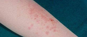
1. Rash
. The rash is a symptom of many diseases, both skin and related to other organs. Rashes can be a symptom of an allergic reaction, stress, gastrointestinal pathology, and even cancer. If the rash is brightly colored and does not go away for more than 48 hours, do not delay visiting a specialist.
2. Warts
. The appearance of warts on the skin is caused by various strains of the human papillomavirus. The virus is transmitted from person to person, so if you find papillomas or condylomas on any part of the body, you should make an appointment with a dermatologist or dermatovenerologist.
3. Peeling
. A seemingly harmless symptom can serve as a signal of a dangerous disease. If peeling skin is accompanied by prolonged itching, redness and severe dryness, it is better to consult a doctor for an accurate diagnosis and treatment. If peeling has become the skin’s response to exposure to frost, sun or wind, then we recommend that you protect your skin more carefully and use special creams.
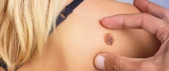
4. Change in skin color
. A change in skin color after tanning or peeling is quite understandable, but if you find suddenly appearing spots on your body or face, this is a reason to make an appointment with a doctor. Red spots can signal an acute allergic reaction or gastrointestinal diseases, yellowing of the skin is one of the symptoms of hepatitis B and C, dark spots can indicate necrotic processes.
Types of fungal skin diseases
Mycoses (mycosis, singular, Greek mykes mushroom; synonym fungal diseases) are a large group of skin diseases caused by pathogenic fungi.
Classification of mycoses is carried out according to various signs of diseases: by the genus and type of fungi, the depth of their penetration into the affected tissues, and their preferential localization. The following types of fungal skin diseases are distinguished:
1. Keratomycosis (for example, pityriasis versicolor). Keratomycosis refers to fungal skin diseases in which the fungus affects only the stratum corneum of the epidermis and does not cause an inflammatory reaction of the skin.
2. Dermatophytosis. These include epidermatophytosis inguinalis, epidermatophytosis of the feet, rubrophytosis, trichophytosis, microsporia, favus.
3. Candidiasis is a disease of the skin, mucous membranes and internal organs caused by fungi of the genus Candida. Candida fungi are considered opportunistic microorganisms, as they are widely distributed in the external environment. The optimal temperature for their growth is 21-27 degrees, but they can grow at a temperature of 37 degrees. The depth of penetration of fungi into the affected tissue is different and depends on the location of the disease: for example, when affecting the vaginal epithelium, fungi penetrate into all its layers, including the basal one, and in the oral cavity they affect only superficial epithelial cells.
4. Deep mycoses, including North American and keloid blastomycosis, sporotrichosis, chromomycosis. This group of diseases is mainly common in the countries of South America, Africa, and the USA. Infection occurs due to skin injuries, scratches, cracks. The clinical picture is different: tubercles and nodes appear on the skin, prone to decay with the formation of ulcers. They can affect the deep layers of the skin, subcutaneous tissue, underlying muscles and even bones and internal organs. This causes severe general symptoms, which do not exclude death.
5. Pseudomycoses are classified into a separate group: erythrasma, actinomycosis. Initially, these diseases were classified as fungal, but a more detailed study of their pathogens made it possible to classify them as microorganisms that occupy an intermediate position between fungi and bacteria.
Stock
Antibody test for coronavirus (COVID-19)
from 2000 a
Rapid test for coronavirus (COVID-19)
Results within 25 minutes from the moment the biomaterial is submitted
1600a
Coronavirus (COVID-19) antibody test at home
from 4200 a
Coronavirus (COVID-19) test at home
from 4950 a
Coronavirus (COVID-19) test
2750a
Acne disease (acne treatment)
Acne (Acne vulgaris), syn. Juvenile acne is a chronic, often recurrent disease characterized by purulent inflammation of the sebaceous glands.
Acne occurs as a result of an imbalance of androgens in the body and, as a result, increased sebum secretion. The ducts of the sebaceous glands become clogged with sebum, and comedones (blackheads) appear. The sebum that has stagnated in the blocked ducts begins to decompose, forming a favorable nutrient medium for pathogenic microflora, mainly coccus, corynebacteria, and propionbacteria.
More often, this dermatological disease occurs at the age of 15-18 years, which is usually associated with puberty, but can occur at any age during exposure to unfavorable factors. The occurrence of acne is facilitated by hereditary predisposition, dysfunction of the endocrine glands, dysfunction of the digestive system, hypovitaminosis, poor nutrition (excessive consumption of food high in fats and carbohydrates). The main clinical manifestation is the appearance of comedones and papulopustular rashes on the skin. Typically, an inflammatory process develops around the comedone: the comedone transforms into a conical papule of bright red or blue-red color. A small pustule may appear in the center of such a papule - then they speak of papulopustular acne.
The inflammatory process is characterized by varying depths, and therefore there are indurative acne, which is extensive infiltrates with an uneven surface, and phlegmonous acne, in which the inflammatory process affects the deep layers of the skin and develops slowly and for a long time. In some cases, acne tends to merge.
Particularly distinguished is necrotic acne, in which necrosis of tissue cells develops deep in the follicle. Presumably, its development is streptococcal in nature. Most often it occurs in men aged 30-50 years, is localized on the skin of the forehead and temples and is a pustule with hemorrhagic contents. After the disappearance of such acne, scars remain on the skin.
Acne keloid is a distinct type of acne that occurs predominantly in men aged 20-40 years. They are usually located in the back of the head and on the back of the neck. The skin around them is sharply compacted, hair in such areas grows in tufts. They leave behind keloid scars.
Diagnostics
To undergo the examination, you must make an appointment with a dermatologist. The doctor will ask the patient about complaints and study medical history to identify risk factors for the disease. The next stage of diagnosis is an initial examination of damaged skin. The dermatologist evaluates the shape and size of the ulcers, and also determines the depth of tissue damage. To clarify the cause of pyoderma and the severity of the patient’s condition, instrumental and laboratory examinations are prescribed.
Additional diagnostic methods:
- Dermatoscopy is a method of visual examination of areas of tissue damage. To determine the type of disease, the doctor uses an optical or digital device that allows you to repeatedly enlarge the image (photo). Dermatoscopy is used to search for specific signs of different types of pyoderma and differential diagnosis.
- Microbiological examination of the contents of blisters, boils and other pathological structures. The doctor carefully punctures the membrane of the bladder and collects the exudate in a sterile container. In the laboratory, specialists inoculate the material on various nutrient media to identify the causative agent of the disease. After specifying the type of causative agent of dermatitis, specialists conduct a test for the sensitivity of microorganisms to antibiotics. Bacterial culture is the most reliable way to diagnose the infectious form of the disease.
- Blood analysis. In the treatment room, a nurse collects venous blood and sends the material to the laboratory. First of all, specialists evaluate the ratio and quantity of blood cells. An immunological test can detect signs of an autoimmune disease. Also, if necessary, serological diagnostics are carried out: specialists look in the blood for specific antibodies produced by the body in response to infection. It is often necessary to exclude a sexually transmitted infection.
In case of nonspecific symptoms, the doctor must exclude the presence of other diseases with similar symptoms. For this purpose, differential diagnosis of toxicerma, epidermolysis bullosa, pemphigus vegetans and fungal skin lesions is carried out. The presence of HIV infection is excluded. If necessary, a consultation with an immunologist, allergist, rheumatologist and infectious disease specialist is scheduled.
Prevention
Preventing infectious and inflammatory skin diseases on your own is not a difficult task.
Basic methods of prevention:
- hygienic skin care;
- timely treatment of cuts and burns;
- treatment of chronic infections;
- regular examinations by a dermatologist for chronic skin diseases;
- control blood sugar levels in diabetes.
Consultation with a dermatologist and immunologist will help the patient learn more about methods of prevention and treatment of pyoderma.