Stroke
37198 11 August
IMPORTANT!
The information in this section cannot be used for self-diagnosis and self-treatment.
In case of pain or other exacerbation of the disease, diagnostic tests should be prescribed only by the attending physician. To make a diagnosis and properly prescribe treatment, you should contact your doctor. We remind you that independent interpretation of the results is unacceptable; the information below is for reference only.
Coagulogram (screening): indications for use, rules for preparing for the test, interpretation of the results and normal indicators.
Indications for the purpose of the study
Coagulogram (hemostasiogram) is a comprehensive hematological study aimed at assessing the state of the hemostasis system (blood clotting), or blood clotting indicators. The hemostasis system includes blood cells (platelets) and specific substances (clotting factors) dissolved in blood plasma and contained in platelets. When the integrity of the vessel wall is violated, the coagulation system is activated and a blood clot is formed, preventing blood loss.
Some time after the bleeding stops, fibrinolysis begins - the process of dissolving the blood clot after restoring the damaged vessel wall to resume blood flow.
Indications for a coagulogram are conditions accompanied by increased bleeding or, conversely, increased thrombus formation.
Increased bleeding leads to hemorrhagic syndrome. It is manifested by the formation of hematomas, pinpoint hemorrhages under the skin (petechiae), bleeding of the mucous membranes (nosebleeds, bleeding from the gums), the appearance of blood in the stool, urine, prolonged menstrual bleeding, as well as possible hemorrhages in the internal organs and body cavities. The most dangerous manifestation of hemorrhagic syndrome is hemorrhage in the brain - hemorrhagic stroke, which can lead to rapid impairment of brain function, including death. A hereditary disease associated with blood clotting disorders (decreased or absent clotting factors) is called hemophilia.
The opposite situation - thrombosis - is the formation of blood clots (thrombi) inside blood vessels, preventing the free flow of blood.
People with obesity, diabetes mellitus, heart rhythm disturbances (the most significant of which are atrial fibrillation and flutter), low physical activity, and patients after major operations (for example, joint replacement, heart valve replacement, emergency operations, etc.) are at risk of increased thrombosis. , with varicose veins, previous myocardial infarctions, strokes, and various autoimmune diseases.
Thrombosis in a certain area of the vascular bed is accompanied by an acute or gradual disruption of the blood supply to any organ with disruption of its functions and subsequent tissue necrosis.
In addition to the above conditions, a coagulogram is performed before operations to assess the risk of bleeding, when prescribing certain medications, and also necessarily in case of liver diseases, since most of the proteins of the blood coagulation system are synthesized in this organ.
References
- Zabolotskikh, I.B., Sinkov, S.V., Lebedinsky, K.M. Clinical guidelines “Perioperative management of patients with disorders of the homeostasis system”, 2022. - 42 p.
- Agibova, N.E., Boeva, O.I., Baykulova, M.Kh. and others. Hageman factor in predicting the risk of thromboembolic complications in atrial fibrillation. — Cardiovascular therapy and prevention, 201. — No. 8. — P.26-27.
- Chaudhry, L., El-Sadek, W., Chaudhry, G. et al. Factor XII (Hageman factor) deficiency: a rare harbinger of life threatening complications. — The Pan African medical journal, 2022. — Vol. 33(39).
Preparation for the procedure
The essence of a coagulogram is to assess the activity of proteins of the blood coagulation system.
Therefore, proper preparation for this analysis is extremely important. Blood is taken on an empty stomach; the last meal should be at least 8-10 hours before the test. It is recommended to exclude excess amounts of fatty, sweet and protein foods. The drinking regime remains normal. The day before it is necessary to avoid stressful loads and intense sports. If the patient is taking any medications that affect coagulogram parameters, then, depending on the goals set for the study, it is necessary to either continue or stop taking them a few days before. This should be discussed with your doctor before taking the test.
If you need urgent results, you can contact the INVITRO offices, where express diagnostics are carried out. In this case, the analysis will be ready in about 2 hours.
Some of the proteins of the blood coagulation system are acute phase proteins. They are produced during stress, which includes not only physical and emotional stress, but also illness. Consequently, any persistent or short-term, but intense stress in the body can lead to changes in coagulogram parameters.
Dehydration (dehydration) is one of the reasons for blood thickening and an increase in the concentration of coagulation factors, which also leads to a distortion of the result.
The state of the blood coagulation system changes during pregnancy, both in the direction of increasing and decreasing the activity of a number of proteins.
The use of many medications affects the blood coagulation system. Thus, the first generations of oral contraceptives lead to an increased risk of thrombosis, and anticoagulants lead to bleeding. The list of drugs that affect the activity of the coagulation system is huge. When receiving any drug therapy, you should first consult with your doctor about the possible effect of a particular drug on the test result.
Taking certain medications by a pregnant woman, for example, warfarin, rifampicin, phenytoin, barbiturates, affects the production of clotting factors in the fetus. The child may subsequently be at high risk of bleeding.
Factor VII. Activation.
Thus, cutting the peptide bond between arginine residue 152 and isoleucine residue 153 leads to the formation of the active form of factor VII (VIIa). As a result, a light chain (molecular weight about 20,000) is formed from the N-terminus of the peptide chain, and a heavy chain (molecular weight about 30,000) is formed from the C-terminus. Both chains remain covalently linked by a disulfide bridge. from N-
Rapid activation occurs after the inactive factor VII combines with its cofactor (tissue factor) in the presence of calcium ions.
Coagulogram (screening)
No. OBS103Comprehensive studies
Hemostasiogram (coagulogram), screening A basic set of tests used for screening assessment of the state of the blood coagulation system.
Up to 1 business day
RUB 1,360 Add to cart
No. OBS109Comprehensive studies
Hemostasiogram (coagulogram) extended This set of tests is used for a detailed assessment of hemostasis disorders: if thrombophilia is suspected, the risk of developing thromboembolism and/or disseminated intravascular coagulation (DIC), or the patient’s severe general condition.
Up to 1 business day
RUB 3,790 Add to cart
You can take a coalogram at the nearest INVITRO medical office. A list of offices where biomaterial is accepted for laboratory research is presented in the “Addresses” section.
The blood coagulation system is primarily an enzymatic system that provides external, internal and general coagulation pathways. These mechanisms are successively replaced, but they can also occur independently of each other.
The screening coagulogram includes the following indicators:
- prothrombin (prothrombin time, prothrombin (according to Quick), INR - international normalized ratio);
- fibrinogen;
- APTT (activated partial thromboplastin time);
- thrombin time.
These indicators are basic and allow you to evaluate all coagulation pathways.
Normal indicators
| Index | Reference value | Unit |
| Prothrombin time | 9,0–15,0 | sec. |
| Prothrombin according to Quick | 78–142 | % |
| INR | depends on the presence of concomitant pathology in the patient for which he receives anticoagulant therapy | |
| APTT | 25,4–36,9 | sec. |
| Thrombin time | 10,3–16,6 | sec. |
| Fibrinogen | 2,00–4,00 | g/l |
| up to 5.6 (2nd and 3rd trimester of pregnancy) | g/l | |
Decoding the result
I. Quick's prothrombin, prothrombin time, and international normalized ratio (INR) reflect the activity of the extrinsic coagulation pathway.
- Prothrombin time
is the time of blood clotting after the addition of substances (thromboplastin with calcium) to the plasma that trigger the extrinsic blood coagulation pathway. Time is measured in seconds (sec.). The longer the prothrombin time, the lower the activity of coagulation factors. - Prothrombin according to Quick
is considered a more objective indicator of the external pathway, since during the analysis the clotting time is studied depending on the different concentration of coagulation factors in the blood obtained by diluting the test material in the laboratory. The indicator is measured as a percentage (%). The longer blood clotting occurs, the lower the Quick prothrombin percentage. - The international normalized ratio (INR)
is a prothrombin test standardized in accordance with international recommendations. It is calculated using a certain formula. The higher the INR, the longer it takes for a blood clot to form.
Prothrombin time, prothrombin according to Quick and INR with normal fibrinogen content and activity also reflect the activity of the prothrombin complex (II, V, VII, X blood coagulation factors).
When prescribing therapy with anticoagulants from the group of vitamin K antagonists (warfarin), there is an increase in INR and prothrombin time and a decrease in prothrombin according to Quick. Other causes of these changes may be vitamin K deficiency (for example, with hemorrhagic disease of the newborn), hereditary deficiency of factors II, V, VII, X, liver and intestinal diseases. The opposite picture, i.e., a decrease in INR, a decrease in prothrombin time and an increase in prothrombin according to Quick, occurs in conditions accompanied by increased blood clotting: thrombosis, blood thickening during dehydration, when taking oral contraceptives, barbiturates.
II. Activated partial thromboplastin time (aPTT)
with normal content and activity of fibrinogen, it characterizes the internal pathway of blood coagulation, which includes the activity of factors II, V, VIII, IX, X, XI, XII. APTT is determined by adding reagents that trigger this clotting pathway to a blood sample and measuring the time it takes for the blood to form a clot. Measured in seconds (sec.). The slower the blood clotting, the longer the APTT.
This indicator is used to monitor therapy with heparin (direct anticoagulant), and an increase in the aPTT value is noted. Other causes of increased time include hereditary and acquired deficiencies of intrinsic pathway coagulation factors, for example, in hemophilia, hemorrhagic disease of the newborn, liver and intestinal diseases, some autoimmune diseases, etc. The reasons for increased aPTT are the same as for Quick's prothrombin.
III. Thrombin time and fibrinogen reflect the overall, or final, pathway of blood clotting.
- Fibrinogen
is blood clotting factor I, a protein synthesized in the liver and converted into fibrin, the basis of the clot during blood clotting. It is also an acute phase protein. It is measured in grams per liter (g/l). With increased thrombus formation and various inflammatory diseases, the synthesis of this protein increases. In case of liver diseases, hereditary fibrinogen deficiency, etc., its concentration decreases. - Thrombin time
is the blood clotting time necessary for the formation of a fibrin clot when thrombin is added to the plasma - an enzyme (factor IIa), which appears when blood coagulation factors interact when a vessel is damaged. Thrombin time depends on the level and activity of fibrinogen. Measured in seconds (sec.).
Changes in thrombin time generally correlate with fibrinogen levels. However, with such a hereditary disease as dysfibrinogenemia, i.e., a violation of the functional activity of fibrinogen, an increase in thrombin time and a coagulation disorder occurs, despite the normal amount of fibrinogen in the blood.
Vitamin K
Vitamin K was discovered by the Danish researcher Henrik Dam in the course of research from 1929 to 1935, who received the Nobel Prize in 1943 together with the American researcher Edward Doisy for the discovery and study of the properties of vitamin K. Dam studied the metabolism of cholesterol and to determine the effect of nutrition on the synthesis of cholesterol in organism conducted experiments on chickens that were given a special low-fat diet. Chickens became ill with an unknown disease if this diet continued for more than 2-3 weeks. They experienced multiple hemorrhages under the skin, in the muscles and internal organs, and the blood clotted very slowly. The disease arose as a result of a lack of an unknown chemical substance that did not coincide with any of the vitamins known at that time. As Dahm himself recounted in his Nobel lecture, it was named vitamin K after the German-Scandinavian word Koagulation (coagulation) and was the first vitamin named from a non-English word.
Vitamin K includes a group of fat-soluble vitamins that are necessary for the post-translational modification of several proteins, most of which are proteins of the blood coagulation and anticoagulation system. Chemically, they belong to the derivatives of 2-methyl-1, 4 naphthoquinolone.
All members of the vitamin K family have a methylated naphthoquinolone ring and a side chain containing varying numbers of isoprene residues. Phylloquinolone (vitamin K1) has 4 isoprene residues in its side chain, one of which is unsaturated. Menaquinolones (vitamin K2) contain varying amounts of unsaturated isoprene residues. They are usually designated as MK-n, where n is the number of spoilen residues. The presence of isoprene residues brings vitamins K closer to other fat-soluble vitamins (A, E and ubiquinolones (coenzymes Q)). Vitamins K1 and K2 are the only naturally occurring vitamin Ks. The remaining vitamins K (K3, K4, etc.) are synthetic.
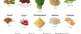
Vitamin K1 is found in green vegetables (spinach, lettuce, cauliflower, cereals, avocados, kiwi, bananas, meat, dairy products, eggs, soybeans, vegetable oils, in particular olive oil. Vitamin K2 is synthesized by intestinal bacteria, therefore vitamin deficiency vitamin K in adults is a rare occurrence, mainly in dysbiosis after treatment with antibiotics. However, in young children, whose intestines are not yet populated with bacteria in sufficient quantities, hypovitaminosis K is often observed, which can lead to bleeding. Vitamin K does not penetrate placenta.
As stated above, vitamin K is required for the carboxylation of glutamic acid residues in some proteins, which are converted to gamma-carboxyglutamic acid (Gla) residues. Proteins that undergo such post-translational modification are called Gla proteins.
There are 14 known Gla proteins in humans:
- Blood clotting proteins: prothrombin (factor II), factors VII, IX, X, proteins C, S and Z
- Bone metabolic proteins: osteocalcin (synonym: bone Gla protein, BGP) matrix Gla protein (MGP)
- Participation in vascular repair (angiogenesis)