Raynaud's syndrome is a serious condition that is caused by spasm of peripheral blood vessels. Because of it, blood flow in the small vessels of the legs (at the level of the feet) and hands is disrupted. This syndrome is usually one of the symptoms of many other diseases. For example, it occurs in rheumatism, endocrine disorders, psychopathy, etc. Only sometimes it is impossible to determine the underlying disease, so Raynaud's disease may be considered independent.
Most often, this syndrome affects young and mature women, as well as residents of regions with cold climates.
Provoking factors
The underlying causes of Raynaud's syndrome have not been identified, but there are a number of factors that, according to doctors, contribute to its development. These include:
- hereditary tendency;
- permanent injury to fingers and toes;
- endocrine system disorders: diabetes, hyperthyroidism, hypothyroidism, etc.;
- disorders associated with hematopoietic functions;
- constant contact with cold water;
- some diseases of the spine;
- alcohol dependence, drug addiction;
- unstable mental state;
- heavy metal intoxication;
- serious stress;
- constant contact with chemicals;
- certain autoimmune diseases.
The disease in an advanced state can manifest itself even without provoking factors - then they talk about spontaneous attacks of Raynaud's syndrome.
Causes of Raynaud's syndrome
Raynaud's syndrome can be a consequence of the following diseases and factors:
- various autoimmune connective tissue pathologies (systemic scleroderma, rheumatoid arthritis, systemic lupus erythematosus);
- inflammatory diseases of blood vessels - systemic vasculitis (granulomatosis or Wegener's disease, polyarteritis nodosa, drug-induced vasculitis, cryoglobulinemic vasculitis);
- blood diseases (thrombophlebitis, thrombocythemia, cryoglobulinemia, leukemia);
- diseases of the endocrine system (pathology of the adrenal glands and thyroid gland);
- hypothermia, stress, emotional overstrain;
- long-term use of medications that have a vasoconstrictor effect (migraine medications).
Symptoms, manifestations
Symptoms of Raynaud's disease include:
- whitening of the skin on the fingers;
- feeling of coldness, numbness;
- burning, pain, feeling of fullness;
- swelling, cyanosis;
- formation of ulcers and necrosis (over time);
- nail dystrophy;
- curvature of fingers.
The attacks begin with the skin turning white, then the situation gradually worsens. The attacks themselves become longer, and the damage becomes more obvious. This depends on the stage of Raynaud's syndrome.
Are you experiencing symptoms of Raynaud's syndrome?
Only a doctor can accurately diagnose the disease. Don't delay your consultation - call
Symptoms of Raynaud's disease
There are three stages of Raynaud's disease:
- The first stage is characterized by a significant increase in the tone of the vascular walls, which causes a short-term spasm of the arteries. In response to cold or stress, the skin turns pale and an acute attack of pain occurs. After the negative factor ceases, the signs of Raynaud's disease disappear, the color of the hands and/or feet acquires a natural shade.
- The second stage is characterized by the addition to the symptoms described above of swelling and numbness of the extremities, cyanosis and marbled skin color. The pain in stage 2 Raynaud's disease is intense and burning. Increases sensitivity to exposure to cold.
- At the third stage, pain and bluishness of the skin are observed constantly. Trophic disorders appear in the form of ulcers on the fingertips and the formation of necrotic areas. Such wounds are difficult to treat because blood circulation in the limbs is impaired. This is fraught with the development of secondary infection and even sepsis. There is intolerance to low temperatures.
Stages
At the angiospastic stage, the patient experiences slight tingling, goosebumps, and chilliness. The skin becomes pale, sensitivity in the fingers is lost, and an aching sensation of pain may appear. After this, hyperemia occurs - that is, the vessels become overfilled with blood, slight swelling and redness appear.
At the angioparalytic stage, exacerbations occur that last longer, cyanosis (blue discoloration) and severe swelling occur. The pain gets worse. Often the patient may experience a fever, cold sweats, and even a feeling of fear and anxiety.
In the third, trophoparalytic stage, the pain becomes less, but if Raynaud's disease is not treated, the consequences for the fingers are much worse. These are the appearance of ulcers, areas of necrosis - the fingers are literally destroyed, and the patient becomes disabled. The condition is serious both from a physical and psychological point of view.
It is the third stage that is truly dangerous, and the first two only lead to it. But treatment and observation by a doctor should begin as early as possible in order to prevent complications.
Treatment methods
The stage affects how Raynaud's disease is treated.
At stages I and II, conservative methods are prescribed. First of all, it is necessary to eliminate the factors that provoke the attack: cold, emotional overload, occupational hazards. In this regard, clinical recommendations for Raynaud's disease include a categorical cessation of smoking (nicotine increases arterial tone), normalization of the emotional background, exclusion of hypothermia and limb injuries, as well as a change of job if it is associated, for example, with vibrations or excessive physical exertion.
An attack of vasospasm should be relieved by warming the extremities. Warm baths, wrapping, and massage of the affected area are suitable for this.
Drugs for Raynaud's disease include vasodilators (for example, phentolamine) - they improve blood circulation and prevent spasms. In case of ulcers and necrosis, local wound healing ointments are used.
Drug treatment can last for years until the vasodilators no longer provide the desired effect. In this case, a sympathectomy is performed - a surgical intervention in which the nerve fibers that cause vascular spasm are stopped or removed. Necrosis requires necrectomy.
Auxiliary therapeutic methods: plasmapheresis sessions, reflexology, physiotherapy, hyperbaric oxygenation, psychotherapy.
How to completely cure Raynaud's disease is unknown, since the exact cause of the disease has not been established. But if you follow the doctor’s recommendations, the prognosis is favorable. There are cases when attacks of vasospasms suddenly disappeared after changing profession or moving to another climate zone.
Diagnostics
As a rule, diagnosing Raynaud's disease is not particularly difficult for professionals, but a comprehensive examination is still required. After examination, cold tests and history taking, patients are prescribed:
- blood biochemistry;
- hemogram;
- coagulogram;
- general urine analysis;
- immunogram;
- Ultrasound – examination of internal organs;
- spine tomography;
- Dopplerography of blood vessels, as well as some other studies.
An important goal is to rule out other diseases that may cause similar symptoms.
Causes
Primary Raynaud's phenomenon is much more common than secondary. There are no signs of other concomitant diseases.
In secondary Raynaud's syndrome, the causes are the presence of the following pathologies:
- connective tissue diseases;
- disturbances in the functioning of the endocrine glands;
- blood diseases;
- neoplasms on the body;
- neurological disorders.
This condition can also be caused by injury (frostbite) or taking certain medications.
Vasospastic reactions often occur at low temperatures and emotional stress. Nicotine also contributes to the development of pathology, but some underestimate its influence.
Treatment
Since stress, as the diagnosis of Raynaud’s syndrome shows, is one of the possible factors in the development of the disease, treatment will be aimed both at working directly with the vessels and at improving the overall quality of a person’s life.
To restore physical condition, vasodilators, antispasmodics, glucocorticosteroids, cytostatics and other medications are used. If medication and additional recommendations do not give the desired result, surgery is prescribed. The endoscopic method makes the operation more gentle when it comes to correcting the functioning of blood vessels. But in extreme cases, patients may require amputation of a limb or part of it.
At the same time, as already mentioned, other methods of work are used to treat Raynaud's syndrome:
- psychotherapy. It aims to reduce stress levels, as well as discover the hidden causes of suppressed stress, if any;
- different types of physiotherapy. These are magnetic therapy, electrophoresis, paraffin baths, etc.;
- physiotherapy;
- mud treatment;
- a diet focused on healthy eating;
- special massages and much more.
As an aid, after consultation with a doctor, for Raynaud's syndrome, you can use some folk recipes, for example, pine baths.
Treatment, as a rule, is long-term, and therefore requires patience from the patient and strict adherence to all recommendations of the attending physician.
Raynaud's syndrome in the practice of a therapist
The syndrome was first described by Maurice Raynaud in 1862 as “local asphyxia of the limbs” [1], and Thomas Lewis subsequently introduced the division into primary Raynaud's disease and secondary Raynaud's phenomenon [2]. Epidemiology The prevalence of SR, according to the results of epidemiological studies, in different countries ranges from 2.1 to 16.8%. Differences in the prevalence of SD found in individual studies are partly explained by the fact that in different studies the results were obtained using postal or telephone surveys, questionnaires distributed randomly to the population, or the inclusion in the study only of people who sought medical advice about subjective sensations, accompanying SR. In Europe, the highest frequency is observed in the UK and France, and the lowest in Spain and Italy. These data confirm the well-known position that in countries with a relatively cold climate, the frequency of SR is significantly higher than in countries with a warmer climate [3]. The peak incidence occurs in the 2nd–3rd decade of life. Among patients with SR, women predominate and, according to various authors, the ratio of women to men ranges from 2:1 to 8:1 [4,5]. The age of onset of SR in women is significantly lower than in men [6]. With age, there is a tendency towards an increase in the proportion of men among people with SR. Thus, in one study, the male/female ratio under the age of 50 was 1:5.2, and in the older age group it was 1:1.1 [7]. Pathogenesis Despite the fact that almost 150 years have passed since the first description, the pathophysiology of Raynaud's syndrome remains not entirely clear and, apparently, SR is a multifactorial disease. It is believed that the cause of excessively pronounced vasospasm in response to provoking stimuli is a defect in the central and local mechanisms of regulation of vascular tone. Among these factors, vascular endothelial, intravascular and neuronal disorders appear to be important. In secondary SR, structural changes in blood vessels are also a significant factor [8]. A key role in the genesis of SR is played by an imbalance between vasodilators and vasoconstrictors due to disturbances in the neurogenic control of vascular tone and mediators produced in the hemocirculation [9]. Among vascular mediators, great importance is attached to changes in the production of nitric oxide, endothelin-1, serotonin, thromboxane and angiotensin, the level of which in patients with SR increases under the influence of cold [10]. In addition, the regulation of vascular tone depends not only on humoral vasodilatory mediators, but also on neuropeptides released from the corresponding nerve endings. Imbalances in a number of neuronal transmitters, including calcitonin gene-related peptide, neuropeptide Y, and α-adrenergic receptor interacting agents, also lead to insufficient vasodilation and increased vasoconstriction. Along with this, platelet activation to oxidative stress is important [11]. The classification of SR is divided into primary (idiopathic, or Raynaud's disease) and secondary, associated with other diseases [12]. To verify the variant, diagnostic criteria for primary [13] and secondary [14] SR have been proposed, based on clinical features, data from laboratory and instrumental studies (Table 1). In most cases, SR is idiopathic, accounting for 80–90% of the total number of patients [15,16]. It should be noted that antinuclear factor (ANF) has a relatively low prognostic value for connective tissue diseases (30%), while the identification of specific autoantibodies significantly increases the likelihood of a secondary nature of SR. Approximately 15–20% of patients with SR who exhibit specific autoantibodies and/or capillaroscopic changes, but do not have symptoms of connective tissue diseases, subsequently (usually within two years) develop one or another connective tissue disease. The clinical significance of SR is due to its high prevalence in the population and frequent association with other, often life-threatening diseases of patients (Table 2). Secondary SR is most often associated with systemic scleroderma, systemic lupus erythematosus, other connective tissue diseases, hematological disorders and certain medications. Clinic Factors that provoke vasospastic reactions, which are also called Raynaud's attacks, are low ambient temperature and/or emotional stress. Clinically, SR manifests itself as clearly demarcated areas of sequentially changed color of the skin of the fingers: pale, blue, red (so-called triphasic SR). The first two phases of color changes reflect the state of vasospasm and hypoxia, and after the end of the vasospasm attack, due to reactive hyperemia, the skin acquires a bright red color. Classic triphasic Raynaud's attacks are observed in only 15% of patients, while in most cases (85%) biphasic color changes are observed [17]. A significant proportion of patients complain of sensory disturbances (numbness, tingling, pain) during a Raynaud's attack. Raynaud's syndrome has some clinical features: • Most often, color changes are observed on the fingers. • Changes begin on one finger, then spread to other fingers and become symmetrical on both hands. • Fingers II–IV are most often involved; the thumb usually remains intact. • Changes in skin color can also be observed in other areas: ears, tip of the nose, face, above the knees. • During Raynaud's attacks, livedo reticularis may appear on the extremities, which resolves after the vasospasm ends. • In rare cases, there is damage to the tongue, which is manifested by numbness and transient speech disturbances (becomes slurred, blurred). Vasospasm usually lasts 15–20 minutes and ends with a rapid restoration of blood flow, as evidenced by the intense pink color of the skin (reactive hyperemia). At the same time, the frequency and duration of episodes of vasospasm can vary both in different patients and in the same patients at different times of the year (more intense in winter than in summer). Diagnosis of SR is established primarily on the basis of complaints and clinical symptoms of the disease. The diagnosis of SR is considered possible if the answer to the following three questions is positive: - Are your fingers unusually sensitive to cold? — Does the color of fingers change when exposed to cold? — Do they turn white and/or bluish? It should be taken into account that sensitivity of the fingers to cold is also observed among absolutely healthy people. Thus, in a survey of approximately 7 thousand people, almost 12% of them answered affirmatively to the question: “Are your fingers or limbs unusually sensitive to cold temperatures?” [18]. In addition, cool skin or undefined mottled discoloration of the skin of the fingers, hands, and extremities is considered a normal response to cold exposure. A sign such as whitening of the fingers is highly sensitive for SR (94–100%) and has high specificity (75–78%) [19]. The British Scleroderma Research Group has proposed the following definition of SR [20]: • Reliable SR – repeated episodes of biphasic changes in skin coloration in the cold. • Probable SR is a single-phase change in skin color accompanied by numbness or paresthesia when exposed to cold. • No SR – there are no changes in skin color under the influence of cold. In all patients with newly diagnosed SR, it is necessary to conduct special studies for the purpose of differential diagnosis of the primary and secondary nature of the pathology. First of all, it is necessary to clarify: • whether the patient has symptoms of connective tissue diseases with which SR is most often associated (arthritis, myalgia, fever, dry syndrome, skin rash, cardiopulmonary disorders); • whether the patient is taking any medications at the time of the study, especially chemotherapy drugs; • whether the patient is exposed to vibration or other mechanical influences that injure the hands; • whether episodes of Raynaud's syndrome are associated with certain positional changes. Along with the diagnosis of SR itself, it is equally important to determine the clinical form. Primary SR in most cases is moderate in nature and only in 12% of patients the symptoms of the disease are significantly pronounced [21]. The average age of onset of primary SR is 14 years, and only 27% of cases develop at the age of 40 years or older [22]. In 1/4 of patients, SR occurs in first-degree relatives [23]. Despite the identity of the clinical manifestations of primary and secondary SR, there are some differences in individual signs between these two conditions. The following signs indicate the likelihood of a secondary nature of SR: • late age of onset, • male gender, • painful episodes of vasospasm with signs of tissue ischemia (ulceration), • asymmetric nature of attacks. It must be taken into account that clinical signs of SR-associated disease may develop several months or years after the onset of SR. Instrumental and laboratory research methods are aimed primarily at clarifying the nature of Raynaud's syndrome - primary or secondary. The most informative among instrumental methods is capillaroscopy of the nail bed (CNL). The method allows you to visually assess the local capillary network of the nail bed and identify structural changes in the capillaries and disturbances in capillary blood flow. In primary SR, there are no structural changes in the capillaries, but functional disorders are detected in the form of a pronounced decrease in blood flow velocity in the capillaries or intracapillary stasis (Fig. 1A). Secondary SR is characterized by changes in the size and shape of capillary loops and reduction of the capillary network (Fig. 1B). In order to differentiate primary and secondary SR, a number of other instrumental studies are also used. Laser Doppler flowmetry – assessment of skin blood flow; the use of provocative tests reveals increased vasospasm and decreased vasodilatory potential. Thermography is an indirect assessment of blood flow based on skin temperature; the time it takes to restore the initial skin temperature after cooling and the gradient tо along the finger reflect the severity of vascular damage [24]. Temperature differences between the fingertip and the dorsum of the hand greater than 1°C at a temperature of 30°C have positive and negative predictive values (70 and 82%, respectively) for identifying SR secondary to systemic scleroderma (SSc) [25]. Plethysmography – measurement of blood pressure in the digital artery; a decrease in pressure by 70% or more, after local cooling, indicates the secondary nature of SR (sensitivity 97%); SSD is characterized by a decrease in pressure to 0 at 30°C (specificity 100%). Color Doppler ultrasound scanning – visualization and measurement of the diameter of the digital artery, assessment of blood flow velocity; allows you to differentiate primary and secondary SR. For early differential diagnosis of primary and secondary CP, it is recommended to adhere to the algorithm shown in Figure 2. Differential diagnosis of Raynaud's syndrome should be carried out first with acrocyanosis, a condition characterized by prolonged cyanosis of the hands or feet, which is aggravated by cold. Some conditions may be mistakenly classified as Raynaud's syndrome. These include carpal tunnel syndrome, reflex sympathetic dystrophy, and superior outlet syndrome. All these syndromes are associated with mechanical damage to the neurovascular bundle of the upper extremities. Particular attention should be paid to taking drugs such as α-interferon, antitumor agents (cisplatin, bleomycin, vinblastine, etc.), β-blockers and bromocriptine. Livedo reticularis is also observed in vasculitis, antiphospholipid syndrome and occlusive diseases of peripheral vessels, in which, unlike SR, this symptom is stable. Patients with peripheral vascular diseases, accompanied by decreased blood flow and ischemia, often complain of freezing of the extremities, numbness and tingling. In SR, unlike peripheral vascular diseases, these symptoms are observed only during vasospasm and disappear completely after restoration of the original blood flow. Course and prognosis Primary SR has a favorable course and prognosis. At the same time, during long-term observation of 307 women with SR (on average 12 years), 38% of them showed no changes, 36% had a decrease in the frequency and severity of Raynaud’s attacks, in 16%, on the contrary, an increase in the severity of clinical symptoms, and in another 10% – manifestations of SR disappeared [26]. In a prospective observation of patients with SR for 3 years, DeAngelis R, et al. established the development of connective tissue disease in 10% of patients with an initial diagnosis of probably secondary SR and in no cases of primary SR [27]. With a longer follow-up, over 12 years, the development of connective tissue disease was observed in 30% of patients with probably secondary SR, as well as in 9% of patients with primary SR [28]. The Framingham Study showed that 81 of 639 (12.6%) patients with RS developed signs of another disease after an average of 10.4 years (0.6–27.9 years) [29]. The most common development was systemic scleroderma (in 53 of 81 or 65% of patients) and mixed connective tissue disease (in 8 of 81 or 10% of patients). In secondary SR, the prognosis is determined primarily by the disease with which the association is noted. Treatment All patients with both primary and secondary Raynaud's syndrome are recommended to avoid cooling, smoking, contact with chemicals and other factors that provoke vasospasm at home and at work. You should avoid stressful situations, sudden changes in temperature, keep your whole body warm and especially your hands and feet (wear warm clothes, a hat, mittens instead of gloves, thermal underwear, etc.). Identification and elimination of provoking factors (cold, vibration, etc.) is the basis of therapy for professional SR. Treatment of the underlying disease, aimed at eliminating vascular disorders and reducing the activity of the pathological process, has a positive effect on the manifestations of secondary SR. In cases of frequent and prolonged episodes of vasospasm in primary SR and in almost all cases of secondary SR, drug therapy is necessary. To treat SR, drugs with a vasodilating effect or agents that affect the rheological properties of blood are used. Among vasodilators, effective means of treating SR are calcium channel blockers, which are considered the first-line treatment for SR; the drug of choice is nifedipine, which is prescribed at a dose of 30–60 mg/day in separate courses or for a long time. A third of patients may experience side effects: refractory tachycardia, headache, facial flushing, swelling of the ankles, etc. Preference is given to long-acting nifedipine due to the reduced incidence of adverse events. If nifedipine is intolerant, it is possible to prescribe other calcium channel blockers (amlodipine, isradipine and felodipine, which are long-acting drugs), but their therapeutic effect is somewhat lower. Amlodipine is prescribed once a day at a dose of 5 mg; if the effect is insufficient, the daily dose can be increased to 10 mg. The most common side effect of amlodipine is swelling of the ankles. Isradipine is prescribed at a dose of 2.5 mg 2 times a day. Side effects in the form of headaches and flushing are usually moderate in nature. Felodipine is used in a dose of 10 mg 1 time per day, mainly in the form of dosage forms that provide gradual release of the drug. Calcium channel blockers inhibit platelet activation, which also has beneficial effects in SR. The effect of the drugs is expressed in a decrease in the frequency of SR attacks and their duration, and its long-term use leads to the reverse development of vascular-trophic disorders. Compared with secondary SR in patients with primary SR, the therapeutic effect is usually manifested to a greater extent. With long-term use of nifedipine in large doses, paresthesia, muscle pain, as well as the development of tolerance and a decrease in the therapeutic effect of the drug are possible. With the progressive nature of SR, it is recommended to use Vazaprostan® (prostaglandin E1), which has a pronounced vasodilatory effect, inhibits the activity and aggregation of platelets, reduces thrombus formation, has a positive effect on the endothelium, etc. The drug is administered intravenously in a dose of 20–40 mcg of alprostadil per 100– 200 ml of saline solution for 1–2 hours daily; 15–20 infusions per course. The initial effect can be detected after 2-3 infusions, but a more lasting effect is observed after the end of the course of therapy and is expressed in a decrease in the frequency, duration and intensity of attacks of Raynaud's syndrome, a decrease in chilliness, numbness and ischemic pain, as well as ulcerative-necrotic changes in the extremities , up to complete healing of ulcers in 1/3 of patients. The positive effect of Vazaprostan® usually lasts for 4–6 months; It is recommended to carry out repeated courses of treatment (2 times a year). If the effect is insufficient, EULAR experts recommend adding infusion prostanoids (iloprost and intravenous epoprostenol - ilomidine) to the treatment of vascular disorders. The drugs have a pronounced vasodilator effect and are used to treat severe Raynaud's syndrome, mainly secondary, associated with systemic scleroderma, with active digital ulcers, and pulmonary hypertension. Other drugs that have a pronounced vasodilating effect include angiotensin II type I receptor blockers (losartan). The effect of the drug was more pronounced in patients with primary SR and consisted of a reduction in the frequency of vasospasm attacks. In the treatment of SR, a number of vasodilators are used (transdermal nitroglycerin, hydralazine, papaverine, minoxidil, nicotinic acid derivatives), which can be effective in some patients, mainly with primary SR. However, the frequent development of side effects (systemic hypotension, headache) limits the use of these drugs. The results of studies on the effectiveness of angiotensin-converting enzyme inhibitors in patients with SR are very contradictory. Currently, drugs in this group have not found widespread use in clinical practice. In the complex treatment of primary SR, angioprotectors and ginkgo biloba can also be used - a plant drug that has a moderate vasoactive effect (reduces the number of WRD attacks with prolonged use). The use of sympatholytic drugs is justified by the fact that adrenergic stimulation plays an important role in vasocontrol. Prazozin reliably reduces the severity and frequency of vasospasm in patients with primary cf. Preparations that improve the rheological properties of blood that reduce viscosity and have an anti -aggregation effect are of great importance in therapy of secondary SR: Dipiridamol of 75 mg or more per day; Pentoxifillin at a dose of 800–1200 mg/day orally and intravenously; Low molecular weight dextrans (reopoliglyukin, etc.) - intravenously dropped 200-400 ml, per course of 10 infusions. It is possible to use anticoagulants, more often with secondary SR, in the presence of signs of thrombosis. In the treatment of SR, it should be taken into account the need for long -term long -term therapy and often the integrated use of drugs of different groups. It is recommended to combine drug therapy with other treatment methods: hyperbaric oxygenation, reflexology, psychotherapy, physiotherapy, digital sympatectomy. Sympathectomy can be used in the absence of a rapid effect of drug therapy as an auxiliary measure in the complex therapy of severe cf. Temporary chemical sympathctomy (lidocaine, etc.) can be preferable as an additional effect on acute vasospasm with critical digital ischemia; Active drug therapy continues and may be more successful. Thus, the doctor has at his disposal a fairly wide arsenal of therapeutic effect on the SR and the associated vascular -trophic disorders. In most cases, SR is a disease characterized by a favorable prognosis and a stable course. In the debut of the disease, especially in the presence of risk factors for its secondary nature, all patients with SR are subject to medical examination and medical examination once a year. Patients should be warned about the need for an additional visit to the doctor if new symptoms appear that indicate the possible development of diseases with which SR is most often associated, primarily systemic connective tissue diseases.
References 1. Raynaud M. London: New Sydenham Society; 1862. Local Asphyxia and Symmetrical Gangrene of the Extremities. 2. Lewis T. Experiments relating to the peripheral mechanisms involved in spasmodic arrest of the circulation in the fingers, a variety of Raynaud's disease. Heart. 1929;15:7–101. 3. Maricq HR, Carpentier, PH, Weinrich, MC, et al. Geographic variation in the prevalence of Raynaud's phenomenon: a 5 region comparison. J Rheumatol 1997; 24:879 4. Harada N, Ueda A, Takegata S. Prevalence of Raynaud's phenomenon in Japanese males and females. J Clin Epidemiol. 1991;44(7):649–655. Voulgari PV, Alamanos Y, Papazisi D, Christou K, et al. Prevalence of Raynaud's phenomenon in a healthy Greek population. Ann Rheum Dis. 2000; 59(3):206–210. 5. Riera G, Vilardell M, Vaque J, Fonollosa V, Bermejo B. Prevalence of Raynaud's phenomenon in a healthy Spanish population. J Rheumatol 1993; 20(1):66–69. 6. Riera G, Vilardell M, Vaque J, Fonollosa V, Bermejo B. Prevalence of Raynaud's phenomenon in a healthy Spanish population. J Rheumatol 1993; 20(1):66–69. 7. Harada N, Ueda A, Takegata S. Prevalence of Raynaud's phenomenon in Japanese males and females. J Clin Epidemiol. 1991;44(7):649–655. 8. Bakst R, Merola JF, Franks AG Jr, Sanchez M. Raynaud's phenomenon: pathogenesis and management. J Am Acad Dermatol. 2008;59(4):633–653. 9. Kaheleh B, Matucci–Cerinic M. Raynaud's phenomenon and scleroderma. Dysregulated neuroendothelial control of vascular tone. Arthritis Rheum. 1995;38:1–4 10. Furspan PB, Chatterjee S, Freedman RR. Increased tyrosine phosphorylation mediates the cooling–induced contraction and increased vascular reactivity of Raynaud's disease. Arthritis Rheum 2004;50(5):1578–1585 11. Cooke JP, Marshall JM. Mechanisms of Raynaud's disease. Vasc Med 2005;10:293–307. Bakst R, Merola JE, Franks AG Jr, Sanchez M. Raynaud's phenomenon: pathogenesis and management. J Am Acad Dermatol. 2008;59:633–653 12. Lewis T. Experiments relating to the peripheral mechanisms involved in spasmodic arrest of the circulation in the fingers, a variety of Raynaud's disease. Heart. 1929;15:7–101. 13. EC, Medsger TR. Raynaud's phenomenon: a proposal for classification. Clin Exp Rheumatol 1992; 10:485–488] and secondary. In 1/4 of patients, SR occurs in first-degree relatives [Freedman RR, Mayes MD. Familial aggregation of primary Raynaud's disease. Arthritis Rheum 1996; 39:1189–1191 23. Herrick AL, Clark S. Ann Rheum Dis 1998; 57:70–78 24. Anderson ME, Moore TL, Lunt M, Herrick AL. The 'distal–dorsal difference': a thermographic parameter by which to differentiate between primary and secondary Raynaud's phenomenon. Rheumatology 2007 46(3):533–538 25. Keberle M et al. Rhematology 2000; 39:1206 26. Gifford RW, Hines EA. Raynaud's disease among women and girls. Circulation 1957; 16:1012–1021 27. DeAngelis R, Del Medico P, Blasetti P, Cervini C. Raynaud's phenomenon: clinical spectrum of 118 patients. Clin Rheumatol 2003; 22(4–5):279–284 28. Ziegler S, Brunner M, Eigenbauer E, Minar E. Long-term outcome of primary Raynaud's phenomenon and its conversion to connective tissue disease: a 12-year retrospective patient analysis. Scand J Rheumatol. 2003;32(6):343–247. 29. Spencer–Green G. Outcomes in the primary Raynaud phenomenon: a meta–analysis of the frequency, rates, and predictors of transition to secondary diseases. Arch Intern Med 1998;158:595–600. 30. Kowal–Bielecka O., Landewe R., Avouac J., Chwiesko S., Miniati I., Czirjak L., Clements P., Denton C., Farge D., Fligelstone K., Foldari I., Furst DE , Muller–Lander U., Seibold J., Silver RM, Takehara K., Toth BG, Tyndall A., Valentini G., van den Hoogen F., Wigley F., Zulian F., Matucci–Cerinic M.; EUSTAR Co-Authors. EULAR recommendations for the treatment of systemic sclerosis: a report from the EULAR Scleroderma Trials and Research group (EUSTAR). Ann. Rheum. Dis 2009 May; 68(5):620–8.
Prevention
To make the consequences of Raynaud's syndrome less likely to affect you, you should think about prevention, which includes:
- protection of hands and feet in the cold season. It is also necessary to avoid regular contact with cold water;
- rejection of bad habits. Smoking has a particularly strong effect on the functioning of blood vessels;
- careful health monitoring, timely treatment of any diseases;
- a healthy lifestyle, including adequate and balanced nutrition;
- avoiding any injury. This also applies to the correct choice of shoes - they should not be small, they should not rub or injure the foot;
- proper skin care. You should avoid aggressive liquid soap, lubricate your hands and feet with nourishing, moisturizing creams, and also protect your hands when working with household chemicals;
- reduction of stress factors. A calm life without unnecessary worries is also an important component for health.
If a person observes all of the above, and is also treated for all the diseases that have already been detected in him, there is a high chance of not encountering the symptoms of Raynaud's disease.
Raynaud's disease
Raynaud's disease (or phenomenon) is a rare disease of the arteries and arterioles of the extremities.
It occurs in approximately 5% of the world's population, mainly in women aged 25–35 years. It is characterized by vasospastic (spontaneous) contraction of the walls of blood vessels, which causes circulatory disorders and contributes to the formation of a bloodless area, i.e. ischemia. With prolonged vasospasm of the arteries, Raynaud's disease is manifested by a change in the color of the tissues of the arms and/or legs (less commonly, the ears, tip of the nose, chin) - this is the main symptom of the disease. The lesion is symmetrical and bilateral. Raynaud's syndrome should be highlighted separately. Unlike the disease, it always occurs as a complication of other vascular, endocrine or autoimmune diseases, often against the background of chronic scleroderma. It is characterized by an asymmetric manifestation of vasospasm and late development of specific symptoms.
Causes and risk factors for developing Raynaud's disease and syndrome
Predisposing factors are:
- heredity,
- constitutional deficiency of vasomotor innervation of terminal vessels,
- psychogenic factors,
- injuries of the central nervous system,
- chronic poisoning with nicotine, alcohol,
- endocrine disorders,
- infectious diseases,
- overwork and overheating.
Meteotropic influences and occupational hazards are of particular importance:
- people living in damp and cold climates,
- chemical workers,
- miners, fishermen, loggers.
Also, a high incidence is observed in people who often overexert their hands and fingers: milkmaids, typists, pianists, etc.
Treatment of Raynaud's disease in Saratov, treatment of Raynaud's disease in Russia
Sarklinik provides comprehensive conservative treatment of Raynaud's disease in Russia on an outpatient basis. Treatment of Raynaud's disease in Saratov includes a variety of reflexology techniques.
Complex differentiated treatment of patients with Raynaud's disease in Russia at Sarklinik with the widespread use of new reflexology techniques allows one to achieve satisfactory results even with a severe clinical picture of the disease.
Sign up for a consultation. There are contraindications. Specialist consultation is required.
Photo: Millymanz | Dreamstime.com\Dreamstock.ru. The people depicted in the photo are models, do not suffer from the diseases described and/or all similarities are excluded.
Related posts:
Somatoform dysfunction of the autonomic nervous system treatment in Saratov
Raynaud's syndrome, Raynaud's disease, symptom, phenomenon, treatment in Saratov, how to treat the disease
Solarite, solar plexitis, treatment of solaritis in Saratov
Migraine, migraine treatment in Saratov, symptoms, how to treat migraine, headache
Comments ()
Diagnosis of Raynaud's phenomenon
The diagnosis of Raynaud's disease is established primarily on the basis of complaints and clinical symptoms and is considered reliable if the answer to the following questions is positive:
- Is there any unusual sensitivity to cold?
- Does the color of fingers change when exposed to cold?
- do they turn white and/or bluish?
Along with the diagnosis of SR, its clinical variant is specified - primary or secondary. For this purpose, such instrumental and laboratory methods as laser Doppler flowmetry, thermography, the QNL method, plethysmography, and color Doppler ultrasound scanning are used.
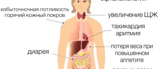
RAYNAUD'S SYNDROME IN PATHOLOGY OF THE ENDOCRINE SYSTEM
SR in association with diseases of the endocrine system has been described in diabetes mellitus, hypothyroidism, hyperthyroidism, hyperparathyroidism, acromegaly and pheochromocytoma. It should be noted that in some cases, systemic scleroderma, in which SR is a mandatory clinical sign, may be accompanied by autoimmune endocrinopathies, such as primary hypothyroidism, Graves' disease, type 1 diabetes mellitus as part of the autoimmune polyglandular syndrome of adults (Fig. 2) [11].
Table 2 also presents possible cases in the practice of an endocrinologist of SR induced by taking combined oral contraceptives and bromocriptine, which was previously actively used in the treatment of acromegaly and hyperprolactinemia, as well as beta-blockers often used to treat concomitant diseases. Interestingly, when taking α-interferon drugs, the development of both SR and cytokine-induced thyroiditis is possible.
Figure 2. Raynaud's syndrome in a patient with autoimmune polyglandular syndrome type 3d: primary hypothyroidism, type 1 diabetes mellitus, limited form of systemic scleroderma, vitiligo (authors' observation).
Acromegaly. SR in acromegaly occurs in approximately 1/3 of cases [12]. There are no data on the dynamics of the course of SR after adenomectomy in the modern literature. However, studies using nail bed capillaroscopy have demonstrated a decrease in the length and number of capillaries in patients with active acromegaly, as well as an increase in the number of tortuous capillaries compared with patients in remission and controls. These changes correlate with disease activity and, being partially reversible, are present in patients with and without diabetes, as well as in patients with arterial hypertension and normal blood pressure [13].
This is consistent with data on decreased vasodilation due to endothelial dysfunction and increased vasoconstriction in response to sympathetic stimulation, which are found in patients with early stage acromegaly and determined using laser Doppler measurements [14].
Two clinical cases of anorexia nervosa are also described, accompanied by Raynaud's phenomenon and acromegaloid appearance features in the absence of increased levels of somatotropic hormone (GH) and insulin-like growth factor (IGF-1) [15]. In addition, similar symptoms may occur with carpal tunnel syndrome, a common clinical manifestation of acromegaly [16]. Differential diagnosis in this case is carried out by carefully collecting anamnesis and according to ultrasound data of the soft tissues of the wrists.
Taking into account the clear clinical picture of acromegaly in the form of changes in appearance, if this disease is suspected in a patient with SR, it is necessary to determine the level of IGF-1 as a primary diagnostic test.
Pheochromocytoma. Data on the prevalence of SR in pheochromocytoma/paraganglioma and its course after surgical treatment are limited. There is a study of 25 patients with pheochromocytoma, two of them (8%) were diagnosed with SR [17]. The mechanism of development of vasospasm in this disease is associated with hyperproduction of catecholamines by a tumor from chromaffin tissue. In addition to catecholamine vasospasm, which is also a cause of vasospastic angina, peripheral ischemia is aggravated by hypovolemia and hypotension outside of catecholamine crises, as well as an increase in platelet aggregation potential. Descriptions of observations note that SR was the initial and for a long time the only manifestation of pheochromocytoma [18, 19].
A clinical case of a combination of pheochromocytoma and systemic scleroderma with the development of ischemic necrosis of the soft tissues of the fingers is described. After surgical treatment of the adrenal tumor, improvement was noted in the form of a decrease in the severity of digital ischemia and healing of soft tissue defects. A feature of this case was also the presence of two episodes of acute coronary syndrome, one of which was induced by the administration of glucocorticoids [20]. There is also a description of a case of SR as the only clinical manifestation of metastasis of an abdominal paraganglioma, removed 3 years before the onset of episodes of vasospasm [21].
Excessive levels of catecholamines in pheochromocytoma/paraganglioma, in addition to vasospasm, usually lead to increased blood pressure, often of a crisis nature - the most characteristic symptom of the disease. Since about 10% of tumors from chromaffin tissue are extra-adrenal, the primary diagnostic test is the determination of catecholamine metabolic products. In the Russian Federation, the most common method is to analyze 24-hour urine for metanephrine and normetanephrine using hydrochloric acid as a preservative.
Hyperparathyroidism. There is a small number of studies on the frequency of SR in primary hyperparathyroidism. The most commonly used group of drugs in the treatment of SR are slow calcium channel blockers. It is assumed that an increase in blood calcium concentration in some cases can lead to a vasospastic effect.
Patients with hyperparathyroidism may exhibit arteriolar calcification and, as a consequence, necrotic lesions of the soft tissues of the distal extremities [22]. Modern sources contain a description of a clinical case of improvement in the course of SR in systemic scleroderma after paradenomectomy for primary hyperparathyroidism [23]. Schwartz et al. described a clinical case of complete regression of SR after surgical treatment of primary hyperparathyroidism in the absence of other possible causes of its development [24].
A decisive role in the diagnosis of hyperparathyroidism, of course, is played by determining the level of parathyroid hormone in the blood, while the initial test is the study of the level of ionized calcium in the blood.
Hypothyroidism. The incidence of SR in patients with hypothyroidism is about 6% [25]. The combination of SR with hypothyroidism was first described in 1976 by Shagan B. and Friedman S. In two patients, the manifestations of SR were leveled out during thyroid hormone replacement therapy [26]. Moreover, in one of them, SR was the only manifestation of hypothyroidism. In the second patient, SR was the first clinical manifestation of primary hypothyroidism, followed by the development of extensive clinical symptoms. Sipila R. described a case of acute myocardial infarction in a 29-year-old woman in the absence of atherosclerotic lesions of the coronary arteries. The pattern of CP has been noted for several years. Clinically manifest hypothyroidism was identified during hospitalization in a cardiology hospital. The probable cause of myocardial infarction in this case was vasospasm associated with hypothyroidism as an analogue of SR [27]. Batthish M. et al. described the development of SR in an 11-year-old girl with hypothyroidism with complete regression after administration of replacement therapy. According to capillaroscopy data, significantly dilated capillary loops were detected with a normal, non-tortuous course [28].
The main feature of the presented clinical cases is the cessation of vasospasm attacks after the administration of thyroid hormone replacement therapy.
It is assumed that such signs of hyperthyroidism as tachycardia, increased cardiac output, increased sweating and tissue catabolism are also due to hypersensitivity to endogenous catecholamines, however, Koehn M. et al. in animal studies found that vascular sensitivity to the action of both norepinephrine, and angiotensin was higher in hypothyroidism than in euthyroidism [29].
It is still unclear whether the relationship between hypothyroidism and CP is a consequence of hormonal deficiency or an immunopathological process. Apparently, remission of SR in patients after administration of levothyroxine therapy indicates the predominant role of thyroid hormone deficiency in the occurrence of SR.
Cases of SR have been described in patients with hypopituitarism. Thus, Al-Deiri M. described the leveling of signs of SR after the initiation of replacement therapy with glucocorticoids and levothyroxine in a patient with panhypopituitarism in Sheehan’s syndrome [30]. Shagan B., presenting a patient with secondary hypothyroidism and adrenal insufficiency, as well as negative levels of antibodies to TPO and TG, emphasizes the key role of thyroid hormone deficiency, rather than an autoimmune process, in the development of SR [31]. It should be taken into account that in patients with systemic scleroderma and other rheumatological diseases, hypothyroidism is more common than in the general population. In one of our own studies, hypothyroidism was detected in 3 (10%) of 30 patients with systemic scleroderma [32].
There are studies that have shown cessation of Raynaud's attacks after thyroid hormone therapy in euthyroid patients with various etiologies of vasospastic disorder. Dessein P. et al [33] showed experience in treating CP with triiodothyronine in 9 patients without cardiovascular disease and in the absence of taking vasoactive drugs. This effect is probably associated with peripheral vasodilation through stimulation of β2-adrenergic receptors in response to increased heat production [34]. Of course, prescribing thyroid hormones solely for the purpose of improving the course of SR in the absence of hypothyroidism is inappropriate and is comparable to prescribing thyroid hormones for the purpose of reducing body weight.
The primary test for diagnosing thyroid disease is the determination of thyroid-stimulating hormone (TSH). If secondary hypothyroidism is suspected (due to damage to the pituitary gland), the TSH level will be uninformative; it is necessary to additionally determine the level of free T4 in the blood. Given the high prevalence of hypothyroidism in the population and similar symptoms, it would probably be advisable to determine the TSH level in all patients with complaints of cold hands.
Diabetes. Data on the frequency of SR in patients with diabetes mellitus (DM) are presented in a few studies. In a study by Grassi W. [35], out of 761 patients with SR, only two had diabetes (0.3%). However, there is a large number of works devoted to capillaroscopy of the nail bed in patients with diabetes.
A close correlation has been described between the presence of neuropathy and impaired thermoregulation of the lower extremities in patients with a short history of diabetes in the form of a delayed response to indirect heating after cooling. Probably, impaired thermoregulation is a consequence of vasospasm. Vasoconstriction under the influence of low temperatures is mediated by sympathetic nervous activity [36]. In cases of short duration of diabetes with autonomic polyneuropathy, only abnormalities in tests based on heart rate (deep breathing test, Valsalva maneuver, orthostatic test) are noted, indicating vagal neuropathy [37]. Increased vascular reactivity to vasopressor agents has also been shown in some patients with diabetes. There is evidence that with the progression of macroangiopathy, the manifestations of vasospasm decrease [38].
As mentioned earlier, capillaroscopy is an important method for diagnosing and differentiating primary and secondary Raynaud's phenomenon in rheumatic diseases. The most specific findings are associated with systemic scleroderma (SSc), the so-called “scleroderma pattern,” which is characterized by dilated capillaries, hemorrhage, avascular areas, and neoangiogenesis. Similar changes are found in patients with dermatomyositis, overlap syndromes, etc. and are called scleroderma-like. The most specific thing at an early stage is the appearance of dilated capillaries. Capillaroscopic changes in systemic autoimmune diseases are specific and differ significantly from the changes found in other diseases. Diseases of social significance, such as diabetes and arterial hypertension, often appear as concomitant diseases in patients with rheumatic pathology. During capillaroscopic examination, dilated capillaries are not observed in patients with diabetes until the late stages of the disease [39].
Microangiopathies are a specific complication of diabetes in both the first and second types of the disease. This primarily concerns the vessels of the retina and kidneys. The walls of microvessels (arterioles, venules and capillaries) are affected due to non-enzymatic glycosylation of proteins, changes in polyol-inositol metabolism, etc. Increased glycosylation of hemoglobin leads to hypoxia, which is one of the prerequisites for microangiopathy [40].
In addition, patients with diabetes may experience a syndrome of limited joint mobility (diabetic stiff hand syndrome, diabetic chiaroarthropathy, diabetic waxy skin). These conditions are characterized by rigidity of the metacarpophalangeal, interphalangeal and wrist joints, flexion contractures with a long history of the disease with a characteristic manifestation in the form of a “prayer sign”, as well as thickening of the skin with loss of fine wrinkles, reminiscent of true sclerodactyly. Skin changes may also affect the metacarpophalangeal joints, as well as the forearm, arms and back [41, 42].
In diabetic stiff hand syndrome, the joints are not directly involved and limited mobility is caused by excess collagen in the periarticular tissue. This syndrome is a common complication of type 1 and type 2 diabetes mellitus, occurring, according to some data, from 8 to 50% of patients with type 1 diabetes mellitus and up to 75% with type 2 diabetes mellitus [43].
The correlation between the development of diabetic upper limb stiffness with blood glucose levels and duration of diabetes is not clear, and autoantibodies and inflammatory markers are usually within normal limits. Some authors point to the association of this syndrome with microvascular changes (retinopathy, nephropathy) [44].
In a study conducted in Poland, 145 children (70 boys, 75 girls) with type 1 diabetes underwent capillaroscopy [45]. Analysis of the results showed an increase in the number of capillaries and their elongation, the presence of megacapillaries, which indicates possible neoangiogenesis. Increased number of capillaries, disturbance in capillary distribution, and the presence of abnormal capillaries were correlated with duration of diabetes. Raynaud's loops were more common in cases with high glycated hemoglobin values.
In another study, the authors performed capillaroscopy with functional tests: occlusion, cold and heat. Changes in the structure of capillaries were noted in patients with a disease duration of more than 5 years, but according to the results of functional tests, there was an increase in the time for recovery of capillaroscopic parameters to the initial ones, only in patients with a short duration of diabetes [46].
Treatment of SR in diabetes is probably no different from that for idiopathic SR. Since persistent hyperglycemia contributes to the progression of vascular damage, individual glycemic targets must be achieved. In type 2 diabetes, drugs for the treatment of SR are often used to treat concomitant conditions (arterial hypertension, dyslipidemia).