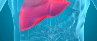
Introduction
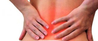
Abdominal pain (AP) is one of the most common reasons for seeking medical help (after headaches, back pain and dizziness) and can be associated with both self-limiting disorders and severe, life-threatening conditions requiring immediate medical intervention.
Establishing a correct diagnosis can be difficult due to the presence of multiple organ systems in the abdominal cavity and the fact that AB can cause many diseases. In this regard, AB is treated worldwide by general practitioners/family doctors, surgeons, internists, emergency physicians, pediatricians, gastroenterologists, urologists and gynecologists. AB usually falls into one of the following categories:
1. The process leading to inflammation of the parietal peritoneum and/or associated with inflammation of the parenchymal organs is usually associated with the occurrence of constant, aching pain, intensifying with changes in the tension of the peritoneum due to pressure/squeezing, stretching or changing position. It is often accompanied by defence, a contraction of the abdominal muscles to reduce tension in the peritoneum.
2. Pain associated with obstruction of a hollow organ, often cramping or colicky in nature, which corresponds to the peristaltic movements of the organ aimed at eliminating the obstruction.
3. Pain associated with vascular disorders in the abdominal cavity (thrombosis or embolism) is caused by hypoxia/ischemia of tissue distal to the site of occlusion; may occur suddenly or gradually, be strong or mild.
4. Functional gastrointestinal disorders such as functional dyspepsia and irritable bowel syndrome IBS [1]. 5. AB may be associated with damage to organs not located in the abdominal cavity (for example, lower lobe pneumonia).
In terms of frequency of occurrence, the leading place in the occurrence of AB is inflammation of hollow and parenchymal organs, accompanied by irritation of the peritoneum. Examples include such common diseases as gastritis, gastroesophageal reflux disease (GERD), peptic ulcers, appendicitis, cholecystitis, hepatitis, intestinal obstruction, urethritis colic, cystitis, pyelonephritis and primary dysmenorrhea.
Links[edit]
- "2-Bromo-2-methylpropane - Compound Summary". PubChem Compound
. USA: National Center for Biotechnology Information. March 26, 2005 Identification. Retrieved June 16, 2012. CS1 maint: discouraged parameter (link) - CRC Handbook of Chemistry and Physics 65th ed.
- Reiners, Matthias; Ehrlich, Niko; Walter, Mark D. (2018). “Synthesis of 1,3,5-tri- tert-
butylcyclopent-1,3-diene and its metal complexes Na {1,2,4-(Me 3 C) 3 C 5 H 2 } and Mg {η 5 -1, 2.4- (Me 3 C) 3 C 5 H 2 ) 2 “.
Inorganic syntheses
.
37
: 199. DOI: 10.1002/9781119477822.ch8. - "2-Bromo-2-Methylpropane 135615." H2NC6H4CO2C2H5, drugs, www.sigmaaldrich.com/catalog/product/aldrich/135615?lang=en®ion=US.
- "Structures of 2-bromo-2-methylpropane". Cambridge Structural Data Center (CCDC), www.ccdc.cam.ac.uk/structures/search?id=doi:10.5517/ccvcqmj&sid=DataCite
Pharmacological properties of hyoscine butyl bromide
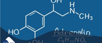
Hyoscine butyl bromide (HBB) is a hyoscine derivative obtained from the leaves of the Duboisia tree, native primarily to Australia. GBB is also known as scopalamine-N-butyl bromide, N-butylscopolammonium bromide and butylscopolamine. The molecular formula is C21H30BrNO4, the molecular weight is 440.37.
GBB contains a nitrogen molecule linked by four different bonds with different chemical groups; thus, it is a quaternary ammonium compound. The GBB molecule is highly polarized and, unlike ammonium and other similar compounds, retains polarity regardless of the pH of the environment. In this regard, when administered orally, only partial absorption of the drug occurs (8%), and systemic bioavailability is less than 1%. However, despite low blood levels immediately after administration, GBB and/or its metabolites are detectable at the sites of action.
The drug does not penetrate the blood-brain barrier and has low binding to plasma proteins. Metabolism is carried out mainly by hydrolysis of the ester bond. After oral administration, excretion of the drug occurs in feces and urine.
As is known, in the parasympathetic nervous system, acetylcholine (ACCh) acts on two types of receptors, muscarinic and nicotinic cholinergic receptors. In most cases, nerve transmission occurs in two stages: upon stimulation, the preganglionic nerve releases ACh into the ganglion, which acts on the nicotinic receptors of postganglionic neurons. The postganglionic neuron then releases ACh to excite muscarinic receptors in the target organ.
Three main types of muscarinic receptors have been well studied:
• M1-muscarinic receptors are found in the nervous system.
• M2-muscarinic receptors, located in the heart.
• M3 muscarinic receptors are located in many parts of the body, especially in the smooth muscle of the blood vessels, bronchi and gastrointestinal tract (GIT), as well as various glands associated with the respiratory tract and GI tract. Stimulation of these receptors leads to indirect vasodilation (via the formation of nitric oxide), bronchoconstriction and increased intestinal motility, as well as increased glandular secretion. M3 receptors are also found in the ciliary body and muscles of the iris, where they are involved in accommodation and allow control of pupil size.
There are also M4 and M5 receptors; the latter are found only in the central nervous system (CNS). The full characteristics of the action and localization of M5 receptors are not fully understood.
Recent studies have also shown that GBB is a potent non-competitive nicotinic receptor blocker in vitro. If such an effect occurs in vivo, it may contribute to the known mechanism of the antispasmodic action of the drug [2].
GBB blocks the action of ACh in the parasympathetic receptors of smooth muscles and secretory glands. This is the basis for both the mechanism of action of GBB and its side effects. Thus, GBB causes a decrease in motility of the gastrointestinal tract and genitourinary tract, and is used to eliminate spasms of these systems, the appearance of which is possible with gastroenteritis, colitis, inflammatory bowel diseases, diverticulitis, biliary colic, cystitis, colic with urethritis and primary dysmenorrhea. It is also used to prevent gastrointestinal spasms before radiological and diagnostic tests, such as endoscopic retrograde cholangiopancreatography and colonoscopy. Based on the mechanisms of action of GBB, side effects/adverse reactions may occur.
The most common of these are: skin reactions (eg, urticaria, rash, erythema, itching) and other types of hypersensitivity, tachycardia, dry mouth, urinary retention. Many of the listed undesirable effects may be associated with the anticholinergic properties of the drug. Anticholinergic side effects are usually mild and self-limiting. It should be noted that, as a quaternary ammonium compound, GBB does not penetrate the blood-brain barrier and therefore does not cause adverse effects from the central nervous system.
Peripheral anticholinergic effects develop due to the ganglion-blocking effect on the walls of internal organs, as well as due to the antimuscarinic effect.
The use of GBB is contraindicated in myasthenia gravis and megacolon/Hirschsprung's disease. In addition, it should not be administered parenterally to patients with uncompensated narrow-angle glaucoma, with tachycardia; prostate hypertrophy with urinary retention; with mechanical stenosis of the gastrointestinal tract.
Other aspects[edit]
tert-
Butyl bromide used to study mass deadenylation of adenine-based nucleosides induced by halogenated alkanes (alkyl halides) under physiological conditions. 2-Bromo-2-methylpropane causes massive deguanylation of guanine-based nucleosides and massive deadenylation of adenine-based nucleosides. [4]
Phase transition from orthorhombic phase III Pmn21 at low temperatures (measured from 95 K) to disordered rhombohedral phase II at 205-213 K. Phase II can exist at 213-223 K, partially coinciding with fcc phase I, which can be observed between 210 -250 K. Phase transitions were also studied at high pressure (up to 300 MPa) [5]
Review of the clinical use of hyoscine butyl bromide
Abdominal pain (nonspecific colic) and IBS
The ability of GBB to eliminate nonspecific colicky ABs is explained by its antimuscarinic effect. Clinically, this effect is manifested by relaxation of the smooth muscles of the gastrointestinal tract, due to which the spasms/colic felt by the patient decrease/disappear. This effect was objectively demonstrated in a study of the electrical and biomechanical activity of the stomach, in which intravenous administration of 20 mg of GBB led to a decrease in mechanical motility by 50.9%, and electrical motility by 36.5% [3].
The main areas of clinical application of GBD in AB were nonspecific abdominal pain of the colic type, IBS, biliary colic and GERD.
The use of GBB in nonspecific AB has been studied in a number of recent studies. A recent large study compared the efficacy and tolerability of 10 mg GBB 3 times daily, 500 mg paracetamol 3 times daily, and a fixed combination of the two compared with placebo in patients with intermittent abdominal cramping pain [4]. All 1637 patients were given a placebo for a week, after which they were randomized to receive one of 4 treatments for 3 weeks. Pain severity (as measured by visual analogue scale and verbal rating scale) was statistically significantly reduced in all treatment groups compared to placebo. Treatment was well tolerated in all groups, and the incidence of side effects did not differ significantly between groups.
Another study of 204 children followed for a week compared GBD with a homeopathic remedy. Both medications were found to provide significant symptomatic relief (severity of spasms, pain/colic, sleep disturbances, difficulty eating and drinking, frequent crying) as assessed by the parent/caregiver, and were well tolerated [5].
Tytgat et al. published two reviews regarding the use of oral/enteral GBD for the treatment of cramping abdominal pain and a number of other clinical scenarios [6, 7]. The first of them contains 10 placebo-controlled studies that examined the effectiveness and safety of oral and rectal use of GBB. The effectiveness of the drug has been established in all studies, which is considered by the authors as evidence in support of the use of the drug for abdominal pain associated with cramps.
The second review provides evidence on the use of GBD for the treatment of colic/spasms; for diagnostic purposes - during visualization; regarding the therapeutic effectiveness and safety of parenteral administration of GBB for the treatment of biliary and renal colic, severe spasms of the genitourinary tract; as well as during childbirth and as palliative treatment. The authors concluded that GBB preparations are fast-acting and highly effective and well tolerated, which supports their use in a number of indications associated with acute abdominal cramps, during childbirth, for palliative treatment, and during diagnostic and therapeutic procedures of the abdominal organs. cavities that can impede spastic contraction [7]. The pharmacological profile of GBB suggests the possibility of its use in conditions accompanied by intestinal irritation. A double-blind, randomized, parallel-group comparative study was conducted, which included 712 patients with IBS. Patients were prescribed: GBB + paracetamol, GBB, paracetamol or placebo for 4 weeks. A visual analogue scale was used to assess symptoms, and by the end of treatment, more than 75% of patients in the GBD group experienced symptom relief [8]. There was also a statistically significant decrease in AB intensity in the GBD group compared to placebo and paracetamol. Other researchers have confirmed the equal effectiveness of GBB and similar drugs, plus their advantage over placebo in the treatment of IBS [9–11].
Renal colic
Renal colic is usually a consequence of the presence of a calculus (stone) in the ureter (less often in the bladder); the severe paroxysmal pain that accompanies this condition is caused by periodic spastic contractions of the blocked hollow organ, aimed at eliminating the blockage. It seems logical that a drug that can eliminate such spasms in the urinary system would have good analgesic effects, which a number of studies have been conducted to evaluate.
Six studies involving 755 patients were identified that assessed GBD as adjunctive pain relief with nonsteroidal anti-inflammatory drugs (NSAIDs) [15–18] and other antispastic drugs [19]. In comparison, the combination of GBB and pethidine was more effective than diclofenac sodium alone [15], although both drugs were more than 90% effective in providing pain relief 30 minutes after administration. Studies have shown that GBB monotherapy is effective in relieving pain, although it is inferior in this regard to therapy using dipyrone and diclofenac [17] or dipyrone and tramadol [19]. Similarly, studies conducted to compare the combination of GBB and dipirone with tenoxicam [16] as well as flurbiprofen [18] confirmed that this combination provided significant pain relief.
Thus, the available data support the effectiveness of GBB in the treatment of renal colic, both as monotherapy and in combination with other drugs.
Dysmenorrhea
Two recent studies examined the use of GBD in primary and secondary dysmenorrhea. In the first double-blind crossover study of 120 women, GBB and paracetamol were compared with lysine clonixinate and proprinox and placebo [20]. Both treatment groups showed a significant reduction in subjective pain ratings compared to placebo.
A second, long-term, open-label study examined the combined use of lysine clonixinate and GBB during three consecutive menstrual cycles in 30 women [21]. All women initially reported very severe (10.7%), severe (42.9%) or moderate (46.4%) pain. By the end of the study, moderate pain persisted in one patient.
Childbirth
Only one study was found that examined GBB primarily as an analgesic for pain associated with childbirth. The study observed 104 primigravida women during labor and delivery; It was found that intravenous administration of GBB leads to a 36% reduction in pain compared with placebo [22].
Some authors have studied the effects of GBB as a labor accelerator, with the argument that if labor pain cannot be safely eliminated, it may be possible to safely reduce the duration of labor (and thus the pain). Five similar studies were found, three of which compared the drug with saline or no treatment [22–24]. One study compared GBB with another antispasmodic drug (drotaverine) [25], and another compared GBB with drotaverine and no therapy [26]. The drug was administered intravenously in 4 studies at doses ranging from 20 to 40 mg, and in one study - rectally [23]. The last 4 studies involved 533 women, divided into almost equal groups; the use of GBB led to a statistically and clinically significant reduction in the period between drug administration and delivery in the absence of significant adverse reactions on the part of the mother or newborn.
Discomfort and pain associated with manipulation
GBB has been studied as a tool to facilitate various studies of the gastrointestinal tract and nearby structures.
In 3 studies, the effectiveness of GBB compared with placebo (2 studies; 208 patients) and glucagon (1 study; 100 patients) was assessed during sigmoidoscopy and/or colonoscopy. One study comparing the drug and placebo showed a reduction in procedure time in the group receiving GBB [27].
Magnetic resonance imaging can be used to obtain high-resolution images of the upper gastrointestinal tract. A study conducted by Wagner et al. [30], showed that intramuscular administration of GBB significantly improves image quality on T2-weighted magnetic resonance imaging of the abdominal cavity. In addition, magnetic resonance imaging of the liver and pancreas has been found to be slightly better at suppressing gastrointestinal motility by GBD.
Propaedeutics of radiation methods for examining the gallbladder
The leading method for diagnosing pathological changes in the gallbladder (GB) is ultrasound examination (US).
Ultrasonography
Its undoubted advantages are non-invasiveness, the ability to quickly and portablely conduct research; absence of ionizing radiation and the need for intravenous administration of contrast agents; independence from the physiological state of the gastrointestinal tract and hepatobiliary system [1–3].
Features of gallbladder syntopy - the proximity of its posterior wall in the body and fundus to the right parts of the large intestine and duodenal bulb - dictate the need to prepare patients for ultrasound examination in order to reduce pneumatization of the corresponding parts of the digestive tract. This requires at least a 6-hour fast on the eve of the diagnostic procedure, and it is optimal to conduct the study on an empty stomach after a night's sleep. Such conditions are also required for the most detailed visualization of the structure of the gallbladder wall and its contents, since it is a hollow organ filled with bile and capable of contracting in response to humoral stimulation during food intake, which leads to a decrease in its size, a sharp thickening of the walls and the inability to detail intraluminal changes [2, 3].
The anatomical structure of the gallbladder is simple. It consists of a narrow neck connecting to the cystic duct, a bladder body with almost parallel walls, and a dome-shaped bottom. At the junction of the neck and the cystic duct, the wall often forms a pouch (or diverticulum) of Hartmann, in which stones can accumulate, blocking the exit of the cystic bile [3–5] (Fig. 1).
To conduct an ultrasound examination of the gallbladder, the scanner sensor (usually with a frequency of 3.5 MHz) is placed in the right hypochondrium of the subject - the site of the anatomical projection. Visualization of the organ can be improved by placing the patient on the left side, which allows the intestinal loops to be somewhat pushed to the left; the same purpose is served by conducting the study while holding the breath with a deep breath. Visualization of the gallbladder through the intercostal spaces is the most consistent, but the least informative and is used mainly in urgent situations in unprepared patients. To better assess the nature of intravesical changes (the presence of small stones, identifying their mobility), it is possible to conduct an examination in a vertical position or with the body tilted forward [2–5].
In the diagnosis of cholelithiasis (GSD), ultrasound scanning rightfully occupies a leading position. The sensitivity of ultrasound in detecting gallstones exceeds 95%, which look like hyperechoic structures with an acoustic shadow [1, 5] (Fig. 2).
In the vast majority of cases, the stones are mobile, but can be fixed to the wall or motionless in the neck of the bladder. Other foreign bodies that mimic cholelithiasis may be blood or pus clots or parasites. Gallbladder polyps are immobile or have limited mobility if they have a pedicle, are always connected to the wall and, as a rule, do not give an acoustic shadow; usually their size does not exceed 5 mm. As a rule, against the background of a long absence of contractions of the bladder, it is possible to visualize the stratification of bile into two components with a clear interface, one of which is anechoic and occupies the upper parts of the bladder in relation to the horizontal plane, the other is more dense, located below. This phenomenon, called stagnation of bile, or sludge, occurs due to the presence of cholesterol and calcium bilirubinate crystals in it and their ability to reversibly form large aggregates (Fig. 3) .
The most common complication of cholelithiasis is acute cholecystitis, the cause of which is almost always a violation of the outflow of bile from the gallbladder. The main ultrasound signs of this pathology are:
- the presence of stones in the lumen of the bladder (the most typical finding is an “impacted” stone in the neck) - Fig. 4;
- thickening of the wall more than 3 mm with the appearance of heterogeneity of its structure, delamination (Fig. 4);
- an increase in the transverse size of the bladder by more than 4 cm; detection of the greatest pain at the site of ultrasound projection of the gallbladder (ultrasound Murphy’s symptom), which may not be detected in gangrenous cholecystitis [3, 5] (Fig. 4).
The combination of these symptoms with the detection of fluid accumulation near the bladder, especially in combination with the identification of a wall defect, indicates its perforation and, possibly, the formation of a peripysical abscess (Fig. 5). The detection of ultrasound signs of gas accumulation in the lumen of the bladder against the background of other signs of acute inflammation indicates such a severe pathology as emphysematous cholecystitis caused by gas-forming anaerobic flora [3, 5] (Fig. 6).
Another complication of cholelithiasis, manifested by a violation of the passage of bile, is choledocholithiasis. Violation of the outflow of bile along the common bile duct causes its expansion of more than 7 mm, expansion of the intrahepatic bile ducts (more than 40% of the diameter of the adjacent branch of the portal vein) (Fig. 7). Indisputable evidence of choledocholithiasis is the detection of stones, which are most often localized in the distal part of the common bile duct, but can be visualized only in 70–80% of cases [1, 4, 5].
Dilation of the intra- and extrahepatic bile ducts is also observed with such a complication of cholelithiasis as Mirizzi syndrome, which consists of obstruction of the common bile duct as a result of the volumetric effect of the inflammatory reaction of tissues to a stone located in the neck of the bladder or cystic duct, with a low confluence it in the common bile duct (Fig.  [5].
[5].
Often the formation of stones in the gallbladder is accompanied by benign changes in its walls, one of the main ones being a violation of cholesterol metabolism. These changes are called hyperplastic cholecystopathy. One of its variants - cholesterol dystrophy is manifested by such ultrasound signs as the detection in the wall of the bladder of hyperechoic deposits no larger than 1 mm in size, which do not give an acoustic shadow, but are accompanied by a reverberation effect (the bubble vaguely resembles a strawberry), which are lipid deposits (Fig. 9) . More pronounced changes in the wall, called adenomyomatosis, are manifested in its thickening, the formation of intramural diverticula (Aschoff-Rokitansky sinuses), containing hyperechoic cholesterol deposits with a reverberation effect (“comet tail”) and microliths that give an acoustic shadow (Fig. 9). Hyperplastic cholecystopathy can affect both the entire bladder and part of its wall [2, 5].
It is known that removal of the gallbladder for cholelithiasis does not relieve patients from metabolic disorders, including hepatocyte dyscholia, which persists after surgery [6]. The loss of the physiological role of the gallbladder, namely the concentration of bile in the liver during the interdigestive period and its release into the duodenum during meals, is accompanied by a violation of the passage of bile into the intestine and indigestion [6, 7].
Changes in the chemical composition and volume of bile, its chaotic entry into the duodenum after cholecystectomy (CE) disrupt the digestion and absorption of fat and other lipid substances, reduce the bactericidal nature of duodenal contents, which leads to microbial contamination and impaired motility of the duodenum, the development of bacterial overgrowth syndrome in the intestine (especially in the ileum), disruption of the enterohepatic circulation and decreased synthesis of bile acids in the liver [6]. As a consequence, a syndrome of impaired digestion, the symptoms of which are often mistakenly interpreted as postcholecystectomy syndrome (PCS), is associated by surgeons primarily with mechanical obstacles to bile outflow that were not recognized before surgery or were not eliminated during cholecystectomy (stones left or emerging again in the common bile duct, stenosis of the papilla of Vater and etc.) [6].
When preparing for a cholecystectomy operation, much attention is always paid to diagnosing mechanical obstacles to bile outflow into the duodenum. The situation is completely different with preoperative verification of extrahepatic biliary dysfunctions. The absence of indirect signs of functional disorders of the sphincter of Oddi in the form of dilation of the common bile duct during ultrasound, increased liver enzymes, pain attacks, etc. does not at all exclude dysfunction of the papilla of Vater, which forms long before the patient’s admission. According to our data, in 45% of patients with cholelithiasis, radionuclide hepatobilis scintigraphy (GBSG) reveals functional disorders in the transport of radiopharmaceuticals from the common bile duct to the duodenum, which do not require retrograde cholangiopancreatography and endoscopic correction.
In some cases, radionuclide methods are simply no alternative due to the strict specificity of the inclusion of radiopharmaceuticals (RP) in various metabolic processes (GBSG). The functional state of the hepatobiliary system in any pathology of the hepatobiliary system, including cholelithiasis, is studied using standard GBSG.
Hepatobiliscintigraphy
GBSG allows you to objectively evaluate the most important processes from the standpoint of the functioning of the digestive transport conveyor: bile synthetic and bile excretory functions of the liver, as well as the transport of bile into the duodenum. The method is based on recording the passage of short-lived radionuclides Tc-99m + bromezide through the biliary tract.
The study is carried out on an empty stomach, in a horizontal position of the patient after the administration of 3 mKM Tc-99m + bromezide intravenously. The duration of the procedure is 60 minutes. As a choleretic breakfast, patients take chicken egg yolks or 200 ml of 10% cream 30 minutes from the start of the study.
Normal indicators of GBSG are considered:
1) the half-life (T1/2) of the radiopharmaceutical (RP) from the liver is less than 35 minutes; 2) the half-life (T1/2) of radiopharmaceuticals from the common bile duct is less than 50 minutes; 3) the time of receipt of radiopharmaceuticals into the duodenum is less than 40 minutes; 4) adequate supply of radiopharmaceuticals to the intestine is the predominance of radiopharmaceutical activity in the duodenum compared to that in the common bile duct by the end of the study.
The generally accepted standard method of radioisotope research with a choleretic breakfast does not always make it possible to specify the nature of functional changes in the bile outflow. This is explained by the fact that the food load exerts its effect both by activating the entry of cholecystokinin (CC) into the bloodstream upon irritation of I-cells of the duodenal mucosa and intramural nerve plexus [8]. The activity of digestive enzymes and the sensitivity of the sphincter apparatus of the biliary tract to intestinal hormones is variable and in practice difficult to determine [9]. The tone of intramural nerve fibers depends on the physiological activity of the organs of the upper digestive tract [10, 11].
In this regard, in the radionuclide diagnosis of extrahepatic biliary dysfunction (IBD), intravenous administration of the hormone cholecystokinin is often used, but the relaxing effect of this drug depends on the state of the central nervous system, the hormonal background of the patient and is disrupted in case of cholesterosis of the gallbladder, since the localization of receptors for cholecystokinin coincides with the sites of deposits cholesterol esters in the bladder wall and bile ducts, which makes it difficult to accurately determine the dose of the administered hormone [8, 10].
To clarify the nature of bile outflow disorders along the common bile duct in the Faculty Surgery Clinic named after. S. I. Spasokukotsky Russian National Research Medical University named after. N.I. Pirogov on the basis of the First City Hospital performs GBSG with amino acid cholekinetic test (GBSG-ACT) and GBSG with Buscopan® test (RF patent No. 2166333).
GBSG with amino acid cholekinetic test
The study is carried out on an empty stomach. 30 minutes after the administration of the radiopharmaceutical and the start of the study, a solution of amino acids Vamin-14 or Freamin, which does not contain glucose and electrolytes, is injected into a peripheral vein. We consider the last condition to be very important, since the hyperglycemia that occurs during glucose infusion completely or partially inhibits the secretion of cholecystokinin [8]. The dose of the drug was chosen at the rate of 1.5–2 ml/kg body weight (80–130 ml). The duration of the infusion is 5–7 minutes, since the administration of an amino acid solution for more than 10 minutes (regardless of the dose) does not lead to an increase in the release of endogenous cholecystokinin, but, on the contrary, reduces the incretion of the hormone [9]. IAP and the cause of delayed excretion of the radiopharmaceutical by hepatocytes are assessed based on the differences in the parameters of standard GBSG and GBSG-ACT (Fig. 10).
GBSG using hyoscine butyl bromide
Hyoscine butyl bromide (Buscopan®) is a derivative of the tertiary ammonium compound hyoscine. Hyoscine is an alkaloid present in plants of the genus Duboisia. It is chemically processed by adding a butyl group to produce a quaternary ammonium structure. This modification forms a molecule that still has anticholinergic properties comparable to those of hyoscine.
But unlike hyoscine, quaternary ammonium compounds such as hyoscine butyl bromide limit systemic absorption and significantly reduce the number of adverse reactions. Hyoscine butyl bromide is an anticholinergic drug with a high degree of affinity for muscarinic receptors located on the smooth muscle cells of the gastrointestinal tract, causing an antispasmodic effect. In addition, the drug binds to nicotinic receptors, which determines the effect of blocking the nerve ganglia, which determines its antisecretory effect.
The technical side of the study differs little from the above-mentioned AChT test. At the end of standard hepatobiliscintigraphy, instead of infusion of an amino acid solution, the patient takes 20 mg of hyoscine butyl bromide per os. After 20 minutes, the data is re-recorded and processed (Fig. 11). Thus, the use of the Buscopan® test reduces diagnostic time for the doctor and simplifies the diagnostic procedure for the patient.
GBSG with the Buscopan® test has proven itself to be most effective in the study of patients after cholecystectomy (Fig. 12).
The leading factors of liver dysfunction after cholecystectomy are the presence and duration of dyskinesia of the sphincter apparatus of the biliary tract. In patients, paradoxical spasm of the sphincter of Oddi significantly predominates as the cause of delay of radiopharmaceuticals in the common bile duct. Functional disorders of bile outflow are caused by cholesterosis of the biliary tract, in particular the sphincter of Oddi (Fig. 13, 14).
X-ray methods
X-ray methods for studying the gallbladder and extrahepatic bile ducts are practically not used today, so we provide a brief description of them as a historical reference.
Survey radiography
Plain radiography of the abdominal cavity is performed much less frequently than ultrasound due to radiation exposure. But, nevertheless, it was a fairly informative method for diagnosing cholelithiasis. On the x-ray you can see the presence, location and number of x-ray stones containing calcium salts (Fig. 15).
Oral cholecystography
Oral cholecystography is performed if X-ray negative (cholesterol) stones are suspected. The method is based on absorption in the gastrointestinal tract and excretion of the contrast agent with bile (Fig. 16).
If absorption in the intestine is impaired, the excretory function of the liver is reduced, the cystic duct is blocked by a stone, etc., oral cholecystography may be negative, i.e., the shadow of the gallbladder is not detected on it.
Intravenous cholecystography
Intravenous cholecystography is performed if the result of the oral radiocontrast method is negative. Using this technique, it is possible to contrast the gallbladder in 80–90% of cases (Fig. 17).
Computed tomography and magnetic nuclear (magnetic resonance) imaging
The shortcomings of classical X-ray studies of the gallbladder are successfully compensated by computed tomography (CT) and magnetic nuclear tomography (magnetic resonance imaging, MRI). In calculous cholesterosis, stones are visualized as shadows of a homogeneous structure (cholesterol stones) (Fig. 18) or are represented by heterogeneous shadows with alternating areas of mixed stones - a cholesterol core with a calcium-bilirubin shell (Fig. 19).
Computed tomography and magnetic nuclear tomography make it possible to suspect cholelithiasis in patients examined for other pathologies of the abdominal organs, since a description of the image of the gallbladder is a mandatory component of the protocol for these studies.
Literature
- Practical guide to ultrasound diagnostics. Ed. V.V. Mitkova. M.: Vidar-M, 2003. T. 1. 720 p.
- Brant WE, Helms CA Fundamentals of Diagnostic Radiology, 2nd ed. Lippincott, Williams and Wilkins, p. 836–841.
- Kurtz AB, Middleton WD Ultrasound: The Requisites. Philadeiphia, Hanley & Belfus, 1996, p. 35–71.
- Parulekar SG Transabdominal sonography of bile ducts // Ultrasound Q. 2002, (18) 3: 187–202.
- Rumack CM, Wilson SR, Charboneau JW (eds). Diagnostic Ultrasound, 2nd ed. St. Louis, Mosby, 1998, p. 172–195.
- Savelyev V. S., Petukhov V. A. Cholelithiasis and indigestion syndrome. M.: BORGES, 2010. 258 p.
- Savelyev V. S., Petukhov V. A., Boldin B. V. Cholesterosis of the gallbladder. M.: VEDI, 2002., 176 p.
- Vysotskaya R. A. Prostaglandins and gastrointestinal hormones in chronic liver diseases. Diss. doc. biol. Sci. M., 1992, 340 p.
- Houda R., Tooli J., Dodds WJ Effect of enteric hormons on sphincter of Oddi and gastrointestinal myoelectric activity in fasted conscious opossums // Gastroenterol. 1983, vol. 84, p. 1–9.
- Weechsler-JG Bedeutung der Gallenblase in der Regulation des duodenogastralen Refluxes // Z-Gastroenterol. 1987, Aug. 25, Suppl 3: p. 15–21.
- Petukhov V. A. Lipid distress syndrome. Ed. V. S. Savelyeva, M.: MAX PRESS, 2010. 544 p.
V. A. Petukhov*, 1, Doctor of Medical Sciences, Professor D. A. Churikov**, Candidate of Medical Sciences
* GBOU VPO RNIMU im. N. I. Pirogova Ministry of Health of the Russian Federation, Moscow ** State Clinical Hospital No. 1 named after. N. I. Pirogova, Moscow
1 Contact information