General blood test and its interpretation
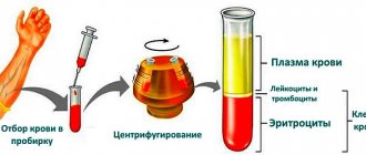
The indicators of a general fasting blood test are assessed (the price of the test is available to everyone) by quantity, percentage and quality. They evaluate “red blood cells” - red blood cells, which, with the help of hemoglobin, carry oxygen throughout the body. Also, the indicators of the general blood test of a child and an adult, which are deciphered in children and adults at prices below average, are carried out by us, help to evaluate all types of white blood cells - leukocytes and blood platelets - platelets. The former are responsible for many processes in the body related to the functioning of the immune system, and the latter are responsible for the ability of blood to clot.
What do violations of norms in a blood test indicate?
Violations of the above OAC norms in children may indicate either a slight illness in the child or the development of a serious and sometimes fatal disease.
Only a doctor can evaluate the totality of all factors, including the full picture of the analysis performed, the general condition of the baby, his age, the presence or absence of somatic diseases or congenital conditions, etc.
Do not try to independently evaluate the results obtained and draw conclusions, much less self-medicate. Delay in receiving quality and professional treatment can cost your child his life.
Leukemia
Leukemia refers to cancers of the blood, the causes of which have not yet been fully determined. Leukemia (or leukemia) is a malignant tumor process in the hematopoietic and lymphatic tissue.
The likelihood of a complete recovery and return of the child to normal life directly depends on the timing of seeking medical help. What could be a signal that there are serious problems? Deviations in the general blood test:
- a significant drop in red blood cell levels;
- decreased platelet levels (2.5 times lower than normal);
- drop in hemoglobin level (sometimes 3 times);
- depending on the form of the disease, the leukocyte count can be highly elevated or, conversely, catastrophically low;
- a sharp increase in ESR.
Allergic manifestations
Any allergic manifestations, from the most minor to those requiring urgent medical attention, are reflected in the indicators of a general blood test.
The presence of foreign allergens in the body does not go unnoticed.
- the number of leukocytes increases;
- ESR increases slightly;
- the number of eosinophils (one of the types of leukocytes), which are considered the main indicator of allergies in a blood test, increases sharply;
- the level of ferritin, a specific protein, increases as the body’s response to the inflammatory process;
- all other indicators remain practically within normal limits.
Photo source: shutterstock.com
Thrombocytosis
A number of diseases, the main indicator of which can be considered a change in the composition of the blood, where platelets are significantly increased (in some cases, the level of platelets can be increased by 3-4 times). Among the most common causes of thrombocytosis may be:
- presence of tumor diseases;
- severe blood loss;
- acute inflammation or infectious diseases;
- disruption (at the gene level) of the production of activated protein C, which ensures the fluid properties of blood.
Worm infestations
Infection with parasites, if not treated in a timely manner, can have very serious consequences. Some specialists from European medical organizations believe that the presence of helminthic infestation (worms) in the human body reduces its lifespan by 10-15 years.
If infection with parasites (for example, Giardia) occurs, this immediately affects the composition of the child’s blood. The child’s body is especially sensitive to this type of infection, so based on the results of a child’s blood test, the following conclusions can be drawn:
- hemoglobin levels decrease;
- the level of eosinophils increases;
- there is a significant increase in ESR.
Atypical leukocytes
The appearance of atypical leukocytes in a child’s analysis (differences in shape, size, color, change in nucleus) may indicate:
- about the presence of a viral or bacterial infection in the child’s body (for example, sore throat);
- about intoxication of the body;
- about the development of pathological tumor processes or the presence of genetic pathology.
Hypochromic anemia
Hypochromia or hypochromic anemia is a violation of the color of a blood smear (red blood cells) taken for analysis. Hypochromia is closely related to another disorder, microcytosis - the predominance of small red blood cells in the blood.
Such violations indicate the presence of a systemic disease that provokes the appearance of one of the types of anemia.
Mononucleosis
Mononucleosis is an acute viral disease that requires urgent medical treatment.
- the level of lymphocytes increases (by more than 40%);
- the level of monocytes increases (by more than 10%).
General blood test: interpretation in adults
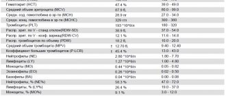
General clinical blood test: the norm of red blood cells is from 3.7 to 5.1 per 10 to 12 cells per 1 liter, the concentration of hemoglobin in them is 120-140 g/l in women and 130-160 g/l in men.
The blood test is general and its interpretation requires an assessment of the shape of the red blood cells, which normally represent a biconcave disc. Changes in these indicators may indicate iron deficiency and anemia, or blood thickening. Indicators of a general blood test and their norm help determine the type of anemia, its possible cause, etc.
The number of “young” red blood cells - reticulocytes - in a general fasting blood test is in the range of 0.2-1.2%. They increase with increased hematopoiesis, accelerated destruction of mature red blood cells (hemolysis) and decrease with anemia, kidney disease, thyroid disease, consequences of radiation exposure, etc.
What do changes in blood parameters indicate?
Deviation of the blood hemogram from the norm in the table:
- A reduced level of hemoglobin is observed in anemia, leukemia, severe blood loss, pregnancy, congenital diseases of the circulatory system, and lack of iron and vitamins.
- Hemoglobin increases with congenital pathologies of the heart or lungs and their insufficiency, while at altitude, with dehydration caused by impaired renal function, diabetes mellitus and diabetes insipidus, vomiting or diarrhea, low fluid intake or heavy sweating.
- An increased level of leukocytes is observed in cases of cancer, purulent-inflammatory processes, burns or other injuries during which the soft tissues of the body are damaged.
- An increased number of lymphocytes indicates infection with any viruses, and neutrophils indicate bacterial infections. Eosinophils are considered to be markers of parasitic infestations and allergic reactions.
- An increased number of platelets occurs in chronic inflammatory diseases (tuberculosis, ulcerative colitis, cirrhosis of the liver), after operations, treatment with hormones, in some types of cancer, and iron deficiency anemia.
- A decrease in the number of platelets indicates heavy metal poisoning, blood diseases, kidney failure, pathologies of the liver, spleen, and hormonal disorders.
In addition, most drugs affect the results of laboratory tests of the above indicators.
We recommend
Therapeutic massage for the back: features, indications and contraindications Read more
Reasons that may affect the tests:
- The tourniquet was applied incorrectly when drawing blood or the application time was exceeded.
- Shaking the test tube.
- Insufficient amount of blood taken for testing.
- Mixed-up tubes with samples from different patients.
- The test tube filling standards were not met.
- Long-term storage of the collected blood sample.
- Biochemical parameters are disturbed.
- Blood is taken after the transfusion procedure.
- Technical failure of the equipment.
If the decoding of the hemogram does not correspond to the patient’s complaints and the results of the physical examination performed by the doctor, then an extended study is prescribed.
General blood test interpretation: ESR and leukocytes
An increase in ESR, or erythrocyte sedimentation rate (normally 1-10 mm per hour in men and 2-15 mm/hour in women), helps detect signs of an inflammatory response.
The total number of leukocytes is 4-9 per 10 to the 9th power of cells per liter. The optimal ratio of fasting blood count indicators is normal for different types of leukocytes:
· For neutrophils - band 1-6% - segmented 47-72%
· For basophils 0-1%
· For monocytes 2-9%
· For eosinophils 0-5%
· For lymphocytes 18-40%
Changing the number of different types of white blood cells can help determine the presence of a viral or bacterial infection, an inflammatory reaction, atypical changes in cells in the bone marrow, a parasitic disease, an allergy, etc.
Blood hemogram - what kind of analysis is it?
It is carried out for the purpose of:
- Detection of anemia, leukemia, lymphoma.
- Diagnosis of bleeding and bleeding disorders.
- Detection of viral and bacterial infections.
- Detection of polycythemia vera (polycythemia vera).
- Studies of lymphadenopathy (enlarged lymph nodes).
- Detection of various allergies.
- Analysis of splenomegaly (enlarged spleen)
.
- Detection of thalassemia.
- Monitoring the treatment of liver diseases.
Currently, venous blood is an ideal material for this type of research. What is this - a blood hemogram? This is a quantitative and qualitative analysis of individual blood structures (erythrocytes, leukocytes, platelets, etc.).
What does a blood hemogram show:
- This examination method makes it possible to identify the nature of the occurrence of certain symptoms of pathology after a specialist has studied the patient’s medical history and physical examination.
- In order to monitor changes in blood counts during chemotherapy.
When to do this analysis:
- After injuries accompanied by internal and/or external bleeding.
- In case of malaise, fatigue, bleeding, bone pain, swelling of the lymph nodes, infection, etc.
- At the preparatory stage before surgery.
- To assess the quality of blood after transfusions.
Decoding a blood hemogram provides information not only about the number of cells. It can also indicate the physical characteristics of some of them, which helps the doctor make an accurate diagnosis.
There are cases when the analysis indicators do not change even in the presence of any disease.
General clinical blood test for a child interpretation
Children's bodies continue to develop and adapt to the changes associated with growing up. Therefore, the data of a general fasting blood test in children of different age groups are different.
A general clinical blood test for a child under one year of age does not differ in price from an adult. Interpretation: Number of erythrocytes - 4.3-7.6 per 10 to 12 cells/l, reticulocytes - 3-51%, hemoglobin concentration - 180-240 g/l, platelets - 180-490 per 10 to 9 cells/ l, leukocyte level – 8.5-24.5 per 10 to 9 degrees, ESR – 2-4 mm/hour.
The ratio of types of leukocytes: eosinophils - 0.5-6%, basophils - 0-1%, band netrophils 1-17%, and segmented ones - 45-80%, lymphocytes 12-36%, monocytes - 2-12%;
General blood test in children (the price is quite affordable) older than 1 year:
The number of erythrocytes decreases - 3.6-4.9 per 10 in 12 cells/l, hemoglobin - 110-135 g/l, reticulocytes - 3-15%, leukocytes - 6.0-12.0 per 10 in 9 degrees. ESR becomes closer to adult levels - 4-12 mm/hour.
MEDICAL CENTER
Interpretation of study results contains information for the attending physician and is not a diagnosis.
The information in this section should not be used for self-diagnosis or self-treatment. The doctor makes an accurate diagnosis using both the results of this examination and the necessary information from other sources: medical history, results of other examinations, etc. Hemoglobin (Hb, hemoglobin) Units of measurement in the medical center STUDIO DOCTOR: g/dl. Alternative units: g/l. Conversion factor: g/l x 0.1 ==> g/dl. Reference values
| Age, gender | Hemoglobin level, g/dl | |
| Children | ||
| 1 day - 14 days | 13,4 — 19,8 | |
| 14 days - 4.3 weeks | 10,7 — 17,1 | |
| 4.3 weeks - 8.6 weeks | 9,4 — 13,0 | |
| 8.6 weeks - 4 months | 10,3 — 14,1 | |
| 4 months – 6 months | 11,1 — 14,1 | |
| 6 months – 9 months | 11,4 — 14,0 | |
| 9 months - 12 months | 11,3 — 14,1 | |
| 12 months - 5 years | 11,0 — 14,0 | |
| 5 years - 10 years | 11,5 — 14,5 | |
| 10 years - 12 years | 12,0 — 15,0 | |
| 12 years - 15 years | Women | 11,5 — 15,0 |
| Men | 12,0 — 16,0 | |
| 15 years - 18 years | Women | 11,7 — 15,3 |
| Men | 11,7 — 16,6 | |
| 18 years - 45 years | Women | 11,7 — 15,5 |
| Men | 13,2 — 17,3 | |
| 45 years - 65 years | Women | 11,7 — 16,0 |
| Men | 13,1 — 17,2 | |
| > 65 years old | Women | 11,7 — 16,1 |
| Men | 12,6 — 17,4 | |
Increased hemoglobin levels:
- dehydration (with severe diarrhea, vomiting, increased sweating, diabetes, burn disease, peritonitis);
- physiological erythrocytosis (in residents of high mountains, pilots, athletes);
- symptomatic erythrocytosis (with insufficiency of the respiratory and cardiovascular systems, polycystic kidney disease);
- erythremia.
Decreased hemoglobin:
- anemia of various etiologies;
- overhydration.
Hematocrit (Ht, hematocrit)
Units of measurement in the medical center STUDIO DOCTOR: %.
Reference values
| Age, gender | Hematocrit indicator, % | |
| Children | ||
| 1 day - 14 days | 41,0 — 65,0 | |
| 14 days - 4.3 weeks | 33,0 — 55,0 | |
| 4.3 weeks - 8.6 weeks | 28,0 — 42,0 | |
| 8.6 weeks - 4 months | 32,0 — 44,0 | |
| 4 months – 9 months | 32,0 — 40,0 | |
| 9 months - 12 months | 33,0 — 41,0 | |
| 12 months - 3 years | 32,0 — 40,0 | |
| 3 years - 6 years | 32,0 — 42,0 | |
| 6 years - 9 years | 33,0 — 41,0 | |
| 9 years - 12 years | 34,0 — 43,0 | |
| 12 years - 15 years | Women | 34,0 — 44,0 |
| Men | 35,0 — 45,0 | |
| 15 years - 18 years | Women | 34,0 — 44,0 |
| Men | 37,0 — 48,0 | |
| 18 years - 45 years | Women | 35,0 — 45,0 |
| Men | 39,0 — 49,0 | |
| 45 years - 65 years | Women | 35,0 — 47,0 |
| Men | 39,0 — 50,0 | |
| 65 years - 120 years | Women | 35,0 — 47,0 |
| Men | 37,0 — 51,0 | |
Increased hematocrit:
- dehydration (with severe diarrhea, vomiting, increased sweating, diabetes, burn disease, peritonitis);
- physiological erythrocytosis (in residents of high mountains, pilots, athletes);
- symptomatic erythrocytosis (with insufficiency of the respiratory and cardiovascular systems, polycystic kidney disease);
- erythremia.
Decreased hematocrit:
- anemia of various etiologies;
- overhydration.
Red blood cells
Units of measurement at the STUDIO DOCTOR medical center: million/µl (106/µl). Alternative units: 1012 cells/L.
Conversion factors: 1012 cells/l = 106 cells/μl = million/μl.
Reference values
| Age, gender | Red blood cells, million/µl (x106/µl) | |
| Children | ||
| 1 day - 14 days | 3,90 — 5,90 | |
| 14 days - 4.3 weeks | 3,30 — 5,30 | |
| 4.3 weeks - 4 months | 3,50 — 5,10 | |
| 4 months – 6 months | 3,90 — 5,50 | |
| 6 months – 9 months | 4,00 — 5,30 | |
| 9 months - 12 months | 4,10 — 5,30 | |
| 12 months - 3 years | 3,80 — 4,80 | |
| 3 years - 6 years | 3,70 — 4,90 | |
| 6 years - 9 years | 3,80 — 4,90 | |
| 9 years - 12 years | 3,90 — 5,10 | |
| 12 years - 15 years | Women | 3,80 — 5,00 |
| Men | 4,10 — 5,20 | |
| 15 years - 18 years | Women | 3,90 — 5,10 |
| Men | 4,20 — 5,60 | |
| 18 years - 45 years | Women | 3,80 — 5,10 |
| Men | 4,30 — 5,70 | |
| 45 years - 65 years | Women | 3,80 — 5,30 |
| Men | 4,20 — 5,60 | |
| 65 years - 120 years | Women | 3,80 — 5,20 |
| Men | 3,80 — 5,80 | |
Increased red blood cell concentration:
- dehydration (with severe diarrhea, vomiting, increased sweating, diabetes, burn disease, peritonitis);
- physiological erythrocytosis (in residents of high mountains, pilots, athletes);
- symptomatic erythrocytosis (with insufficiency of the respiratory and cardiovascular systems, polycystic kidney disease);
- erythremia.
Decreased red blood cell concentration:
- anemia of various etiologies;
- overhydration.
MCV (mean erythrocyte volume) Determination method: calculated value. Units of measurement at the STUDIO DOCTOR medical center: fl (femtoliter). Reference values
| Age, gender | Mean erythrocyte volume, MCV, fl | |
| Children | ||
| 1 day - 14 days | 88,0 — 140,0 | |
| 14 days - 4.3 weeks | 91,0 — 112,0 | |
| 4.3 weeks - 8.6 weeks | 84,0 — 106,0 | |
| 8.6 weeks - 4 months | 76,0 — 97,0 | |
| 4 months – 6 months | 68,0 — 85,0 | |
| 6 months – 9 months | 70,0 — 85,0 | |
| 9 months - 12 months | 71,0 — 84,0 | |
| 12 months - 5 years | 73,0 — 85,0 | |
| 5 years - 10 years | 75,0 — 87,0 | |
| 10 years - 12 years | 76,0 — 90,0 | |
| 12 years - 15 years | Women | 73,0 — 95,0 |
| Men | 77,0 — 94,0 | |
| 15 years - 18 years | Women | 78,0 — 98,0 |
| Men | 79,0 — 95,0 | |
| 18 years - 45 years | Women | 81,0 — 100,0 |
| Men | 80,0 — 99,0 | |
| 45 years - 65 years | Women | 81,0 — 101,0 |
| Men | 81,0 — 101,0 | |
| 65 years - 120 years | Women | 81,0 — 102,0 |
| Men | 83,0 — 103,0 | |
Increasing MCV values:
- B12 deficiency and folate deficiency anemia;
- aplastic anemia;
- liver diseases;
- hypothyroidism;
- autoimmune anemia;
- smoking and drinking alcohol.
Reducing MCV values:
- Iron-deficiency anemia;
- anemia of chronic diseases;
- thalassemia;
- some types of hemoglobinopathies.
It should be taken into account that the MCV value is not specific; the indicator should be used to diagnose anemia only in combination with other indicators of a general blood test and biochemical blood test.
RDW (Red cell Distribution Width, distribution of red blood cells by size)
Method of determination: calculated value Units of measurement in the medical center STUDIO DOCTOR: %
Reference values
< 6 months — 14.9 – 18.7
> 6 months — 11.6 – 14.8
Increasing RDW values:
- anemia with heterogeneity of red blood cell size, including those associated with nutrition; myelodysplastic, megaloblastic and sideroblastic types; anemia accompanying myelophthisis; homozygous thalassemia and some homozygous hemoglobinopathies;
- a significant increase in the number of reticulocytes (for example, due to successful treatment of anemia);
- condition after red blood cell transfusion;
- interferences – cold agglutinins, chronic lymphocytic leukemia (high number of leukocytes), hyperglycemia.
There are also a number of anemias that are not characterized by an increase in RDW:
- anemia of chronic diseases;
- anemia due to acute blood loss;
- aplastic anemia
- some genetically determined diseases (thalassemia, congenital spherocytosis, presence of hemoglobin E).
It should be taken into account that the value of the RDW indicator is not specific; the indicator should be used to diagnose anemia only in combination with other indicators of a general blood test and biochemical blood test.
MCH (average amount of hemoglobin in 1 red blood cell)
Determination method: calculated value.
Units of measurement and conversion factors: pg (picograms).
Reference values
| Age, gender | Average hemoglobin content in 1 red blood cell, MCH, pg | |
| Children | ||
| 1 day - 14 days | 30,0 — 37,0 | |
| 14 days - 4.3 weeks | 29,0 — 36,0 | |
| 4.3 weeks - 8.6 weeks | 27,0 — 34,0 | |
| 8.6 weeks - 4 months | 25,0 — 32,0 | |
| 4 months – 6 months | 24,0 — 30,0 | |
| 6 months – 9 months | 25,0 — 30,0 | |
| 9 months - 12 months | 24,0 — 30,0 | |
| 12 months - 3 years | 22,0 — 30,0 | |
| 3 years - 6 years | 25,0 — 31,0 | |
| 6 years - 9 years | 25,0 — 31,0 | |
| 9 years - 15 years | 26,0- 32,0 | |
| 15 - 18 years old | Women | 26,0 — 34,0 |
| Men | 27,0 — 32,0 | |
| 18 – 45 years old | Women | 27,0 — 34,0 |
| Men | 27,0 — 34,0 | |
| 45 – 65 years | Women | 27,0 — 34,0 |
| Men | 27,0 — 35,0 | |
| 65 years - 120 years | Women | 27,0 — 35,0 |
| Men | 27,0 — 34,0 | |
Increasing MCH values:
- B12 deficiency and folate deficiency anemia;
- aplastic anemia;
- liver diseases;
- hypothyroidism;
- autoimmune anemia;
- smoking and drinking alcohol.
MCH Downgrade:
- Iron-deficiency anemia;
- anemia of chronic diseases;
- some types of hemoglobinopathies.
It should be taken into account that the MCH value is not specific; the indicator should be used to diagnose anemia only in combination with other indicators of a general blood test and biochemical blood test. MCHC (mean erythrocyte hemoglobin concentration) Determination method: calculated value
Units of measurement at the STUDIO DOCTOR medical center: g/dl. Alternative units: g/l. Conversion factor: g/l x 0.1 ==> g/dl.
Reference values
| Age, gender | Average hemoglobin concentration in erythrocytes, MSHC, g/dl | |
| Children | ||
| 1 day - 14 days | 28,0 — 35,0 | |
| 14 days - 4.3 weeks | 28,0 — 36,0 | |
| 4.3 weeks - 8.6 weeks | 28,0 — 35,0 | |
| 8.6 weeks - 4 months | 29,0 — 37,0 | |
| 4 months – 12 months | 32,0 — 37,0 | |
| 12 months - 3 years | 32,0 — 38,0 | |
| 3 years - 12 years | 32,0 — 37,0 | |
| 12 years - 15 years | Women | 32,0 — 36,0 |
| Men | 32,0 — 37,0 | |
| 15 years - 18 years | Women | 32,0 — 36,0 |
| Men | 32,0 — 36,0 | |
| 18 years - 45 years | Women | 32,0 — 36,0 |
| Men | 32,0 — 37,0 | |
| 45 years - 65 years | Women | 31,0 — 36,0 |
| Men | 32,0 — 36,0 | |
| 65 years - 120 years | Women | 32,0 — 36,0 |
| Men | 31,0 — 36,0 | |
Increased MSHC values: hereditary microspherocytic anemia. Decrease in MCHC values:
- Iron-deficiency anemia;
- anemia of chronic diseases;
- some types of hemoglobinopathies.
It should be taken into account that the MCHC value is not specific; the indicator should be used to diagnose anemia only in combination with other indicators of a general blood test and biochemical blood test.
Platelets Determination method: conductometry using the hydrodynamic focusing method.
Determination method: conductometry using the hydrodynamic focusing method. Units of measurement at the STUDIO DOCTOR medical center: thousand/μL (103 cells/μL). Alternate units: 109 cells/L. Conversion factors: 109 cells/l = 103 cells/µl = thousand/µl. Reference values:
| Age | Platelet concentration, thousand/µl (103 cells/µl) | |
| Children | boys | girls |
| 1 day - 14 days | 218 — 419 | 144 — 449 |
| 14 days - 4.3 weeks | 248 — 586 | 279 — 571 |
| 4.3 weeks - 8.6 weeks | 229 — 562 | 331 — 597 |
| 8.6 weeks - 6 months | 244 — 529 | 247 — 580 |
| 6 months - 2 years | 206 — 445 | 214 — 459 |
| 2 years - 6 years | 202 — 403 | 189 — 394 |
| Age | Platelet concentration, thousand/µl (103 cells/µl) | |
| 6 years - 120 years | 150 — 400 | |
Increased platelet concentration:
- physical stress;
- inflammatory diseases, acute and chronic;
- hemolytic anemia;
- anemia due to acute or chronic blood loss;
- conditions after surgical interventions;
- condition after splenectomy;
- oncological diseases, including hemoblastosis.
Decreased platelet concentration:
- pregnancy;
- B12 deficiency and folate deficiency anemia;
- aplastic anemia;
- viral and bacterial infections;
- taking medications that inhibit platelet production;
- congenital thrombocytopenia;
- splenomegaly;
- autoimmune diseases;
- conditions after massive blood transfusions.
Leukocytes Determination method: conductometry using the hydrodynamic focusing method. Units of measurement at the STUDIO DOCTOR medical center: thousand/μL (103 cells/μL). Alternate units: 109 cells/L. Conversion factors: 109 cells/l = 103 cells/µl = thousand/µl. Reference values:
| Age | Leukocyte concentration, thousand/µl (103 cells/µl) |
| 1 day – 12 months | 6,0 – 17,5 |
| 12 months – 2 years | 6,0 – 17,0 |
| 2 years – 4 years | 5,5 – 15,5 |
| 4 years – 6 years | 5,0 – 14,5 |
| 6 years – 10 years | 4,50 – 13,5 |
| 10 years – 16 years | 4,50 – 13,0 |
| 16 years – 120 years | 4,50 – 11,0 |
Increased leukocyte concentration:
- physiological leukocytosis (emotional and physical stress, exposure to sunlight, cold, food intake, pregnancy, menstruation);
- inflammatory processes;
- viral and bacterial infections;
- conditions after undergoing surgical interventions;
- intoxication;
- burns and injuries;
- heart attacks of internal organs;
- malignant neoplasms;
- hemoblastoses.
Decreased leukocyte concentration:
- viral and some chronic infections;
- taking medications (antibiotics, cytostatics, non-steroidal anti-inflammatory drugs, thyreostatics, etc.);
- autoimmune diseases;
- exposure to ionizing radiation;
- wasting and cachexia;
- anemia;
- splenomegaly;
- hemoblastoses.
Features of a general blood test depending on the age of the child
The laboratory has its own standards for each blood indicator. Of course, they don't vary much. But you must always evaluate the standards that the laboratory describes. In modern results, next to each child’s blood indicator, the normal limits are indicated.
But it is important to understand that the norms are not adjusted to the age of a particular patient. Therefore, it is necessary to remember that, for example, at the age of up to 5 years, lymphocytes and neutrophils change places as a percentage of each other. This physiological phenomenon is described in more detail above.
A special age, which differs significantly in the norms of blood parameters, is the newborn period. It must be remembered that most cells are above normal (leukocytes, red blood cells, platelets, hemoglobin). Such saturated blood is characterized by a compensatory reaction to hypoxia before and during birth. And also such blood contains a large number of young cells - precursors, which then, if not needed, die.
Is it possible to take a general blood test for a child for a fee, where and how much will it cost?
There are situations when you need to take a general blood test, but there is no time to wait for a coupon from the clinic. Of course, in this case you can donate blood to the laboratory for a fee. Today, quite a few paid clinics and laboratories have opened in every city.
As a rule, there are no queues and there is no need to sign up. You just need to come to the clinic’s opening hours in the morning and get tested. But no one will give advice after receiving the results, so it is advisable to contact a specialist for decoding. The average price of a general blood test in the Russian Federation is 500 rubles.
Description of the analysis
The mcv value is calculated based on data obtained from a general blood test. This is one of the most widespread and frequently prescribed laboratory tests. It allows you to comprehensively assess the condition of the entire hematopoietic system.
Erythrocyte indices, which are part of a general blood test, allow one to diagnose and classify various anemias with fairly high accuracy. Thanks to this, the doctor will be able to make an accurate diagnosis and prescribe the correct treatment regimen.
Since the analysis is simple, inexpensive and publicly available, it is used as a screening technique for identifying disorders in the hematopoietic system. Another advantage of the technique is the possibility of dynamic monitoring of the effectiveness of the treatment and timely adjustment of the dosages of drugs prescribed to the patient.
The result of the study may be affected by the presence of severe leukocytosis, a large number of reticulocytes, and a significant excess of normal glucose levels. All these conditions lead to an increase in the average size of the red blood cell.
It should also be taken into account that a simultaneous increase in macrocytic (large) and microcytic (small) red blood cells can create the appearance of normality. This is due to the fact that mcv is a calculated indicator that does not always reflect the objective state of blood cells. To identify abnormalities in such cases, direct microscopy of a blood smear taken from the subject is required.
What can influence the distortion of the results?
As recommended above, you should not eat before drawing blood. Eating can affect the number of white blood cells, they will be increased. Leukocytosis can also be observed after physical exertion or emotional stress. Therefore, it is better to exclude these provoking factors. And if it is impossible, postpone donating blood if the situation allows.
Medicines can affect the results of many blood parameters, therefore, after consultation with a doctor, it is advisable to stop taking them for the period of the analysis. Girls during menstruation should inform the doctor about this, because the result of the blood tests will be distorted and create a false picture of their health status.
MCV reduced
A decrease in the average volume of red blood cells most often occurs with the development of iron deficiency anemia. They can develop as a result of exposure to various factors, such as:
- Chronic blood loss.
- Lack of iron in the diet.
- Impaired absorption of iron in the intestines.
A decrease in the volume of red blood cells is observed in a variety of chronic diseases - long-term infections, oncological pathologies. Rarely, a congenital decrease in mcv is possible, for example, with thalassemia. This is a group of genetic pathologies that occur when the synthesis of hemoglobin globin chains is disrupted.
If the synthesis of the alpha chain is impaired, alpha thalassemia develops. This is a relatively mild pathology, characterized by a decrease in hemoglobin to 70-90 g/l and a decrease in the mcv index. This form of the disease often does not manifest itself in any way and is detected by chance, when studying laboratory blood parameters.
Beta thalassemia, which occurs when the synthesis of the hemoglobin beta chain is impaired, is much more severe. If defective genes were inherited from one of the parents, it is manifested by a decrease in the mcv index and hemoglobin level. When defective genes are passed on from both parents, Cooley's anemia develops. It is characterized by an extremely severe course; red blood cells not only sharply decrease in volume, but are also quickly destroyed. This leads to enlargement of the liver and spleen and disruption of their functions. To maintain life, such patients require regular blood transfusions, and a complete cure is possible with a bone marrow transplant.
