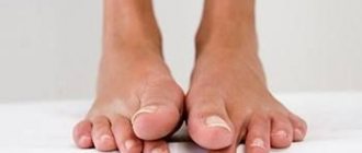
Seborrhea develops in the so-called seborrheic zones - areas of the skin in which there are especially many sebaceous glands, about 400-600 per 1 cm2 1. These include:
- scalp;
- T-shaped area of the face (forehead, nasolabial folds, chin);
- the central part of the chest and the upper part of the back between the shoulder blades.
Seborrheic dermatitis is a chronic inflammatory disease that develops when the yeast-like fungus Malassezia furfur begins to actively multiply in the affected areas. About 3% of the population suffers from manifestations of this disease, and young people are more susceptible to them, among whom the number of patients increases to 5%.2
The disease is considered quite common. It manifests itself especially often in the first years of a child’s life, as well as in people after 40 years of age. This age-related feature of the occurrence of seborrheic dermatitis is explained by the fact that it is during these periods of a person’s life that the sebaceous glands on the body work with high intensity.
Causes of occurrence and development
The development of seborrhea and seborrheic dermatitis is based on dysfunction of the sebaceous glands of the skin, which manifests itself in the fact that the glands begin to produce either too little secretion (sebum) or too much, in addition, the composition and consistency of the secretion also changes. Excess secretions are mixed with dust, dead skin cells and can serve as a substrate for the active reproduction of opportunistic microorganisms, most often fungi of the genus Malassezia, which are normally present on the skin of 90% of the population3. The metabolic products of this pathogen cause skin irritation and its inflammatory reaction, manifested in the form of redness and the appearance of papules covered with greasy crusts.
Why does the functioning of the sebaceous glands malfunction? Doctors identify many factors that can play a role in dysfunction of the sebaceous glands. Among them:
- genetic predisposition;
- changes in hormonal levels (increased levels of progesterone and androgens, decreased levels of estrogen - therefore, the disorder is often found in men), including in infancy, puberty, pregnancy, and chronic diseases;
- decreased immunity due to chronic or infectious diseases, taking potent medications;
- a lot others.
The ducts of the sebaceous glands open into the mouth of the hair follicle, so with seborrhea and seborrheic dermatitis, not only the skin, but also the patient’s hair suffers.
The problem may worsen as a result of prolonged exposure to heat or due to increased sensitivity of the skin to external irritants. At the same time, seborrheic dermatitis in children and adults can be severe, regardless of the number of active fungi on the skin.
Causes of seborrhea on the face
- hormonal imbalance in the body during the transition period in adolescents;
- unbalanced diet;
- disruption of the gastrointestinal tract;
- diabetes;
- thyroid diseases;
- chronic infectious diseases;
- avitaminosis;
- stress;
- weakened immunity.
Men are more susceptible to seborrhea on the face due to the presence of androgens in their blood - hormones that stimulate the sebaceous glands. An excess of these hormones leads to a high level of sebum secretion by the skin.
How does seborrheic dermatitis manifest?
The disease affects certain parts of the human body. Typically this is:
- scalp skin;
- face;
- behind-the-ear areas;
- upper body (most often the area of the chest, shoulder blades);
- large skin folds on the body (for example, groin area, armpits).
Seborrheic dermatitis in adults and children manifests itself in the form of local redness and papules covered with greasy scales.
The disease can also be recognized at an early stage in adults by the appearance of a sign such as dandruff. As the situation worsens, the scalp gradually begins to become covered with red spots; later they can merge, forming lesions with characteristic peeling of the skin. In advanced cases, when oily crusts peel away from the surface of the skin, a moist secretion is discovered underneath. If you do not take any measures to treat seborrheic dermatitis, then this problem may be complicated by the addition of an infection.
In addition to external signs of seborrheic dermatitis, patients often complain of itchy skin. As a result of constant scratching of the inflammation, even greater tissue injury occurs with a high risk of further infection.
The disease not only causes serious discomfort, but also worsens a person’s quality of life as a whole, provokes self-doubt, makes it difficult to establish contacts with other people, and prevents them from building close relationships with the opposite sex.
Symptoms of seborrhea
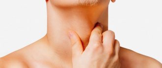
A characteristic manifestation of seborrhea is peeling and inflammation of the skin, accompanied by itching. This process can affect the scalp, the growth boundaries of eyelashes, eyebrows, and mustaches.
With mild seborrhea, small white scales appear on the scalp, dandruff appears in the hair, and patients complain of severe discomfort.
With severe seborrhea, the disease progresses, the skin begins to peel off more and more. The affected areas include the forehead and skin behind the ears. Red spots and plaques covered with scales appear on the skin, which then turn into bloody crusts. The patient begins to experience severe itching that does not stop day or night. If the disease is not treated, plaques appear on the face, back and chest, complicated by a bacterial infection.
Symptoms and forms/Types of seborrheic dermatitis
If we consider seborrhea and seborrheic dermatitis in children and adults, we can distinguish the following varieties:
- Dry seborrhea. With this nature of the disease, the sebaceous glands produce an insufficient amount of secretion. As a result, the skin does not receive enough moisturizing components, dries out, and microcracks appear on its surface. Typically, dry seborrhea occurs without the formation of acute inflammatory foci. Instead, the patient is concerned about the separation of dry dandruff particles. Most often, this form of the disease is observed in adolescent children.
- Oily seborrhea. The form differs from the previous one in that the sebaceous glands, on the contrary, work too actively and secrete a large amount of thick secretion. The substance glues together dead skin cells and is deposited on the hair, causing it to take on an unkempt appearance. The problem often affects young women.
- Combined type. It is characterized by the presence of signs of both dry and oily seborrhea. The disease develops in various parts of the body and therefore causes serious discomfort. In most cases, this form is diagnosed in males.
Depending on the location of the foci of inflammation, seborrheic dermatitis of the scalp, face and torso is distinguished.
The first sign of damage to the scalp is the appearance of dandruff. In this case, the scalp can be either excessively dry or oily, and the scales can range from silvery-white in mild cases to dense fatty crusts in severe cases.
In patients with seborrheic dermatitis of the scalp, over time, the hair becomes sparse, thin, brittle and often falls out.
Focal inflammation on the face is considered no less common. This form of the disease mainly affects teenagers and young people under the age of 25. The cause of seborrheic dermatitis in this case is the high activity of the sebaceous glands in the facial area. During puberty, they begin to work more actively, which can provoke the development of the disease. On the face, rashes are often symmetrical, located in the forehead, nasolabial folds, wings of the nose, and eyebrows.
At the initial stage, the disease manifests itself in limited areas of inflammation, the surface of which is covered with white-yellow scales. The rash is accompanied by itching and constantly peels off. As the disease progresses, areas of inflammation may merge. In some cases, inflammation is aggravated by the addition of a secondary infection.
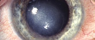
Sometimes the inflammation spreads to the skin of the eyelids. This leads to the development of seborrheic blepharitis. It is characterized by the formation of severe swelling of the eyelids, redness of the eyes, and the formation of cracks in the delicate skin in this area. At the eyelash growth line, inflammatory foci gradually begin to form with a characteristic separation of small scales and crusts. Because of this, patients have to literally “pry” their eyes out in the morning, since the eyelids stick together so much.
On the torso, seborrheic dermatitis is usually localized in the area of the sternum and shoulder blades. The disease occurs with the formation of foci of inflammation with clear boundaries, covered with yellowish scales.
The severity of seborrheic dermatitis varies from mild to severe and depends on the extent of skin lesions, severity of inflammation and intensity of itching.
Infantile seborrheic dermatitis is one of the first skin diseases that a child encounters. It develops against the background of immature functions of the skin and sebaceous glands in infants and manifests itself in the form of fatty yellow crusts covering the head (the so-called “baby cap”), sometimes the area of the eyebrows and cheeks.
Introduction, history and epidemiology
Seborrheic dermatitis (SD) is a common, chronic inflammatory disease that affects approximately 1-3% of the general population in the United States, 3-5% of young patients.
The prevalence of diabetes in HIV-infected people ranges from 20-83%. The incidence has 2 peaks: one in newborns up to 3 months of age, and the other in adults 30-60 years old. It is assumed that these peaks are related to sex hormones. Men are affected more often than women in all age groups and there is no preference for any ethnic group. Malassez was the first to describe the yeast-like fungal elements found in scalp dandruff. In 1952, Leone associated the fungus Pityrosporum Ovale (later called Malassezia
SPP.) with pityriasis of the scalp, seborrheic eczema and various other dermatoses.
In the 50s of the 20th century, the focus of researchers on diabetes was to study its relationship with vitamin B2, B6, B12 and biotin deficiency. However, to date, the relationship between diabetes and nutritional deficiencies has not yet been confirmed. Recent studies have shown the importance of Malassezia
SPP. , present in normal human skin flora, in the occurrence of diabetes? The main argument in favor of this is that diabetes can be treated with antifungal drugs.
In addition to HIV, seborrheic dermatitis often develops in some neurological diseases, such as Parkinson's disease, which improves with treatment with levodopa. The high incidence of diabetes in patients with Parkinson's disease is associated with an increase in the secretion of male sex hormones and the effect of these hormones on the sebaceous glands, and not with autonomic dysfunction (Dysautonomia), as previously thought. A high prevalence of diabetes has also been found in cases of antipsychotic-induced parkinsonism, craniosynostosis, familial amyloid polyneuropathy, traumatic brain injury, traumatic spinal cord injury, cerebrovascular accidents, epilepsy, and facial palsy. DM exclusively on the affected side in patients with paralysis has also been described in patients with stroke, after Chiari decompression type-1 malformations or in skin areas affected by syringomyelia. In 1996, Erci? et al. reported that 30.9% of patients with Down syndrome had diabetes. However, Daneshpazhooh et al. reported the prevalence of diabetes in only 3%. Other systemic diseases in which the incidence of diabetes is higher include acute myocardial infarction, alcoholic pancreatitis, and alcoholism.
Etiopathogenesis
Malassezia
SPP.
Malassezia
SPP.
is a lipophilic fungus that is part of the flora commonly found on human skin. It was first described in the mid-1840s by Eichsted and Sluyter, who associated it with pityriasis versicolor. In 1853 Robin named it Microsporum furfur and in 1874 Malassez described this fungus in dandruff samples taken from the scalp. The genus Malassezia
was first described by Baillon in 1889 and has taxonomic priority over the genus Pityrosporum, used by Sabouraud in 1904 to designate the same group of microorganisms. The current classification includes the genus
Malassezia
, which belongs to the family Cryptococcaceae of the class Basidiomycetes.
Malassezia
is a dimorphic fungus that is highly pleomorphic.
Its original name was based on morphological criteria: Pityrosporum Ovale (oval yeast cells) and Pityrosporum orbiculare (round yeast cells). It was later determined that both forms were morphological variants of the same species. Currently, after evaluation using serological and genetic methods, the genus Malassezia
has been divided into 7 species: M. furfur, M. pachydermatis, M. sympodialis, M. globosa, M. obtusa, M. restricta and M. Slooffiae.
Although Unna was the first to describe diabetes as a disease in 1887, Malassez first observed the fungus in dandruff taken from the scalp in 1874. Later Moore and Kile tied Malassezia
SPP. directly from the SD.
Malassezia
SPP.
associated with both infectious diseases, in which the microorganism is a direct etiological agent, and with inflammatory diseases of multifactorial etiology, in which there is an excessive amount of Malassezia
SPP.
appears as a potentiating or aggravating factor in susceptible patients. The first group includes pityriasis versicolor, Malassezia
(Pityrosporum) folliculitis, pneumonia caused by
Malassezia
spp. , sepsis associated with deep venous catheterization in patients receiving parenteral nutrition, and peritonitis in individuals receiving peritoneal dialysis. The second group includes confluent and reticular papillomatosis (Gougerot-Carteaud syndrome), psoriasis and atopic dermatitis.
Each of these diseases has different clinical and histological manifestations that cannot be explained by the mere presence of a particular Malassezia
on the skin. A number of contributing factors play a role, including immunological individuality and genetic profile.
The exact pathophysiology of DM has not yet been fully established. However, the association of diabetes with the presence of Malassezia
SPP.
on the skin of affected individuals. The fungus is known to be present on all human skin, but may be present to a greater extent in individuals with diabetes. However, in 1989, Bergbrant and Faergman were unable to find any difference in the rate of Malassezia
SPP colonization between individuals with DM and healthy individuals or between DM-affected skin and healthy skin.
These results indicate that there are other pathophysiological mechanisms associated with abnormal responses to Malassezia
SPP.
and that they are not necessarily related to its significance. In 1984, Bourlond et al. showed that P. ovale (i.e. Malassezia
spp.) can be found on any scaly surface (DM, actinic keratosis, nevus and viral warts).
Various studies have been conducted to determine the most common Malassezia
in DM. Nakabayashi et al reported that they found 35% of M. furfur and 22% of M. globosa in individuals with diabetes. Rendic et al found M. globosa in 67%, followed by M. furfur and sympodialis. Gupta and Gaitanis reported more M. globosa. Tajima was the only author to report M. restricta as the most common in diabetes.
Nakabayashi and Sei found that children with DS had more Malassezia
in the skin, which is not a determining factor for the inflammatory response, for which the amount of lipids on the surface of the skin and the human immune response to the presence of the fungus are more important. These researchers also studied the relationship between diabetes and heredity, seasonality, mental stress and decreased T-cell function.
To support the evidence that Malassezia
spp.
contributes to skin inflammation in diabetes, Plotkin in 1996 and De Angelis in 2007 described the production of lipase from these fungi, which is important for its growth in vitro and in vivo. Based on this, it was hypothesized that the fungus uses lipids from the surface of the skin to produce unsaturated and saturated fatty acids that cause an inflammatory response in the skin. Sebum promotes the growth of P. ovale (i.e. Malassezia
) and therefore the development of DM.
Thus, the maintenance of reservoirs of residual sebum (for example, due to poor hygiene) may predispose to the onset of disease, as occurs in patients with neuropathology. The fact that DM responds to treatment with antifungal drugs provides concrete evidence of a link between Malassezia
and DM.
In 2007, Dawson states that the development of DM depends on three factors: sebum production, Malassezia
, and the individual's susceptibility.
Histopathology
The histopathology of DM depends on the clinical stage of the disease. In the acute and subacute phase, the inflammatory infiltrate, consisting mainly of lymphocytes and histiocytes, is accompanied by mild to moderate spongiosis and psoriasiform hyperplasia associated with parakeratosis around the follicular ostia. On the other hand, in the chronic phase, in addition to the above findings, there is marked psoriasiform hyperplasia with dilation of the capillaries and venules of the superficial plexus, making it very similar to psoriasis.
Immunogenetics
Faergemann described an increase in the number of natural killer (NK) T cells, as well as low titers of IgG antibodies in patients with diabetes compared to controls. When exposed to Malassezia
SPP.
The activity of lymphocytes in individuals with diabetes decreases and there is a decrease in the production of IL-2 and IFN-γ and an increase in the production of IL-10. In a subsequent study, the same author reported an increase in the number of NK1 + and CD16 + cells associated with complement activation in diabetes compared with the skin of unaffected areas in the same patients or on the skin of individuals without diabetes, indicating the presence of an intense, irritative, non-allergic immune response. In 1988, Parry and Shape failed to detect either circulating antibodies against the fungus or an increase in systemic susceptibility, leading to the conclusion that there is no change in the humoral response to Malassezia
spp. and changes in the cellular immune response are possible. Neuber et al also report changes in cellular immunity in diabetes.
Watanabe et al. showed that M. furfur does not result in the production of keratinocyte cytokines, whereas this occurs with other Malassezia .
In addition, these researchers reported that depending on the
Malassezia
, the production of pro-inflammatory interleukins of a specific profile is stimulated, thus characterizing various diseases. For example, the production of IL-8 causes neutrophil chemotaxis, which is clinically manifested as
Malassezia
folliculitis. Moreover, in the absence of production of monocyte chemotaxis protein-1 (MCP-1) is clinically determined by diabetes.
Malassezia
was found in patients with diabetes .
This observation was significant in patients with atopic dermatitis and suggests the absence of anti- Malassezia
SPP. humoral immune response in diabetes. Passi et al found decreased levels of serum vitamin E, polyunsaturated fatty acids, and erythrocyte glutathione peroxidase activity in patients with diabetes (both HIV-infected and HIV-negative individuals) and proposed to link these findings to the pathogenesis of the disease. In 2007, Ianosi described a decrease in CD20+ and an increase in CD45RO in the inflammatory infiltrate of diabetes.
Human histocompatibility gene system (HLA)
Although there is evidence that heredity is a predisposing factor for DM, it was not until 1976 that Tsuji published a description of HLA typing in patients with seborrheic dermatitis, in which he found an increased frequency of HLA-AW30 and/or AW31 and HLA-B12 in seborrheic dermatitis.
Clinical diagnosis
Diabetes has different characteristics depending on the age group of those affected: it is a pediatric form of self-limitation, while in adults the disease is chronic. The lesions consist of erythematous scaly plaques of varying size and intensity. In infancy, DM is more common during the first 3 months of life (10% in boys and 9.5% in girls), with scaling of the scalp being the most common clinical manifestation (42%). It is characterized by yellowish lesions that appear shortly after birth. They can also develop on the face and body, in the area behind the ears, neck, armpits and groin area. A child with diabetes may suffer from a rare, generalized form, which is often associated with immunodeficiency. In adults, DM is a chronic, relapsing dermatosis that can vary from mild to moderate erythema with papular and exudative lesions with periods of exacerbation. The most commonly affected areas are the face (87.7%), scalp (70.3%), chest (26.8%), lower extremities (2.3%), upper extremities (1.3%) and other areas (5.4%). ), such as large folds. The lesions consist of papules or thin scales with well-defined borders that may be pink, light yellow or erythematous, with small, dry white or even moist or oily yellowish lesions. They may be limited to small areas of the body. However, there are reports of common forms and even erythroderma. The presence of itching is not a constant sign. The main factor complicating diabetes is the addition of a secondary bacterial infection, which increases erythema and exudation, causing local discomfort and lymphadenopathy near the lesions. Lesions develop mainly in areas where sebum production is high, such as the scalp, face, ears, postauricular folds and areas of the sternum, eyelids and body folds. Lesions on the scalp range from mild flaking to honey-colored crusts firmly attached to the scalp and hair. Involvement of the malar areas, nasolabial folds and eyebrows is typical. Involvement of the eyelids leads to blepharitis. In men, the beard area is affected. From the folds, the armpits, navel, inguinal, inframammary and anogenital areas are affected, where there may be a wet surface, cracks develop, which later become the entrance gate for secondary infection.
Differential diagnosis
DM is differentiated from psoriasis, atopic dermatitis (mainly in the childhood form of DM), ringworm, cutaneous lymphoma and cutaneous Langerhans histiocytosis. There is also a type of dermatitis that is similar to DM and occurs with the use of drugs (gold-containing drugs, buspirone, aminazine, ethionamide, griseofulvin, haloperidol, IL-2, interferon-α, lithium drugs, methoxalene, methyldopa, phenothiazines, psoralens and stanozolol and others) or nutritional deficiencies (riboflavin, pyridoxine, niacin and zinc deficiencies). Childhood DM is similar to atopic dermatitis (AD). However, the affected areas (body folds in DM and extensor surfaces in AD) and the absence of itching in DM make it possible to distinguish these dermatoses. Diaper dermatitis does not affect the folds of the body, while for diabetes they are favorite. Pediatric psoriasis is very similar to DM in this age group, and it is almost impossible to differentiate between the two dermatoses. There is a debate about the difference between SD of the scalp and a disorder called simple pityriasis capillitii, with mild, dry flaking of the scalp, which may only represent a physiological change in the stratum corneum or may be a consequence of excessive use of cosmetics such as hair creams or gels. In addition, it is difficult to differentiate between DM and scalp psoriasis. Psoriasis is more characterized by thick plaques with dry white crusty scales. At first glance, skin lesions in diabetes are similar to acute cutaneous lupus erythematosus (bilateral malar rashes) and rosacea. DM in body folds must be differentiated from primary contact dermatitis, from inverse psoriasis, dermatophytosis and erythrasma. Langerhans histiocytosis can affect the body folds and scalp, resulting in a clinical picture that is very similar to DM. However, the presence of a purple component in the lesions makes it possible to distinguish histiocytosis.
Seborrheic dermatitis and HIV
Eisenstat first described the association of DM and acquired immunodeficiency syndrome (AIDS) in 1984. The prevalence of DM in HIV-infected individuals varies among different authors. In Soeprono et al., 85% of AIDS patients had diabetes. Berger has 36%, Goodman 32%, and Blanes et al. 31%. No increase in Malassezia
SPP in AIDS patients compared with immunocompetent individuals.
Malassezia
to be more important in AIDS than the degree of presence of the fungus on the skin.
Rinkon et al. The predominance of Malassezia
globosa was reported in individuals with lichen versicolor (67%) and in HIV-infected patients with diabetes (85%), while in HIV-negative patients with diabetes, the predominant
Malassezia
were M. furfur and M. restricta. found in 72% and 26% of cases, respectively. Vidal et al. studied the differences in the lipid profile of the skin of individuals with AIDS from individuals without AIDS. However, these researchers did not determine its relationship with the development of diabetes. Passi et al. (1991) reported that total skin surface lipid concentrations were similar in HIV-positive and HIV-negative patients with diabetes. However, they reported significant changes in the lipid fractions of HIV-infected patients, including a decrease in squalene and an increase in cholesterol and cholesteryl esters. DM usually develops in HIV-infected individuals with CD4+ T-cell counts of 200–500 and is considered an early cutaneous manifestation of AIDS. Response to antiretroviral therapy is variable and there are conflicting reports in the literature: some investigators have reported improvement in diabetes after initiation of antiretroviral therapy, while others have reported exacerbation. The histopathology of DM in HIV-positive patients is similar to that in HIV-negative patients. However, there is greater involvement of follicles in the process and a larger number of plasma cells in inflammatory infiltrates in HIV-positive patients. The first appearance of diabetes or its exacerbation in HIV-infected people who previously had a mild form of the disease may mean seroconversion from the latent phase to the symptomatic stage. DM in AIDS may also be more extensive, more intense, and resistant to conventional treatment. Use oral antifungals or combination topical medications with antifungals and corticosteroids. There have also been reports of clinical improvement with the use of antiretroviral drugs.
Treatment
Because it is a chronic inflammatory disease that likely occurs in response to the presence of a fungus ( Malassezia
SPP.) and its metabolites through the use of skin lipids, the purpose of the treatment is to control inflammation, microorganism proliferation and skin oiliness. Various classes of drugs are used. The therapeutic arsenal for the treatment of diabetes is very extensive. The first rule is to inform patients about the chronic, relapsing nature of the disease. Such a patient, who knows about the course of the disease, treats treatment with greater confidence and responsibility. According to a survey by Peyri et al., the most commonly used drugs were corticosteroids (59.9%) and imidazole drugs (35.1%). Moisturizers were also reported in 30.7% of cases, topical calcineurin inhibitors in 27.2% and other pharmacological treatments such as systemic antihistamines and various natural treatments in 5.1%. Soaps: include those containing ketoconazole 2% and sulfur with or without salicylic acid. Oil-based soaps with Tea Tree (Melaleuca alternifolia) have proven effective against diabetes due to their antifungal potential. They are also available in the form of shampoos. Shampoos: Shampoos used in diabetes are classified according to their effect. Antiproliferative: Shampoos based on coal tar and its derivatives have an antimitotic and cytostatic effect, causing a decrease in cell division in the epidermis, which is the cause of the spread of lesions. Other examples are shampoos with selenium sulfate (1 and 2.5%) and zinc pyrithione (1 and 2%). Antifungal shampoos include shampoos based on ketoconazole 2%, ciclopirox 1%, as well as selenium sulfide and zinc pyrithione. With a keratolytic effect: based on salicylic acid (2-6%) with or without sulfur (2-5%), they help remove crusty scales. Anti-inflammatory drugs: containing corticosteroids (clobetasol propionate). Combining different classes of drugs in one product or using alternate therapy are the most effective regimens in reducing the number of relapses.
Topical drugs
Topical antifungals: ketoconazole, as well as other imidazole derivatives, and antifungal agents from other pharmacological classes, such as ciclopirox, can be used in the form of lotions, creams or ointments for recurrent diabetes. Although the anti-androgenic effect of these drugs remains a matter of debate, the dose required for this effect is very high (equivalent to 600-800 mg/day of oral ketoconazole) and is unlikely to be achieved with topical treatment. On the other hand, there is some evidence that antifungals have anti-inflammatory effects comparable to those of hydrocortisone. For example, ciclopirox olamine has been shown to inhibit the action of 5-lipoxygenase and cyclooxygenase in vitro.
Topical corticosteroids: These can be used in the form of lotions, hair tonics, shampoos and foams. They lead to rapid improvement of symptoms (erythema, desquamation and itching). However, relapses are common. They should be used for the shortest possible time due to possible side effects that occur with long-term use.
Anti-inflammatory calcineurin inhibitors - tacrolimus 0.03 and 0.1% and pimecrolimus 1%. They are an alternative to topical corticosteroids. These are tacrolimus and pimecrolimus, which have an anti-inflammatory effect in diabetes that is equal to or greater than that of low-potency TGCS, without the side effects inherent in TGCS. They can be used 1-2 times a day. They are well tolerated and can be used for resistant forms of facial diabetes. They also cause longer-lasting remission than TGCS.
Other therapeutic options described in the literature include:
- Metronidazole 1% gel is commonly used to treat rosacea;
- tacalcitol (1,24-(R)-dihydroxyvitamin D3) cream, a drug used to treat psoriasis;
- topical lithium succinate, which has an anti-inflammatory effect;
- benzoyl peroxide, a bactericidal drug used to treat inflammatory acne.
Orally administered drugs can also be used, mainly for extensive and resistant cases of diabetes. The following antifungal agents are indicated:
- Ketoconazole 200 mg/day for 14 days
- Itraconazole 100 mg/day for 21 days
- Terbinafine 250 mg/day for 4 weeks.
Several years ago, the use of isotretinoin as a seborrhea regulator was reported and for this reason its use was proposed in diabetes. The study used very low doses of the drug (2.5 mg 3 times a week and up to 5 mg/day) with good efficacy. A case of sebaceous hyperplasia with a good response to isotretinoin has recently been reported. However, scientific studies on the use of isotretinoin in diabetes are becoming rarer. In addition, there has been a report of facial rash similar to SD in individuals using isotretinoin to treat acne.
Treatment of seborrheic dermatitis: basic approaches and methods
The tactics and treatment regimen for seborrheic skin dermatitis are selected by the doctor based on the form in which the disease occurs and in which area the inflammation is localized.
If the disease is mild, then this form is characterized by the presence of a small amount of dry or greasy scales, moderate redness of the skin, and occasional mild itching. With this course of the disease, the doctor, as a rule, limits himself to external therapy. For example, with mild damage to the scalp, or dandruff, the doctor may recommend the use of Skin-cap shampoo. Apply shampoo to damp hair, then massage the scalp until foam forms and rinse thoroughly. After this, you need to reapply the shampoo, leave for 5 minutes, and rinse thoroughly with plenty of water. Skin-cap shampoo can be used both for exacerbation of seborrheic dermatitis according to the scheme 2-3 times a week, and for the prevention of further relapses 1-2 times a week.
When the disease occurs in moderate to severe form, the symptoms of seborrheic dermatitis become more pronounced. At this stage, brighter red spots appear on the skin, sometimes noticeable swelling forms on the skin, and the surface is covered with dense gray-yellow crusts. All this occurs against the background of severe itching, which is present almost constantly. Due to discomfort, the patient cannot work fully, it is difficult for him to concentrate on anything, and often in the acute phase of the disease a person experiences problems with sleep.
In this case, there are also generally accepted recommendations on how to treat seborrheic dermatitis. The measures taken are comprehensive; patients may be prescribed hormonal medications. Treatment of seborrheic dermatitis is prescribed by a doctor, based on the severity of the disease and the individual characteristics of the patient. One of the non-hormonal drugs for the treatment of seborrheic dermatitis is the drug Skin-cap. It inhibits the activity of the Malassezia fungus and helps reduce signs of inflammation and also helps reduce flaking. Depending on the location of the inflammatory foci, Skin-cap shampoo, cream or aerosol may be prescribed. Skin-cap aerosol, for example, is suitable for treating the scalp, and the cream is convenient to apply to the face. The duration of treatment for an exacerbation varies, usually it takes from two weeks to 1 month to completely eliminate the symptoms of the disease.
The use of shampoo, cream and aerosol Skin-cap helps to significantly alleviate the condition of seborrheic dermatitis. The active ingredient of the drug Skin-cap has the following effects:
- provides anti-inflammatory effect;
- suppresses the growth and reproduction of pathogenic fungi, which are the main cause of exacerbation of the disease;
- inhibits the activity of bacteria that can increase inflammation.
The drug can be recommended to patients with any form of seborrheic dermatitis; treatment is possible for both adults and children over the age of one year. Products from the Skin-Cap line do not just eliminate the signs of the disease, they help keep the disease under control, preventing relapses.
Symptoms of seborrheic dermatitis on the face
With seborrheic dermatitis on the face, symptoms appear:
- the appearance of pink, red or burgundy spots on the skin;
- the formation of white or yellowish scales and hemorrhagic crusts;
- peeling of the skin;
- irritation and increased sensitivity of the skin;
- itching and burning of varying intensity.
The course of the disease worsens in the autumn. In summer, unpleasant symptoms usually disappear.
If the ducts of the sebaceous glands become clogged, acne appears.
The disease is especially severe in men who grow beards and mustaches. Large plaques form under the hairline, which eventually transform into papules. If an infection occurs, then erythroderma is also detected. When papules coalesce, swollen areas colored red form.
The symptoms of seborrheic dermatitis are similar to those of other dermatological diseases. Therefore, only a doctor can make a correct diagnosis after a thorough examination of the patient.
Features of SKIN-CAP release forms
The cream is quickly absorbed and softens and moisturizes the skin well and does not leave greasy marks on it. It is suitable for facial skin and affected surfaces that require moisturization.
The aerosol can be applied even to the scalp using a special nozzle, which is included in the kit, without touching the hair. It helps eliminate itching and dries out wet rashes.
Seborrhea shampoo effectively7 fights its manifestations on the scalp. Actively helps eliminate dandruff and itching, fights the root cause of the disease - pathogenic microorganisms. Ideal for preventing relapses.
Skin-cap shower gel is a cosmetic product that is indispensable for daily hygiene in cases where seborrhea affects the skin of the chest and back. It cleanses, soothes and moisturizes the skin, helps eliminate inflammation and restores the protective properties of the skin.
Prevention of seborrheic dermatitis
Patients with this disease need to know not only how to treat seborrheic dermatitis, but also learn the basic rules for preventing exacerbation of the disease. The main task in this case is to exclude those factors that can trigger the triggering of pathology development mechanisms.
Thus, as part of the prevention of seborrheic dermatitis, it is worth paying attention to several aspects:
- following a certain diet;
- compliance with personal hygiene rules;
- quality skin care;
- maintaining an active and healthy lifestyle;
- timely treatment of concomitant diseases.
Seborrheic dermatitis on the face photo
Photo 1. Seborrheic dermatitis on the face of an adult
Photo 2. Seborrheic dermatitis on the face of an adult
Photo 3. Seborrheic dermatitis
Photo 4. Seborrheic dermatitis on a child’s face
Diet for dermatitis
The diet for seborrheic dermatitis should be based on healthy foods containing a small amount of fat. These include a variety of cereals, lean meats and fish, and always fresh vegetables and fruits.
It is important not only to eat well and properly, but also to follow some recommendations for cooking. Thus, the most preferred options for heat treatment of products are boiling, baking, steaming or grilling without adding oil.
How to prevent seborrhea
To prevent seborrhea you need:
- strengthen your immunity;
- eat properly and balanced;
- observe the rules of personal hygiene;
- take vitamins “A” and “B”;
- spend more time walking outdoors and being in the sun;
- reduce consumption of sweet, fatty and spicy foods;
- pay great attention to the treatment of the gastrointestinal tract;
- strengthen your immunity;
- choose the right shampoos and creams for your skin type.
Maintaining personal hygiene
Patients with seborrheic dermatitis are recommended to injure the inflammation as little as possible. Personal hygiene rules include cleansing the skin twice a day using special products with a low pH. To cleanse the skin, it is prohibited to use aggressive agents such as laundry or tar soap.
General recommendations for maintaining personal hygiene:
- the use of washcloths or sponges with hard fibers is excluded;
- For a while, you should avoid cleansing your skin with scrubs and peels containing abrasive particles and aggressive chemical compounds;
- During water procedures, it is recommended to use dechlorinated water; for this purpose, it is recommended to first settle or filter it;
- the duration of water procedures should be reduced to 10-15 minutes;
- when choosing shower products, choose products without dyes and fragrances, with a low pH;
- use towels with soft bristles, and it is better to blot your skin with them rather than rub them;
- to reduce the likelihood of skin injury when combing, try to cut your nails as short as possible;
- Avoid using other people's personal hygiene items.
Diagnosis and treatment of seborrhea
To effectively treat the disease, correct diagnosis is necessary. A dermatologist may recommend that the patient undergo the following tests:
- blood chemistry;
- sebometry;
- ultrasound examination of the thyroid gland;
- Ultrasound examination of the abdominal organs.
The MedicCity clinic employs doctors of the highest category who know how to help you! Diagnostics and treatment are carried out using modern equipment. In addition, neurologists will help you overcome depression, and endocrinologists and nutritionists will select proper and balanced nutrition.
Skin care rules
Skin care prone to symptoms of seborrheic dermatitis is primarily focused on normalizing the functions of the sebaceous glands. It is important not only to reduce sebum production, but also to ensure skin hydration.
General care recommendations:
- Try to reduce the use of comedogenic makeup, as it can cause clogged pores.
- Use care products that do not contain alcohol. It irritates the skin, dries it out, causing the sebaceous glands to work more actively.
- For care, choose special cosmetic products designed for seborrheic skin.
Treatment of oily and dry seborrhea on the face
In the NEARMEDIC network of clinics, individuals have a comprehensive approach to the treatment of seborrhea. Therapy includes:
- use of special cosmetics to care for problem skin;
- restoring the balance of the body by taking vitamins and minerals;
- diagnosis and treatment of the causes of the disease;
- treatment of complications that arise after seborrhea.
The main goals of treating seborrhea on the face are:
- normalization of the nervous system;
- restoration of the endocrine system;
- activation of metabolic processes.
In each case, our specialists select individual treatment tactics.