Dermatitis is a skin disease that has many causes. Often skin lesions develop against the background of constant exposure to an allergen. This disease is called allergic contact dermatitis. It is very common and occurs as a result of the contact effect of an irritating substance on the epidermis. This dermatitis can appear on any part of the body, but most often affects the hands and face.
There are no statistics on the prevalence of the disease, but it is known that it occurs very often and is also often not properly diagnosed. In most cases, the allergic reaction goes away after contact with the irritant stops. There is evidence that at least 2% of the population suffers from allergic dermatitis. The incidence rate is greatly increased in some chemical industries, and therefore the disease is included in the list of occupational diseases. More and more patients are being registered every year - this may be due to both the spread of new allergens and the improvement of diagnostic methods.
At CELT you can get advice from a gastroenterologist.
- Initial consultation – 3,500
- Repeated consultation – 2,300
Make an appointment
Mechanism of disease development
An allergen is any substance that can cause an allergic reaction in a susceptible person. Almost anything can act as such a substance: from chemical compounds to food products. As a rule, at the first contact it takes at least two weeks for a specific reaction to occur. And it can be difficult for patients to associate the first symptoms of the disease with any specific irritant.
If sensitivity has already manifested itself, it means that the reaction occurs more and more quickly, and the symptoms increase. This phenomenon is called sensitization: the more and longer the allergen affects the body, the faster and more pronounced the response appears. In case of repeated contact, the rate of development of symptoms is reduced from several hours to three days.
In order for this disease to begin to develop, prolonged contact of the allergen with open skin is required. Currently, scientists name several thousand substances that can cause allergic dermatitis. And their number is increasing every year.
How to care for skin with dermatitis?
Helping herbs
Many plants have an amazing property - when absorbed into the skin, they reduce inflammation and normalize its structure. Therefore, a common remedy for the treatment of dermatitis is herbal compresses, wraps, and herbal ointments for dermatitis.
You can use a single plant or herbal mixtures that contain several herbs.
- Grind the celandine and squeeze the juice out of the resulting mass. Dilute the juice with boiled water in a ratio of 1 to 2. Use the solution for compresses: soak gauze in it and apply to the affected skin for 10-15 minutes.
- 1 tbsp. Pour 100 ml of boiling water over a spoonful of dry crushed string and leave until a dark brown solution is obtained. Moisten gauze or bandage in the prepared concentrate and apply it to the irritated skin until completely dry (not with weeping dermatitis, otherwise the bandage may dry to the skin). The procedure can be carried out 3 times a day.
Recipe with tar soap
The healing properties of tar have been known for a very long time. This thick oily liquid has a positive effect on the condition of the skin. Birch tar has an anti-inflammatory and antiseptic effect, improves blood circulation and stimulates skin regeneration.
You can find tar soap on sale and use it to wash skin affected by dermatitis, or prepare soap according to a folk recipe.
- Mix 100 g of crushed and melted baby soap with 2 tbsp. spoons of any oil, add 2 tbsp. fly in the ointment and 100 ml of water. Mix the resulting mixture until smooth.
Causes
The most common causes of the disease are the following allergens:
- Food
- some medications
- pollen and plant sap
- cleaning products (can cause skin irritation even in the absence of an allergic reaction; it is not recommended to use household chemicals without gloves)
- dust
- Pets
- insect saliva
- varnishes, paints
It is important to remember that the causes are not always obvious, so it is important to pinpoint the source before starting treatment.
Atopic dermatitis: issues of etiology, pathogenesis, methods of diagnosis, prevention and treatment
General information
Atopic dermatitis (AD) is a common, persistent dermatosis, occupying 50–60% of the structure of allergic diseases, and this figure is steadily growing (Balabolkin I.I., Grebenyuk V.N., Williams HC et al. 1994) For the first time the term “ “atopic dermatitis” was proposed by Sulzbeger in 1923 for skin lesions accompanied by increased sensitivity to various allergens, manifested by instability of the cell membranes of skin vessels, combination with other atopic diseases (bronchial asthma, hay fever, rhinitis, etc.).
Atopic dermatitis (AD) is a common, persistent dermatosis, occupying 50–60% of the structure of allergic diseases, and this figure is steadily growing (Balabolkin I.I., Grebenyuk V.N., Williams HC et al. 1994) For the first time the term “ “atopic dermatitis” was proposed by Sulzbeger in 1923 for skin lesions accompanied by increased sensitivity to various allergens, manifested by instability of the cell membranes of skin vessels, combination with other atopic diseases (bronchial asthma, hay fever, rhinitis, etc.).
Currently, AD is considered as an independent nosological form
, clearly different from contact allergic dermatitis, microbial and seborrheic eczema, limited neurodermatitis. AD occurs most often in early childhood against the background of exudative diathesis, eczematous process, often with complicated heredity due to poor nutrition, intoxication, metabolic disorders, disorders of the nervous and endocrine systems (hypofunction of the adrenal cortex, gonads, hyperfunction of the thyroid gland), but can also form in adulthood.
Leading signs of atopy
are severe itching, chronic recurrent course, white dermographism, increased levels of IgE in the blood serum, decreased IgM and IgA, a sharp increase in IgG, indirectly indicating delayed-type hyperreactivity (Samsonov V.A. 1985, Suvorova K.N. 1998, Sanford AJ 1995 ). The impact of unfavorable, exogenous (physical, chemical, biological) and endogenous (genetic predisposition, immune disorders) factors aggravate the clinical picture of the disease. However, the etiology remains unclear, the pathogenesis has not been fully studied, and a clear classification has not been developed.
Pathogenesis
Psychosomatic disorders play a certain role in the development of AD. Severe itching, irritability, restless shallow sleep, inappropriate reactions, white dermographism are classic manifestations of psychosomatic pathology. When assessing the psychosomatic status of patients, a high degree of anxiety, the development of reactive depression, and asthenovegetative syndrome were revealed. (Revyakina V.A., Ivanov O.L., Belousova T.A. 2000).
It has been shown that the main substrate in psychoneuroimmune interaction is neuropeptides (substance P, calcitonin gene-like peptide), which ensure the relationship between nerve fibers, mast cells and blood vessels. Under the influence of the “axon reflex,” vasodilation develops, manifested by erythema. Substance P ensures the release of histamine from skin mast cells and has a direct effect on blood vessels, increasing their permeability, which may explain the poor effectiveness, in some cases, of antihistamines. Thus, a direct relationship is visible between the central and autonomic parts of the nervous system. Improvement in psycho-emotional status under the influence of therapy was correlated with the positive dynamics of the skin process. (Ivanov O.L., Belousova T.A. 2000).
Hereditary predisposition in the pathogenesis of atopic dermatitis is confirmed by the high frequency of occurrence of the association of HLA antigens: A3, A9, B7.8, B12, B40. Clinical data also indicate the role of heredity in the transmission of pathological symptoms from parents to children. Thus, from an allergic father, signs of atopy develop in a child in 40–50% of cases, from a mother – in 60–70%. If both parents are carriers of atopy, then the incidence of the disease in the child reaches 80%. (Mazitov L.P. 2001).
Research by Toropova N.P. the possibility of transplantental transfer of ready-made antibodies from mother to fetus and its hypersensitization is shown, this apparently explains the development of allergic reactions to mother's milk in the first months of life. Such mothers are recommended to follow a strict diet, limiting the consumption of nitrogenous extractives, chlorides, and proteins.
A certain number of children develop latent sensitization, which manifests itself in the form of allergic reactions at the age of 19–20 years. It is not the disease that is inherited, but a set of genetic factors that contribute to the formation of an allergic factor in the body (Fedenko E.S. 2001).
The functional state of the gastrointestinal tract is of great importance in the formation of blood pressure. Dysfunction of the gastrin regulation link was revealed, consisting of imperfect parietal digestion, insufficient activity of enzymes in the processing of chyme, accumulation in the lumen of the small intestine of a huge amount of protein allergenic complexes, their free absorption and the creation of preconditions for sensitization and aggressive course of the skin process. (Toropova N.P., Sinyavskaya O.A. 1993).
The risk of developing food allergies increases due to non-compliance with the diet of a pregnant woman, children in the first months of life who are bottle-fed, as well as the use of food additives containing xenobiotics. Thus, in children of the first year of life, chicken eggs, cow's milk proteins, and cereals are common causes of AD. The course of AD is aggravated by the development of dysbiosis, due to the often uncontrolled use of antibiotics, corticosteroids, the presence of foci of chronic infection, allergic diseases (asthma, rhinitis), dysmetabolic nephropathies, and helminth infections. The waste products of the latter activate immunocompetent cells that carry out the synthesis of IgE and immune complexes.
In the development of exacerbation of blood pressure, inhaled allergens play an important role. The possibility of forming complex associations with bacterial, fungal, viral and drug allergens has been shown, causing the formation of polyvalent sensitization (Maksimova A.E. 1997).
According to Fedenko E.S. (2001) a causally significant allergen in the development of exacerbation of blood pressure are nonsteroidal anti-inflammatory drugs, sulfonamides, and B vitamins. We also observed the development of allergic reactions such as toxicoderma, urticaria, to B vitamins in patients with diffuse neurodermatitis, true eczema (Zheltakov M.M., Skripkin Yu.K., Somov B.A., Butov Yu.S. 1969).
Considerable attention has recently been paid to the polygenic type of inheritance, the characteristic features of which are immune disorders at the level of differentiation of the T-lymphocyte subpopulation. It has been established that null T-helpers (Th0) under the influence of antigens differentiate into T-helpers of the first (Th1) or T-helpers of the second type (Th2), which differ from each other in the secretion of cytokines, PGE. The first type controls apoptosis of mutated cells through a-TNF, and g-IFN inhibits the development of viruses. The second type provides protection against bacterial allergens and activates antibody formation due to IL-4, IL-5 and IL-13.
In AD, lymphocyte differentiation proceeds through Th2, activating b-cells and the synthesis of allergic IgE antibodies. The process of sensitization occurs with the participation of mast cells with the release of histamine, serotonin, kinins and other biologically active substances, which corresponds to the early phase of the hyperergic reaction. This is followed by an IgE-dependent late phase, characterized by infiltration of skin T lymphocytes, determining the chronicity of the allergic process.
It has been shown that the development of the inflammatory process in patients with AD occurs in the presence of dendritic cells, Langerhans cells with a constantly high level of eosinophils, IgE, cytokines and mediators. The ability of eosinophils to live long and produce neurotoxins and enzymes in the tissue ensures a chronic process accompanied by severe itching, damage to keratinocytes and an even greater release of cytokines and inflammatory mediators, creating conditions for a “vicious circle”.
Thus, the analysis shows that exogenous (physical, chemical and biological) and endogenous (the role of the nervous system, gastrointestinal tract, genetic predisposition and immune disorders) factors take part in the development of AD.
Clinical aspects of blood pressure
Typical clinical picture of blood pressure
characterized by: itching of the skin, persistent hyperemia or transient erythema, papulovesicular rashes, exudation, dry skin, peeling, excoriation, lichenification, widespread or limited in nature. The disease usually begins in the first months of life, then taking a recurrent course with the possibility of complete or incomplete remission of varying frequency and duration.
Atopic reactions in childhood occur:
- often in the form of acute inflammatory exudative reactions;
- localized on the face, in folds, outer surfaces of the limbs;
- there is a clear relationship with nutritional factors;
- followed by a chronic, wave-like course of inflammation, vegetative dystonia and lichenification.
At subsequent stages, patients develop: - persistent lechinization;
- less significant reactions to allergenic stimuli;
- less clear seasonality.
Possible clinical forms of manifestation:
– erythematous-squamous;
– vesiculocrustosis;
– erythematous-squamous with weak or moderate lichenification in the elbow and popliteal folds;
– lichenoid with a large number of lichenoid papules;
– prurigo-like (Suvorova K.N. 1998).
Based on studies conducted in children with AD, Korotky N.G. identified a number of clinical and pathogenetic options for the development and course of the disease:
1. True, allergic variant
AD with a predominance of a specific IgE-mediated immune mechanism
2. Mixed version of blood pressure
, where both specific and nonspecific mechanisms are expressed.
3. Pseudo-allergic variant
with a predominance of nonspecific mechanisms.
In true, allergic and mixed variants of AD, the severity of the process depends not only on skin damage, which may not always be significant, but also on other organ manifestations of atopy, in particular, bronchial asthma and gastrointestinal pathology. In the pseudoallergic variant of AD, the leading place in the development of the pathological process is given to neurovegetative and microcirculatory disorders.
Diet therapy
Due to severe dysfunction of the gastrointestinal tract, timely and adequately prescribed diet therapy, in most cases, promotes remission of the disease or even complete recovery. The elimination diet is based on the reliably proven sensitizing role of certain foods in the development of exacerbations of blood pressure and their exclusion.
Products containing food additives (dyes, preservatives, emulsifiers), as well as strong meat broths, fried foods, spices, spicy, salted, smoked, canned foods, liver, fish, caviar, eggs are excluded from the diet of patients suffering from AD. , cheeses, coffee, honey, chocolate and citrus fruits.
The diet should include fermented milk products, cereals (oatmeal, buckwheat, pearl barley), boiled vegetables and meat. The developed diets must be optimal in protein and vitamin content and are compiled in close cooperation with an allergist and nutritionist.
Drug therapy
When choosing a systemic drug, the patient’s age, period of illness, and the presence of concomitant diseases are taken into account.
, nutritional and psychotropic drugs are prescribed
.
Among herbal preparations, it is preferable to use tincture of peony, motherwort and valerian root, novo-passit. Antidepressants are also used in therapy. Amitriptyline
is prescribed orally 0.025–0.05 g;
nialamide
orally 0.025–0.01 g. Among tranquilizers,
diazepam
0.005–0.015 g per day,
lorazepam
0.001–0.0025 g per day are used.
Indications for the use of antihistamines
justified by the critical role of histamine in the mechanism of skin itching and the development of inflammation in AD.
Due to the presence of a sedative effect, 1st generation antihistamines are not advisable to prescribe to school-age children. For planned long-term use, it is more rational to choose any 2nd generation antihistamine (loratadine, terfenadine, cetirizine, ebastine). Ebastine (Kestin)
does not cause pronounced anticholinergic and sedative effects; it is prescribed in a daily dose of 10 mg, and if symptoms are severe, the dose can be increased to 20 mg.
Cetirizine
is prescribed in tablets of 0.01 g for 7 days, at a rate of 0.25 mg/kg 1-2 times a day. 2nd generation drugs are currently not used in children under 2 years of age.
Diazolin, chloropyramine, clemastine
It is preferable to use during periods of severe skin itching, for 7–15 days, if not only an antipruritic, but also a sedative effect is required.
Cyproheptadine
has antiserotonin activity, which expands its scope of application. Clemastine from 6 to 12 years, 0.5 – 1.0 mg, over 12 years, 1 mg 2 times a day. Chloropyramine is prescribed to children under 1 year old, 6.25 mg (1/4 tablet), from 1 to 6 years old, 8.3 mg. (1/3 tablet), from 6 to 14 years, 12.5 mg. 2–3 times a day. In therapy, it is often necessary to combine the use of 1st and 2nd generation drugs.
Membrane stabilizing agents
.
From this group, ketotifen
and
sodium cromoglycate
. They stabilize mast cell membranes, antagonize H1-histamine receptors, inhibit the development of the allergic process and can act as a calcium channel blocker. The therapeutic effect appears after 2–4 weeks. Sodium cromoglycate additionally affects the gastrointestinal mucosa, preventing the development of allergic reactions at this level. The drug is prescribed in the acute and subacute period of blood pressure in combination with antihistamines. Children from 1 to 3 years old at a dose of 100 mg (1 capsule) 3-4 times a day; from 4 to 6 years – 100 mg 4 times a day; from 7 to 14 years – 200 mg 4 times a day. The duration of the course of therapy is on average from 1.5 to 6 months.
It is advisable to prescribe drugs that improve digestion
, to correct the breakdown of allergenic food substances (Festal, Mezim-Forte, Hilak-Forte).
The effectiveness of enzyme preparations
, taking into account violations of the enzyme systems of the gastrointestinal tract in patients. (Korotky N.G. 2000). Dysbacteriosis is an indication for the full use of probiotics, which normalize the microbial landscape of the intestine.
of vitamin preparations helps to increase the effectiveness of treatment
. Of the B vitamins, preference is given to calcium pantothenate (B15), which is prescribed 0.05–0.1 g 2 times a day for a month, and pyridoxal phosphate (B6), which is prescribed 0.1–0.2 g per day. day. It is advisable to prescribe b-carotene, it increases the resistance of lysosome and mitochondrial membranes to the action of metabolic toxins, stimulates the immune system and regulates lipid peroxidation.
Immunomodulatory therapy
is carried out in cases where blood pressure occurs in combination with clinical signs of immunological deficiency and the presence of defects in the immunogram. In the form of a decrease in B-cell level, phagocytic cells, an increase in IgE, an imbalance of Th1-Th2 cells. Clinical signs include: the presence of foci of pyogenic infection; frequent exacerbations of the skin process; frequent acute respiratory viral infections with low-grade fever and lymphadenopathy; lack of clinical effect from adequate standard therapy for blood pressure.
The use of systemic
antibiotics
is advisable for low-grade fever and lymphadenitis. With preliminary determination of microflora sensitivity to antibiotics. In empirical therapy, preference is given to the use of macrolides and 2-3rd generation cephalosporins.
Systemic glucocorticosteroids
(GCS) are most often prescribed in particularly severe, persistent cases of blood pressure, used in a hospital setting and in short courses under the cover of antacid drugs (Almagel) and calcium supplements (calcium gluconate, calcium glycerophosphate). Prednisolone and dexamethasone 20–25 mg per day are used; adults are prescribed betamethasone injections. The mechanism of the anti-inflammatory activity of GCS is to block the activity of phospholipase A, inhibit the synthesis of leukotrienes and prostaglandins, reduce the activity of hyaluronidase and lysosomal enzymes, and activate the synthesis of histaminase (Grebenyuk V.N., Balabolkin I.I. 1998).
External therapy
is an integral part of the complex treatment of blood pressure, occupying a leading place in it. With the help of local treatment, a number of effects are achieved: suppression of signs of skin inflammation; eliminating dryness; restoration of damaged epithelium; improving skin barrier functions.
The choice of drug is determined by the stage of the disease, the phase of inflammation and the severity of skin manifestations. In order to achieve success, it is necessary to follow a certain sequence in the administration of local treatment. For acute weeping processes, lotions and dermatological pastes are used. As the inflammation subsides, non-fluorinated corticosteroids are prescribed in the form of a cream or ointment. Ointments have a more pronounced anti-inflammatory effect and are prescribed for the treatment of subacute and chronic skin lesions. Creams are the form of choice for acute processes.
In cases of pyoderma, erythromycin, lincomycin, geoxyzone ointments, and aniline dyes are prescribed. Among other anti-inflammatory drugs that have long been used in the treatment of blood pressure, one should mention products containing tar, naphthalan, and sulfur.
Correction of concomitant diseases includes examination, identification and sanitation of foci of chronic infection, parasitic infestations, dysbacteriosis, restoration of cortical neurodynamics and autonomic disorders.
Forecast
The course of blood pressure and the quality of life of the patient and his family largely depend on the reliable knowledge he has received about the causes of the development of skin rashes, itching, and on the careful implementation of all doctor’s recommendations and prevention.
Main directions of prevention
– this is compliance with the diet, especially for pregnant and nursing mothers, breastfeeding children.
Particular attention should be paid to limiting exposure to inhaled allergens, reducing contact with chemicals in everyday life, preventing colds and infectious diseases and conditional prescription of antibiotics. Literature:
1. Balabolkin I.I., Grebenyuk V.N., Williams HC etal.
2. Vorontsov I.M. Atopic dermatitis in children. M.–p.263.
3. Grebenyuk V.N. Balabolkin I.I. Progress of external corticosteroid therapy for AD//Pediatrics – 1998. No. 5 pp. 88–91.
4. Zheltakov M.M. Skripkin Yu.K. Somov B.A. Butov Yu.S. Allergic reactions caused by B vitamins. VDV 1969, No. 1, p. 62–65
5. Korotky N.G. Pathogenetic role of disturbances in hormonal regulation of cavity digestion and absorption in hypertension and correction with enzyme preparations. Russian Journal of Skin and Venereal Diseases. 2000 – No. 1 – pp. 12–17.
6. Yazdovsky V.V. HLA and allergic diseases. Pulmonology 1994, 4, 6–9.
7. Maksimova A.E. Features of skin microflora in patients with AD. Author. diss. Ph.D. 1997.
8. Mazitov L.P. Modern aspects of pathogenesis and treatment of allergic dermatoses in children. Russian medical journal 2001 v.9. No. 11 p. 457–459.
9. Naumov Yu.N., Kotenkov V.I., Alekseev L.P. Structure of human HLA genes and antigens of classes 1–2./ Immunology 1994,2,4–8.
10. Revyakina V.A. The role of etiologically significant allergens in the development of AD in children // Allergology-1998 No. 4 pp. 13–14.
12. Samsonov V.A. Neurodermatitis and bacterial allergies. Abstract. Doctor of Medical Sciences M. 1984.
13. Skripkin Yu.K. Somov B.A. Butov Yu.S. Allergic dermatoses. M.1975. 234 p.
14. Smirnova G.I. Allergic dermatoses in children//M.BUK, ltd. 1998, p.299.
15. Suvorova K.N. Atopic dermatitis: immunopathogenesis and immunotherapy strategy. Russian honey magazine. 1998, vol. 6, 368–367.
16. Toropova N.P. Sinyavskaya O.A. Eczema and neurodermatitis in children. Ekaterenburg. 1993, 147 p.
17. Fedenko E.S. – Atopic dermatitis: Rationale for a step-by-step approach to therapy. Consilium medicum 2001 vol. 3 no. 4 pp. 176–183.
18. Khaitov R.M., Luss L.V., Aripova T.U. Prevalence of symptoms of asthma, AR and AD in children. /Allergy, asthma and clinical immunology. – 1998. – No. 9. – pp. 58–69.
19. Leung DYM Role of IgE in atopic dermatitis//Curr. Opinion Immunol–1993–Vol.5–P.956.
20. Sanford AJ “Genetic map of cromosome llg, including the atopy locus. Eur Hum Genet 1995, no. 3 p. 188.
21. Casale TB, Bowman S. Induction of human cutaneous mast cell degranulation by opiates peptides//Immunol–1984–Vol.73.
Symptoms
There are two forms of the disease: acute and chronic. The chronic form differs in the frequency of contact with the allergen and the severity of symptoms.
Typical symptoms of the acute form include:
- redness
- edema
- weeping wounds
- bubbles
- inflammation
- intense itching
Symptoms can appear either directly in the contact area or a little further. Their intensity depends on the age of the patient, on the chemical activity of the allergen, and on the duration of contact.
Symptoms of the chronic form:
- skin changes: the skin pattern is more pronounced than in healthy areas
- dry skin, cracks
Since the symptoms are nonspecific, the disease is easily confused with other dermatological pathologies. Therefore, it is often necessary to undergo examination by a dermatologist.
Diagnostics
Diagnosis includes an examination by an allergist and special tests for allergens, which allow you to determine which substance contributes to the development of the disease. The patient is not always able to identify the allergen on his own.
The examination includes a history, physical examination (sometimes dermatitis can be ruled out by objective visual signs indicating the development of another dermatological pathology), and skin tests.
Currently, the most commonly used patch tests are strips of paper that are glued to the skin. Allergens are applied to the paper in a non-hazardous concentration, but sufficient for the development of a local reaction. There are tests in which there are several active ingredients on one plate at once. As a rule, the paper is glued to the skin of the back and left for a day or two. If there is sensitivity to a certain substance, then redness occurs at the site of contact. Since the allergen concentration is small, the reaction goes away after the application is removed. The CELT clinic uses .
Consultation with a specialist is especially relevant if it is not possible to exclude contact with the allergen, since specific desensitization measures are required.
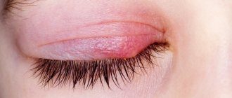
Red dermatitis, also known as lichen planus
This is a chronic itchy skin disease, which is accompanied by the appearance of pink-red-violet rashes, merging into large plaques, up to 10 cm in size, with the formation of garlands and rings.
Locations: the lateral part of the body, the inner surface of the arms in places of flexion, mucous membranes of the mouth and genitals.
There are several forms of damage to the skin and mucous membranes. Manifestations of the disease are very diverse: papules, spots, erosions, blisters, etc.
Our doctors
Orlova Tatyana Vladimirovna
Doctor - allergist-immunologist, pulmonologist, doctor of the highest category
Experience 38 years
Make an appointment
Shundeva Oksana Veniaminovna
Allergist, doctor of the highest category
Experience 39 years
Make an appointment
Treatment
Treatment depends on the severity of symptoms, the form of the disease, and the duration of symptoms. Therapy is prescribed taking into account the patient’s ability to stop contact with the allergen.
The basis of symptomatic treatment is antihistamines to get rid of itching, burning and swelling. From a medical point of view, this group of drugs does not fight the cause, but only the consequences of allergies. Therefore, the course of treatment is usually short - no more than 2-4 weeks. II, III and IV generation drugs are prescribed, focusing on individual experience and effectiveness for a particular patient.
In the presence of severe allergic reactions - inflammation, weeping wounds - local treatment is prescribed. Local remedies include ointments and gels with corticosteroids - they are effective in the chronic course of the disease. The course of treatment is also short: no longer than two weeks. Most often, doctors prefer fluoride-free drugs of the latest generation. Despite their controversial reputation, corticosteroids are safe when used in short courses and are very effective for chronic allergies.
In the most severe cases, oral corticosteroids are prescribed. In this case, the course duration and dosage are selected individually. When prescribing corticosteroids, constant monitoring of intake by the attending physician is required, as well as regular testing.
In treatment, not only medication is important, but normalization of lifestyle and diet is also important. First of all, the patient is required to completely eliminate or minimize contact with the allergen.
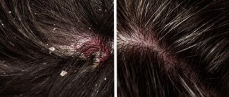
How to distinguish psoriasis from seborrheic dermatitis?
Diseases are often confused. Although both diseases share common features, they also have many differences. Common characteristics include a chronic type of disease and an undulating course, the presence of rashes, itching, and peeling of the skin. What is the difference between psoriasis and dermatitis?
- Prerequisites for development. If psoriasis appears under the influence of autoimmune, endocrine, and genetic disorders, then seborrheic dermatitis is caused by increased activity of the sebaceous glands and yeast-like fungi.
- Symptoms. Despite the fact that in both cases the skin is covered with scales, their structure and appearance are different. When oily yellow crusts appear, we are talking about seborrheic dermatitis, and when dry gray, white or silvery scales form, psoriasis can be diagnosed.
- Localization. Psoriasis can affect almost all areas of the skin, and seborrheic dermatitis is localized mainly on the scalp, neck and face.
If you experience skin rashes that could be a sign of both dermatitis and psoriasis, be sure to consult a dermatologist. Usually, a qualified doctor can determine what it is by the external manifestations of the disease. If mixed symptoms occur, the dermatologist will write a referral for additional examinations.