General information
Elephantiasis is usually called swelling and chronic stagnation of lymph, manifested in the form of deformation and persistent diffuse increase in the size of any part of the human body, most often the limbs and scrotum. Elephantiasis is usually accompanied by painful growth - hyperplasia of the skin and subcutaneous fat.
These changes are fibrotic and irreversible, affecting the skin, subcutaneous tissue and fascia. The pathology most often affects the lower extremities, which become similar to the legs of an elephant, hence the origin of the name of the disease. The causes of elephantiasis can be different, from parasitic agents to congenital defects of the lymphatic system. These include blockage of the lymphatic capillary system and impaired lymph circulation.
What kind of disease is this
Elephantiasis (other names: elephantiasis, elephantiasis or lymphedema) is a pathology in which there is a violation of the outflow of lymph through the lymphatic vessels, which leads to its stagnation, accumulation and subsequent increasing swelling, as a result of which the affected limb increases in volume and in the extreme stage becomes disfigured . Due to stagnation of lymph, metabolic products and toxins accumulate in soft tissues, which causes protein breakdown and the formation of fibrin fibers. Subsequently, rough connective tissue forms at the site of edema, which disrupts muscle function and leads to disability of the patient. Externally, the affected limb looks like a cylinder and resembles an elephant's leg. Cracks, ulcers and warts appear on the skin.
Most often, elephantiasis of the legs develops, and, as a rule, one limb is affected.
Classification
According to the development mechanism there are:
- primary lymphedema (congenital), which is caused by congenital pathology of the lymphatic system, the development of the disease occurs in childhood and adolescence;
- secondary lymphedema (acquired), occurs more often than the primary form of the disease and is caused by previous diseases and injuries/surgeries.
There are three stages in the course of the disease:
- Stage 1 mild edema;
- Stage 2 of dense edema;
- Stage 3 elephantiasis.
Pathogenesis
Elephantiasis begins with slight swelling and swelling, which may subside or reappear. Eventually, after about 1-3 years, there is a two to threefold thickening of the lower leg and possibly the thigh if the leg is affected. The shape of the leg is awkward and cylindrical, turning into thickenings, for example, the deepening of the ankle joint can be smoothed out.
Stages of development of elephantiasis
The mechanism of excessive thickening of the affected parts is based on stagnant processes affecting the lymphatic system. They can arise as a result of factors such as:
- blockage of lymphatic vessels by parasites - nematodes , for example, filaria , chyluria , etc.;
- suppuration and subsequent scarring of the lymph glands, for example, the inguinal glands;
- inflammation of the lymphatic vessels with lymphangitis , phlebitis , erysipelas , varicose ulcers, chronic eczema , syphilis , lupus , with repeated frostbite and other chronic or recurrent processes leading to inflammatory edema.
The resulting stagnation of lymph leads to its penetration into tissue crevices and provides excessive nutrition to the structures of adjacent tissues, which causes hyperplasia as a result of the proliferation of connective tissue in the subcutaneous tissue. This process is chronic, lasts for many years, and therefore is practically undetectable in adolescents.
Attention! Wuchereriosis and other filariasis are caused by parasitism in the human body of very thin worms (no more than 0.3 mm in diameter, up to half a meter long) that can block the lymphatic system. They are spread and dispersed by blood-sucking insects, so it is extremely important to protect yourself with repellents and mosquito nets when on vacation in tropical countries, because they can not only cause elephantiasis, but also penetrate into the eyes, causing blindness .
Skin diseases in diabetes mellitus
Severe metabolic disorders underlying the pathogenesis of diabetes mellitus (DM) lead to changes in almost all organs and tissues of the body, including the skin. The etiology of skin lesions in diabetes is certainly associated with impaired carbohydrate metabolism and the accumulation of corresponding products of impaired metabolism, which leads to structural changes in the dermis, epidermis, follicles and sweat glands. In combination with diabetic polyneuropathy, micro- and macroangiopathies, impaired local and general immunity, this leads to the appearance of various types of rashes, age spots, ulcerations, as well as purulent-septic complications.
The skin of patients with diabetes undergoes peculiar general changes. In severe cases of the disease, it becomes rough to the touch, its turgor decreases, and significant peeling develops, especially on the scalp. Hair loses its shine. Calluses and cracks appear on the soles and palms. A pronounced yellowish coloration of the skin often develops. Nails become deformed and thicken due to subungual hyperkeratosis. Diffuse hair loss may be a symptom of poorly controlled diabetes.
Often, dermatological manifestations can act as “signal signs” of diabetes: itching of the skin, dry mucous membranes and skin, recurrent skin infections (candidiasis, pyoderma).
Currently, more than 30 types of dermatoses have been described, which either precede diabetes or develop against the background of a manifest disease. Conventionally, they can be divided into 3 groups:
- Primary - caused by diabetic angiopathy and metabolic disorders (diabetic dermatopathies, necrobiosis lipoidica, diabetic xanthomatosis, diabetic blisters, etc.).
- Secondary - fungal and bacterial infections.
- Dermatoses caused by drugs used in the treatment of diabetes (eczematous reactions, urticaria, toxicoderma, post-injection lipodystrophy).
As a rule, diabetic skin lesions have a long and persistent course with frequent exacerbations and are difficult to treat.
Diabetic dermatopathy. The most common lesion in diabetes is the appearance on the anterior surface of the legs of symmetrical reddish-brown papules with a diameter of 5–12 mm, which then turn into pigmented atrophic spots (more often detected in men with a long duration of diabetes). There are no subjective symptoms, the course is long, and they can disappear on their own within 1–2 years. Pathogenesis is associated with diabetic microangiopathy. There is no specific treatment for dermatopathy.
Diabetic bladder. Refers to rare skin lesions in diabetes. Blisters appear suddenly, without redness, on the fingers, toes, and feet. Dimensions vary from a few millimeters to several centimeters. The vesicular fluid is clear, sometimes hemorrhagic and always sterile. In most cases, blisters heal without scarring after 2–4 weeks of symptomatic treatment.
Rubeose. In childhood and adolescence, in patients with insulin-dependent diabetes, hyperemia in the form of a slight blush is observed on the skin of the forehead, cheeks (less often, chin), which is sometimes combined with thinning of the eyebrows.
Diabetic erythema. It occurs as ephemeral erythematous spots, which are observed mainly in men over 40 years of age who have had diabetes for a short time. These spots are characterized by large sizes, sharp boundaries, rounded outlines and a rich pink-red color. They are localized mainly on open skin - the face, neck, dorsum of the hand. Subjective sensations are either absent, or patients complain of a slight tingling sensation. The spots have a very short lifespan (2–3 days) and disappear spontaneously.
Acanthosis nigricans. It is characterized by villous hyperpigmented growths, mainly in the folds of the neck and armpit. Patients complain of “dirty skin” that cannot be washed. There are sometimes also small papules on the most prominent points of the finger joints. The pathogenesis is based on the liver's production of insulin-like growth factors, which interact with epidermal receptors and cause thickening of the epidermis and hyperkeratosis.
Diabetic xanthoma. It develops against the background of hyperlipidemia, with the main role played by an increase in triglycerides in the blood. Yellowish plaques are localized mainly on the flexor surfaces of the limbs, on the chest, face, neck and consist of an accumulation of triglycerides and histiocytes.
Necrobiosis lipoidica. A relatively rare chronic dermatosis characterized by focal disorganization and lipid degeneration of collagen.
Insulin-dependent diabetes is the most common cause of necrobiosis lipoidica and occurs in 1–4% of such patients. Skin manifestations may be the first - and for a long time the only - manifestation of diabetes. It is believed that in 18–20% of patients, necrobiosis lipoidica can occur 1–10 years before the development of typical symptoms of diabetes, in 25–32% of patients it develops simultaneously with this disease, but in the majority (55–60%) diabetes precedes skin lesions. There is no direct relationship between the severity of clinical manifestations of necrobiosis lipoidica and the severity of diabetes.
The disease can occur at any age, but more often it affects people between 15 and 40 years of age (mainly women). It occurs against the background of insulin-dependent diabetes and is characterized by large single lesions on the skin of the legs. The disease usually begins with the appearance of small bluish-pink spots or smooth flat nodules of rounded or irregular shape, prone to peripheral growth, followed by the formation of clearly demarcated, elongated oval or polycyclic indurative-atrophic plaques. Their central part (yellowish-brown) is slightly sunken, and the marginal part (bluish-red) is slightly raised. The plaques have a smooth surface, sometimes flaking along the periphery. Gradually, the central part of the plaques atrophies, telangiectasias, mild hyperpigmentation, and sometimes ulcerations appear on it. As a rule, there are no subjective sensations. Pain occurs with ulceration.
The appearance of the lesions is so characteristic that usually no additional research is required. In atypical forms, a differential diagnosis is made with granuloma annulare, sarcoidosis, and xanthomatosis.
There is currently no effective treatment. Drugs that normalize lipid metabolism are used (Lipostabil, Clofibrate, Benzaflavin); improving microcirculation (Curantil, Trental, Teonicol). Drugs such as Aevit, Dipromonium, Nicotinamide, Angiotrophin are indicated. Intralesional administration of corticosteroids, insulin, and Heparin is effective. Externally: applications of 25–30% Dimexide solution, application of Troxevasin, Heparin ointments, application of occlusive dressings with fluoride-containing corticosteroid ointments. Physiotherapy: phonophoresis of hydrocortisone, electrophoresis of Aevit, Trental. Laser therapy: for ulceration, surgical intervention is sometimes used (removal of lesions followed by skin grafting).
Itchy dermatoses (skin itch, neurodermatitis). They are often the first signs of diabetes. There is no direct relationship between the severity of diabetes and the intensity of itching. On the contrary: it has been noted that the most severe and persistent itching is observed in latent and mild forms of diabetes. In most patients, skin itching precedes the development of not only skin lesions in diabetes, but also the diagnosis itself (from 2 months to 7 years). Less commonly, itching develops against the background of established and treated diabetes.
The predominant localization is the folds of the abdomen, inguinal, intergluteal, and ulnar folds. The lesions are often unilateral.
Fungal skin lesions. The most common candidiasis is usually caused by Candida albicans. It is more common in old age and in obese patients with predominant localization of lesions in the genital area and large folds of skin, interdigital folds, mucous membranes (vulvovaginitis, balanopastitis, angular cheilitis). Candidomycosis may play the role of a “signal symptom” of diabetes.
Candidiasis of any localization begins with severe and persistent itching, which is later accompanied by objective signs of the disease. First, a whitish strip of macerated stratum corneum appears deep in the fold, and surface cracks and erosions form. The surface of the erosions is moist, shiny, bluish-red in color, bordered by a white rim. Around the main focus, “dropouts” appear, represented by small superficial vesicles and pustules. Opening up, these elements turn into erosions, also prone to growth and fusion. The diagnosis is confirmed by microscopic or cultural examination.
For local treatment, time-tested, simple and affordable means are used - alcohol or aqueous (the latter is better for large folds) solutions of aniline dyes: methylene blue (2-3%), brilliant green (1%), as well as Castellani liquid, ointments and pastes , containing 10% boric acid. Almost any local antimycotic can be used in the form of 1–2% creams, ointments, and solutions. External agents are used until the skin lesions are completely resolved, and then for another week. Systemic antimycotics include fluconazole, itraconazole or ketoconazole. Fluconazole is prescribed at a dose of 150 mg/day once; in case of torpid course, 150 mg/day once a week for 2–3 weeks. Itraconazole is prescribed at 100 mg/day for 2 weeks or 400 mg/day for 7 days. Ketoconazole is prescribed 200 mg/day for 1–2 weeks. The advisability of prescribing systemic antimycotics is determined by the effectiveness, previous therapy, the motivation of the patient who wants to get rid of the manifestations of the disease as soon as possible, as well as the availability of drugs.
Infectious diseases. Bacterial skin lesions occur in patients with diabetes much more often than in the general population and are difficult to treat. Diabetic foot ulcers are the most serious complication and can lead to amputation and even death.
Pyoderma, boils, carbuncles, cellulitis, erysipelas, paronychia and panaritium are most often caused by staphylococcal and streptococcal flora. The addition of infectious and inflammatory skin diseases, as a rule, leads to severe and prolonged decompensation of diabetes and increases the body's need for insulin. The diagnosis must be confirmed by obtaining a culture to determine sensitivity to antibiotics. The patient is prescribed oral dicloxacillin or erythromycin (if allergic to penicillin). Taking dicloxacillin is the main method of treating outpatients, since 97% of microorganisms are sensitive to it. Non-festering lesions can also be treated by applying heat locally. When fluctuating, the boil must be opened and drained. Large abscesses sometimes require incision and drainage.
In conclusion, it should be noted that skin lesions in diabetes are common conditions today, quite often encountered in clinical practice. Their treatment has certain difficulties and should begin with effective control of blood sugar levels and development of an adequate regimen for taking antidiabetic drugs. Without correction of carbohydrate metabolism in this group of patients, all therapeutic measures are ineffective.
Literature
- S. G. Lykova, O. B. Nemchaninova. Skin lesions in diabetes mellitus (pathogenesis, pathomorphology, clinical picture, therapy). Novosibirsk: Novosibirsk Medical Institute. 1997. 44 p.
- A. S. Mashkilleyson, Yu. N. Perlamutrov. Skin changes in diabetes mellitus // Bulletin of Dermatology and Venereology. 1989. No. 5. pp. 29–31.
- A. Yu. Sergeev, Yu. V. Sergeev. Fungal infections. Guide for doctors. M., 2003.
- I. I. Dedov, V. V. Fadeev. Introduction to Diabetology: A Guide for Physicians. M., 1998. 404 p.
- M. I. Martynova, E. E. Petryaykina, V. F. Pilyutik. Features of skin disorders in insulin-dependent diabetes mellitus. "Attending doctor".
I. B. Mertsalova, Candidate of Medical Sciences RMAPO, Moscow
Causes of elephantiasis of the feet
There are two main causes of elephantiasis:
- congenital or acquired disorders in the lymphatic system, including overproduction of lymph, defects, aplasia, insufficiency of the valve system, stenosis and occlusion of lymphatic vessels, as well as diseases such as Nonne-Milroy syndrome, Shereshevsky-Turner syndrome ;
- infectious, more often parasitic agents, which include Bancroft's filamentum - causes wuchereriosis , but there may also be a streptococcal infection;
- mechanical injuries and thermal burns (usually repeated frostbite);
- tumors that can compress the lymphatic capillaries;
- Iatrogenesis - can be caused by radiation or surgery;
- disorders of the circulatory system - chronic venous insufficiency and postthrombophlebitic syndrome ;
- damage to the central nervous system;
- endocrine disorders.
Life cycles of various representatives of filariae
Elephantiasis, caused by various parasitic roundworms (filariasis), is more common among residents of tropical zones (Indian archipelago, Arabia, the west coast of Africa, Central America), where mosquitoes spread and transmit the infection. In the former CIS countries it is very rare.
Elephantiasis of the lower extremities: causes
There are two types of elephantiasis of the feet:
- The primary form of the disease develops under the influence of lesions and dysfunctions of the lymphatic system. The prerequisites for the occurrence of pathology can be either congenital or acquired. In the first case, we are talking about genetically determined underdevelopment of lymphatic vessels, ducts and nodes, excessive lymph synthesis, and the presence of genomic pathologies such as Shereshevsky-Turner syndrome and Milroy-Mage disease. With a congenital tendency to elephantiasis, the first signs of the disease usually appear quite early: in early childhood or adolescence (in rare cases, the congenital form of the disease may appear at a later age).
- The secondary (acquired) form of the disease is more common than the primary one. It develops under the influence of parasitic infestations, injuries, streptococcal infections, and vascular pathologies.
Since the acquired form of the disease is more common, let us consider the causes of secondary elephantiasis of the lower extremities in more detail:
- Penetration of parasites into the body. The disease is provoked by infection with filaria - extremely thin (up to 0.3 mm) threadworms. Infestation occurs as a result of the bite of mosquitoes, which are carriers of these parasites. That is why filaria infection mainly occurs in countries with hot and humid climates - Africa, Central America and Southeast Asia, since these regions are habitats for mosquitoes. This localization explains the fact that the overwhelming number of cases of elephantiasis are registered in these regions. After entering the host’s body, the parasites concentrate in the lymph nodes, where they form tangles that interfere with normal lymphatic drainage. As a result, lymph accumulates, stretches the walls of lymphatic vessels and ducts, and fluid leaks into the surrounding soft tissue. As a result of the course of the disease, an inflammatory process may develop, followed by the growth of adipose and connective tissue.
- Tumor processes. The process of development of malignant tumors is often accompanied by metastasis of altered cells in the lymph nodes. This occurs due to the ability of cancer cells to travel throughout the body with lymph flow. As a result, lymphatic drainage in one or more nodes is disrupted, lymph accumulates in nearby vessels and ducts, the pressure in them increases, some of the fluid seeps into the surrounding soft tissues and elephantiasis develops.
- Lymphadenectomy (surgical removal of a lymph node). If a lymph node has been removed (most often as a result of oncology treatment), there is a risk of developing lymphedema with the subsequent transition of edema to elephantiasis.
Expert opinion
The development of lymphedema or elephantiasis of the lower extremities due to lymphadenectomy during surgical treatment of pelvic cancer is quite common. The degree of swelling in this case can vary greatly and can be either insignificant or pronounced.
Vascular surgeon, phlebologist
Osipova Ekaterina Yakovlevna
- Streptococcal infection. Erysipelas of the skin, tonsillitis, pharyngitis, scarlet fever and other diseases caused by streptococci cause additional stress on the lymphatic system. As a result, lymphadenitis develops - enlargement and hardening of the lymph nodes, which may be accompanied by soreness, redness of the skin, and an increase in local and general body temperature. The load on the lymphatic system caused by infection causes damage to blood vessels and ducts, as a result of which some of the lymph accumulates in the surrounding soft tissues and edema forms.
- Various injuries. Fractures of the limbs, frostbite, extensive burns and other injuries can cause damage to the integrity of the lymphatic vessels, ducts and nodes. This leads to disruptions in lymphatic drainage processes and accumulation of protein-rich fluid in soft tissues.
- Vascular pathologies. The lymphatic and cardiovascular systems are closely interconnected. Therefore, if one of them does not function well enough, the second may also fail. Varicose veins, thrombosis, phlebitis, thrombophlebitis, obliterating endarteritis and other vascular pathologies of the lower extremities can provoke an increase in the load on nearby lymphatic vessels, resulting in lymphatic edema, which can lead to the development of elephantiasis.
Elephantiasis of the foot: symptoms
Symptoms of elephantiasis
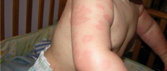
In addition to hypertrophy and hyperplasia - swelling, changes in shape and increase in size of the pathologically altered part of the body, patients may experience symptoms such as:
- the occurrence of papillomatous growths, ulcers and warts (as in the photo below, elephantiasis of the legs);
- the skin may become shiny, pale or bluish, and does not fold;
- lymphatic and venous stasis;
- edema;
- pain;
- stiffness of movements;
- the development of hypertrophic processes in the layers of skin and subcutaneous tissue, caused by an increase in intermuscular space as a result of the proliferation of connective and bone tissue, which ultimately causes thickening of the bone.
Patient with elephantiasis
If the disease is caused by erysipelas , then in the affected area of a sharply elevated painful ridge, a burning sensation, redness (erythema), increased temperature, pinpoint hemorrhages, erosions, trophic ulcers, blisters may occur, which may subside, forming brown crusts.
Erysipelas on the leg
Stages of the disease
At an early stage, lymphedema is quite difficult to recognize: it appears in the evening and disappears after a night's rest. The limbs feel soft to the touch, and patients may confuse them with swelling that occurs after exercise. As the disease progresses, lymphedema increases, becomes permanent, and becomes dense. The skin in the area of the second toe cannot be folded (positive Stemmer's sign). Swelling begins on the foot and spreads higher up the leg.
When the disease reaches the third stage, doctors talk about elephantism, or elephantiasis. The disease leads to the fact that one leg is seriously different in size from the other (if lymphedema is observed only on one limb), it is difficult, and sometimes impossible, for the patient to choose clothes or shoes, and it is difficult to move. The swelling becomes dense, since the lymph contains a lot of protein, and the penetration of protein molecules into the tissue leads to the formation of fibrous fibers there. Elephantiasis is accompanied by the appearance of papillomas on the skin, vesicles from which lymph may leak.
Tests and diagnostics
To confirm the diagnosis of filariasis, clinical data and epidemiological history information, results of laboratory and instrumental research methods are studied. The most important factor is the detection of microfilariae in blood tests using microscopy at low magnification. In addition, immunological techniques can be used, but they do not give strictly specific results.
If the nature of the pathology is not parasitic, then a more complete picture can be given by ultrasound of the lower extremities, rheovasography, lymphography, duplex scanning of blood vessels, radiography and MRI. In addition, general blood tests, urine tests, and a blister test may be performed during the examination.
Diagnosis of elephantiasis of the lower extremities
If the symptoms described above appear, do not delay visiting your doctor. The treatment of diseases of the lymphatic system is carried out by a specialized specialist - a lymphologist. If you do not have the opportunity to visit this doctor, make an appointment with a phlebologist or angiosurgeon: since the lymphatic and circulatory systems function interconnectedly, a vascular specialist can tell you how to treat elephantiasis in the legs.
To make an accurate diagnosis, not only a medical examination is important, but also additional research. This is especially necessary in the initial stages of elephantiasis, when its symptoms may be mistaken for manifestations of vascular diseases (in particular, thrombosis). Before starting treatment for elephantiasis, the following diagnostic procedures are recommended:
- Dopplerography of blood vessels.
- Lymphangiography.
- CT.
- MRI.
- Laboratory tests: general and biochemical blood test, serological test, blood test after taking Diethylcarbamazine. The latest study reveals the presence of filaria in the human body.
- Performing the McClure-Aldrich test.
Methods for treating elephantiasis of the feet
In men
In addition to enlargement of the lower extremities, the pathology can affect the external genitalia. In men, this is the scrotum, which as a result resembles a sac-like tumor and can reach colossal sizes - up to 54 kg of weight, hanging down to the level of the knees and even the floor. In this case, elephantiasis develops in the form of dropsy of the membranes of the testicles - hydrocele. Microfilariae are found in the fluid punctate.
Elephantiasis of the scrotum
Elephantiasis can also spread to the penis, but does not affect the cavernous bodies, urethra and testicles. As with elephantiasis of other parts of the body, the scrotum, perineum and foreskin may become covered with warty and papillomatous growths, and hyperkeratosis , ulcerations, excoriations or eczematous dermatitis If a secondary staphylococcal or streptococcal infection has been associated, the pathology may acquire a purulent-septic character.
Elephantiasis of the scrotum is extremely dangerous, because hypertrophy of the foreskin makes urination difficult, disrupts sexual function, making erection painful and causing impotence .
Diagnostics
Diagnosis of the disease consists of examining the affected limb or part of the patient’s body, collecting complaints and medical history from a phlebologist. The specialist measures the volume of the diseased limb and compares it with the circumference of the healthy one, then prescribes instrumental research methods.
Lymphangiography
The study involves injecting a special dye (methylene blue) into the space between the fingers. The dye begins to spread upward and stains the lymph vessels. Then the skin is incised and a large lymph vessel is isolated, into which a contrast agent is injected. After this, X-rays are taken.
Signs of lymphovascular damage:
- the shape of the vessels resembles beads or a spindle;
- narrowing or blockage of blood vessels;
- areas of dilated lymph vessels;
- release of contrast into tissue;
- the walls of blood vessels are thinned;
- the vascular pattern is poorly expressed - not all lymph vessels are visible.
Doppler examination of blood vessels
The examination is painless and is carried out using an ultrasound machine. Signs of elephantiasis:
- blockage and narrowing of blood vessels in places;
- the presence of varicose areas;
- detection of blood clots or accumulations of parasites in lymph vessels;
- detection of connective tissue between muscles and under the skin;
- damage to vein valves.
Laboratory research
If the parasitic nature of elephantiasis is suspected, the following laboratory tests are carried out:
- blood serology - detection of antibodies to filariae;
- general blood test - increased eosinophils (a sign of the presence of helminths in the body);
- blood clotting – increased clotting rates;
- blood microscopy - examination of a blood smear under a microscope to detect filariae;
- provocative test - taking diethylcarbamazine stimulates the release of parasites into the capillaries and their detection by microscopy of a blood smear.
Other methods
Additional diagnostic methods are also used:
- X-ray of the limb (bone thickening, calcium deposits, osteoporosis);
- thermography;
- MRI;
- lymphoscintigraphy;
- CT scan;
- McClure-Aldrich test - intradermal injection of 0.1 ml of saline solution - the resulting blister resolves almost immediately, which indicates edema.
Among women
Women can also suffer from elephantiasis and, like men, the external genital area, that is, the labia majora and minora, can be affected. Such rare cases are more typical for tropical latitudes; in our climate, elephantistic enlargement of the external genital organs occurs only in women providing paid sexual services. The affected parts can become deformed and reach the size of a fist, sometimes become covered with bubbles that can easily burst and release a clear liquid - lymph, which coagulates in the air. This usually occurs as a result of dilation of the lymphatic vessels and may be accompanied by massive discharge - lymphorrhea .
Related disorders
Symptoms of the following disorders may be similar to those of elephantiasis. Comparisons may be useful for differential diagnosis.
- Hereditary lymphedema is an inherited disease of the lymphatic system characterized by abnormal swelling of certain parts of the body. The lymphatic system is a circulatory network of vessels, ducts and nodes that filter and distribute certain fluid (lymph) and blood cells throughout the body. Lymphatic fluid accumulates in the soft tissues within and under the skin due to obstruction, malformation, or underdevelopment (hypoplasia) of various lymphatic vessels. There are three forms of hereditary lymphedema: congenital hereditary lymphedema or Nonne-Milroy disease, subcutaneous lymphedema or Meige's disease, and lymphedema of Tarde. Symptoms include swelling of the affected areas and thickening and hardening of the skin in the affected areas. In most cases, hereditary lymphedema is inherited as an autosomal dominant trait.
Diet for elephantiasis
Diet for lymphostasis of the lower extremities
- Efficacy: no data
- Timing: as prescribed by the doctor
- Cost of products: 1500-1600 rubles. in Week
Swelling and impaired lymphatic drainage require compliance with a special drinking regime and diet. The main recommendations include:
- eating easily digestible foods;
- enriching the menu with dishes containing rice, peas, lentils, as well as vegetable soups and, in general, boiled food;
- maximum limitation of salt and white sugar consumption; for an afternoon snack, ripe fruits and salads made from them are best suited;
- ban on fried and fatty foods;
- eliminating coffee, black tea, and tobacco completely;
- Your daily diet should include a glass of light cow's or coconut milk, water with honey, orange, pomegranate or grape juice.
Treatment
How to treat elephantiasis? Therapy for elephantiasis is not an easy task, and the process itself takes a long time, sometimes a lifetime. The main thing in the treatment of elephantiasis is the patient’s mood for success. Conservative treatment methods are used only at the early stage of the pathology; if the disease progresses to the second/third stage, surgical treatment in combination with drug therapy is recommended.
Therapy for elephantiasis has the following goals:
- reduce lymph production;
- improve tissue nutrition;
- remove metabolic products and toxins from the body (affected area);
- normalize the condition of lymph vessels and lymph circulation;
- prevent the proliferation of connective tissue.
Conservative therapy
Used at an early stage of the process or as an addition to surgical treatment:
- Diet
The patient should avoid eating fatty, smoked and salty foods, canned food, fast food and marinades, strong tea and coffee, and also completely eliminate alcohol and “quit” smoking. The food should consist of fruits and vegetables, preferably fresh, and some seasonings (ginger, turmeric, garlic, coriander).
- Lymphatic drainage massage
It can be done manually (only by a specialist) or using hardware. Lymphatic drainage massage improves lymph outflow, promotes activation of lymph nodes, and strengthens lymphatic vessels. The technique involves stroking and shaking the affected area/limb. The massage begins from a place remote from the center of the pathological process (for example, from the fingers). Smooth movements of the massage therapist along the lymph vessels “drive” the lymph to the body.
- Bandaging or elastic bandaging
Used after a massage session or in the morning without getting out of bed (in case of damage to the legs). You can tightly bandage the limb with an elastic bandage or wear special compression stockings (stockings, tights).
- Physiotherapy
Prescribed for cases of limb disease. The specialist develops a set of exercises that the patient must perform daily for 15–25 minutes. Therapeutic exercises force the muscles to contract, and they, in turn, affect the blood and lymphatic vessels, normalizing blood and lymph flow.
- Physiotherapy
Physiotherapeutic procedures that are effective are: electrophoresis with lidase and magnetotherapy, phonophoresis with enzymes (longidase, streptokinase) and laser therapy. Physiotherapy normalizes blood circulation, returns interstitial fluid to tissues, improves lymph circulation, reduces swelling and promotes the resorption of connective tissue.
- Medications
For elephantiasis caused by filaria, anthelmintic drugs (diethylcarbamazine, albendazole, ditrazine) are prescribed in combination with antihistamines (suprastin, claritin) to relieve allergic reactions that have developed to toxins and waste products of parasites.
In case of erysipelas or the addition of a secondary infection, the use or parenteral administration of antibiotics (penicillins, cephalosporins) is indicated. Antihistamines, vitamins and immunomodulators are also prescribed to stimulate the immune system, angioprotectors to strengthen the vascular wall, normalize blood and lymph flow, improve the elasticity of the walls of blood vessels (troxerutin, rutoside, trental). In addition, it is necessary to take NSAIDs (butadione, indomethacin) and glucocorticoids (dexamethasone, hydrocortisone), which stop inflammation, reduce swelling, and eliminate pain.
Surgical treatment
The goal of surgery for elephantiasis is to create new pathways for lymph flow and reduce the volume (swelling) of the affected area. Indications:
- progressive stagnation of lymph;
- proliferation of fibrous tissue;
- formation of lymphatic sacs;
- relapse of erysipelas;
- severe pain syndrome.
Various surgical techniques are used:
- restoration of patency of venous and lymphatic vessels;
- creation of lymphovenous anastomoses (crossed lymphatic vessels are sutured to the accompanying branches of the saphenous veins);
- excision of overgrown skin, subcutaneous fat and fascia in order to reduce the circumference of the limb.
Consequences and complications
thrombophlebitis or erysipelas (usually with streptococcal infection) can develop against the background of elephantiasis All these changes can lead to:
- lymphostasis or in other words - lymphedema ;
- possible addition of purulent-septic infections;
- malignancy of papillomas ;
- lymphorrhea , ulcers , eczema and muscle atrophy, since the upper integument and muscle tissue also suffer due to lack of trophism.
Elephantiasis can significantly limit freedom of movement, since it is much more difficult to walk on “heavy legs”; this may also be partly due to the degeneration of muscle tissue. As a result, all this can lead to complete immobilization and disability.
Causes
The causes of elephantiasis vary depending on the form of the pathology:
Primary (congenital) elephantiasis:
- dysplasia or underdevelopment of lymph vessels;
- Milroy-Mage disease (gene pathology associated with sex chromosomes);
- Shereshevsky syndrome (chromosomal abnormality);
- excessive production of interstitial fluid.
Secondary (acquired) elephantiasis:
- obstruction of the lymph nodes or its disruption (compression by the tumor or its metastases, chemotherapy, removal of lymph nodes);
- streptococcal infection caused by hemolytic streptococcus (cellulitis, erysipelas) – bacteria multiply in the lymph capillaries, where they secrete toxins that cause allergic reactions and narrowing of lymphatic vessels;
- damage to lymph vessels caused by extensive trauma, frostbite or burns;
- venous diseases that lead to malnutrition of the soft tissues of the limb and later to disruption of the patency of the lymphatic vessels (varicose veins, post-thrombophlebitis syndrome, phlebitis, thrombophlebitis);
- infection with filamentous helminths - filaria, in particular Bancroft's threadworm (Wuchereria bancrofti) - filariasis or wuchereriosis - parasites are transmitted through the bite of a mosquito/sandfly, the disease is common in the tropics and subtropics (helminths live and multiply in lymph vessels, where they intertwine into balls, which leads to blockage blood vessels; in addition, the development of a toxic-allergic reaction of the body to parasites “spurs” the proliferation of connective tissue fibers).
- autoimmune diseases - lead to damage to the blood and lymphatic vessels (systemic lupus erythematosus);
- chronic eczema;
- radiation;
- syphilis.
Drug therapy
At the first stage of elephantiasis the following are used:
- antihistamines to relieve allergic reactions (Loratadine);
- anthelmintic drugs that prevent parasites from multiplying (piperazine);
- angioprotectors to normalize tissue nutrition (Rutoside);
- pyridoxine to improve metabolism.
At the second stage of the disease, the following is prescribed:
- angioprotectors for relaxing vascular muscles (Troxerutin);
- enzymes to normalize the state of fiber (Lidase);
- non-steroidal anti-inflammatory drugs (Reopirin);
- desensitizing drugs to relieve inflammation (Claritin);
- biostimulants that soften connective tissue;
- vitamins.
At the third stage, to maintain the body, the following is prescribed:
- angioprotectors to reduce edema (Troxerutin);
- antibiotics to destroy infection in diseased tissues (Azithromycin);
- venotonics to improve fluid circulation in blood vessels (Detralex).