- Idiopathic (classical) type
- Immunosuppressive (iatrogenic) type
- Endemic (African) type
- AIDS-associated (epidemic) type
- Diagnostics
- Treatment
- Prevention
Kaposi's sarcoma is a malignant tumor with a multifocal growth pattern, originating from blood and lymphatic vessels.
The causative factor of Kaposi's sarcoma is the human herpes virus type VIII, and the most important mechanism of development is a violation of antitumor immunity.
The course of various types of this disease depends on the nature of immune disorders.
Clinically, there are 4 types of Kaposi's sarcoma: idiopathic , immunosuppressive , endemic and AIDS-associated .
Idiopathic (classical) type
It develops in people over 50 years of age, although in recent years there has been a trend toward a decrease in the age of those affected. Men get sick 9-15 times more often than women.
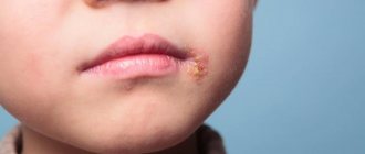
Primary manifestations of the idiopathic type of Kaposi's sarcoma are mainly localized in the area of the feet and legs and look like irregularly shaped red-violet or red-brown spots or nodules.
Hemorrhages, warty growths, areas of pigmentation, etc. appear on the surface of the lesions.
These changes can develop at the site of injury. The rash may be preceded or accompanied by swelling of the lower part of the limb, as a result of which the skin thickens and becomes bluish in color. The lesions are usually symmetrical and initially asymptomatic.
Then burning, tingling, discomfort and pain may appear.
As the disease progresses, more and more areas of the skin are involved in the process: the upper limbs, torso, face, as well as the mucous membranes of the mouth, eyes and genitals.
In the terminal stage of the disease, internal organs are affected.
The idiopathic type of Kaposi's sarcoma can occur acutely, subacutely and chronically.
The acute form is characterized by a rapid onset and rapid course, intoxication, high body temperature, multiple lesions of the skin and mucous membranes, enlarged lymph nodes, and involvement of internal organs.
This form of the disease is more common in young or very old people. Life expectancy with it ranges from 2 months to 2 years. Death occurs due to intoxication and exhaustion (cachexia).
In the subacute form, clinical manifestations are less pronounced, the process develops more slowly and leads to death on average within 2-3 years from the onset of the disease.
The most common chronic form of Kaposi's sarcoma is characterized by a relatively benign course.
It is characterized by a slow development of the disease and a limited nature of the lesion. The disease can last for 6-10 and even 15-20 years. Only in the later stages is it accompanied by the appearance of nodules, enlarged lymph nodes and damage to internal organs.
Death usually occurs from concomitant diseases or complications associated with the treatment.
Diagnosis of Kaposi's sarcoma
Examination is a very important moment for any patient. The more thoroughly it is carried out, the more information doctors receive about the number of affected tissues, general health and other disorders. They need this data to make an accurate diagnosis, determine its stage, assess prognosis and prescribe the correct treatment.
You can undergo a complete diagnosis of Kaposi's sarcoma at our oncology department. We quickly and efficiently carry out any research, take tests and receive their results from our own laboratory in the shortest possible time. All procedures in our Center are performed by specialists with many years of experience in large federal clinics and extensive experience in identifying and combating dangerous tumors.
The examination begins with a question about symptoms and the date of their onset, existing diseases, surgeries, sexual activity and possible contacts with the herpes virus. Then the skin, the inside of the mouth, and in some cases the rectum are examined, after which a series of procedures are prescribed:
-
occult blood tests - Biopsy
- taking tissue samples and sending them to a laboratory for careful examination of the cells. As a rule, the collection of skin particles is carried out under local anesthesia - using drugs that numb the nerves that transmit pain signals to the brain. If internal organs such as the lungs or intestines are affected, material is obtained through the bronchoscopy and endoscopic examinations described below. - Chest X-ray –
performed to detect damage to the respiratory system.
If unusual areas are identified, other tests are prescribed, for example, computed tomography , which creates a clear image of the internal tissues. - Bronchoscopy –
indicated for patients with symptoms such as shortness of breath or coughing up blood. During this procedure, a bronchoscope is inserted into the trachea, which is a tube with a light and a small camera at the end. It allows not only to examine the tissues of the respiratory system, but also to perform their biopsy. - Endoscopic examinations of the gastrointestinal tract
using special equipment - a thin flexible device with a video camera and a light source:
Esophagogastroduodenoscopy
- examination of the inner lining of the esophagus, stomach and the first part of the small intestine to identify ulcers, infections and other lesions. - Colonoscopy
– assessment of the condition of the colon and rectum. - Capsule endoscopy
. The patient swallows a capsule the size of a large tablet containing a light source and a camera. It travels through the entire digestive tract and takes thousands of pictures, which are sent to a device worn around the waist. The images are transferred to a computer and examined by a doctor, and the device is removed from the body during a bowel movement. - Double balloon enteroscopy
. Conventional endoscopic procedures cannot look deep into the small intestine because it is very long and has many curves. This method allows you to circumvent the above limitations using a special device consisting of 2 tubes located one inside the other. It is inserted either into the mouth or anus, reaches the desired point, and then a balloon is inflated at the end, allowing it to remain in place. The outer tube is then pushed forward and secured in place by a second "ball". The procedure is repeated over and over again until the specialist examines all areas of interest and takes a tissue sample.
: are not prescribed to make an accurate diagnosis or to identify Kaposi's sarcoma - they are necessary to assess the general state of health and the quality of the internal organs.
Immunosuppressive (iatrogenic) type
This type of Kaposi's sarcoma is caused by exposure to immunosuppressive (immune suppressive) drugs used to prevent the rejection of transplanted internal organs, or immunosuppressive therapy for chronic diseases.
This type of sarcoma occurs in relatively young people (35-50 years old). The ratio of men to women is 2:1. The tumor is diagnosed on average 30 months after the start of immunosuppressive therapy.
The immunosuppressive type of Kaposi's sarcoma is characterized by severe disturbances in cellular and humoral immunity.
On the other hand, with this type of disease, the functional activity of the immune system remains, which may explain the spontaneous disappearance of tumor manifestations after cessation of immunosuppressive therapy.
Skin manifestations with this type of Kaposi's sarcoma are initially quite limited (the lower extremities are less often affected), but then quickly become widespread, involving the mucous membranes, lymph nodes and internal organs, which can lead to death.
Endemic (African) type
Along with the AIDS-associated type, it is the most common tumor in Central Africa. Occurs in adults and children. The ratio of men to women varies from 3:1 to 10:1.
There is a benign , nodular type of the disease, which occurs in adults, and in its clinical picture and course is not different from Kaposi's sarcoma of the idiopathic type, as well as fulminant , lymphadenopathy, affecting mainly children, accompanied by a high frequency of damage to the lymph nodes, internal organs, bones, and minimal skin manifestations and death after 2-3 years.
The average life expectancy of patients varies from 6 to 26 months.
Kaposi's disease: stages of the disease
Kaposi's sarcoma is divided into several typical stages.
- Spotted. The initial degree, manifested by red spots with a bluish or dark brown (brown) tint and a smooth surface. Their diameter varies from 1 to 5 cm; individual elements can merge into large spots. The main location is the hands and feet. Subjectively, they may not manifest themselves at all or have scant symptoms (minor itching, tingling, etc.).
- Papular. It is characterized by the appearance of so-called papules - nodules, ranging in size from 2 to 10 mm, which have the shape of a sphere or hemisphere. They are located singly or grouped into clusters shaped like arcs or rings. Color - from pink to red with a brown-brown tint. Also, occasionally, a plaque with a bumpy or smooth surface may form from the spot. Horny or papillomatous growths may be observed on the surface.
- Tumor - a severe stage, accompanied by the appearance of tumor nodes. They are soft or dense, up to 50 mm, at the edges they can have bluish or brown tints. Quantity - from single to massive distribution, up to hundreds. They can merge into large lumpy neoplasms. The surface may become covered with ulcers or foul-smelling discharge. Over time, it can penetrate deep into the tissues, right down to the bones, which also affects.
AIDS-associated (epidemic) type
This type of Kaposi's sarcoma develops in conditions of severe immune deficiency and affects young people (on average 38 years), usually homosexual men.
Early lesions on the skin are characterized by small bright pink or bluish spots, similar to insect bites, dense brownish or reddish nodules, which can develop into ulcerated and painful nodules. Tissue swelling can occur in the area of the extremities, scrotum, penis, face, eyelids.
Important clinical features of the AIDS-associated type of Kaposi's sarcoma are: primary lesions of the face, mucous membranes and upper extremities. The favorite localization of the pathological process is the tip of the nose and the hard palate.
If left untreated, this type of sarcoma is characterized by rapid spread of skin rashes.
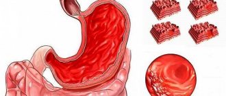
In 75% of patients, internal organs are affected, primarily the stomach and duodenum, as well as the lungs. Less commonly, the liver, spleen, adrenal glands, pancreas, testicles and very rarely the brain are affected.
Only in 5% of patients the disease occurs without skin lesions. Death is more often associated with other manifestations of AIDS rather than with Kaposi's sarcoma.
What examination can the doctor prescribe? How is the diagnosis made?
If a patient has HIV/AIDS and has characteristic spots on his skin, the doctor may suspect Kaposi's sarcoma. The final diagnosis is made after a biopsy. During laboratory tests, human herpesvirus type 8 protein – LANA –
(
latency-associated nuclear antigen
).
To diagnose lesions of the digestive and respiratory systems, endoscopic examinations are prescribed.
Treatment
The choice of treatment depends on the type of Kaposi's sarcoma and the clinical course of the disease.
Radiation therapy can be successfully used in the treatment of patients with single tumor elements in all types of sarcoma. In this case, the total radiation dose is 30-40 Gray.
For limited lesions, the following are used: surgical method , cryotherapy with liquid nitrogen , photodynamic therapy , intratumoral administration of drugs .
In the chronic form of the idiopathic type of Kaposi's sarcoma with widespread rashes, treatment should be combined with the use of antitumor drugs and interferon.
In the subacute form of the idiopathic type of sarcoma, the use of interferon .
Therapeutic tactics for the immunosuppressive type of the disease are based on the maximum possible reduction in doses or complete abolition of immunosuppressive drugs, the prescription of interferon or antitumor drugs (as indicated).
For AIDS-associated Kaposi's sarcoma, antiretroviral drugs (zidovudine, stavudine) are used, which in the early stages of the disease (up to 8-12 weeks) can lead to the disappearance of the manifestations of the disease.
If such therapy is ineffective, as well as in later stages of the disease, antitumor drugs are used
What chemotherapy regimens are used?
At the first stage, the most effective cytostatics with a lower likelihood of severe complications are used - these are anthracycline antibiotics, doxorubicin or daunorubicin, enclosed in special microcapsules. For tumors sensitive to cytostatics, 6 courses with administrations every two to three weeks are sufficient.
Both drugs act aggressively on the myocardium and blood cells, but due to the liposomal form, these side reactions are less pronounced than with conventional forms of cytostatics.
If the first line is ineffective - the continued growth of sarcomatous elements - the patient is transferred to paclitaxel, which has a similar spectrum of toxicity, but with more intense manifestations. The drug is administered with preliminary preparation every 2-3 weeks, the number of courses is determined by the effect.
If there is no result from the second line of chemotherapy, an alternative remains in the form of courses of drugs from other groups, including in combination with glucocorticoids; in domestic practice, a drug from the “Soviet past”, prospidin, which selectively accumulates in the skin, is often used.
Prevention
Primary prevention of Kaposi's sarcoma consists of actively identifying patients and groups at increased risk for developing this disease.
Particular attention should be paid to patients receiving immunosuppressive therapy. In these groups, detection of individuals infected with human herpes virus VIII (HHV-8) is important.
Secondary prevention includes clinical observation of patients in order to prevent relapse (recurrence) of the disease, complications after treatment and rehabilitation of patients.
Causes and risk factors
It is not possible to accurately determine the causes of the disease at this time, however, there are clear patterns that increase the likelihood of developing the disease, such as:
- Human herpesvirus type 8 - There is now some evidence that this viral disease is the main cause of sarcoma.
- Impaired immune status due to damage to the body by HIV infection.
- Some malignant tumors increase the likelihood of Kaposi's sarcoma (myeloma, Hodgkin's and others).
- Decreased immune status during the treatment of a number of diseases with drugs that have an immunosuppressive effect. These are autoimmune diseases, chemoradiotherapy for tumors.
There is an increased risk of developing the disease in men hailing from areas of central Africa and Mediterranean countries.