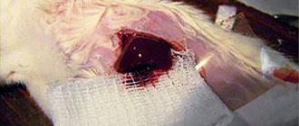
Pharmacological properties of the drug Tachocomb
Absorbent hemostatic agent for local use. The drug consists of a collagen plate coated on one side with fibrin glue components (highly concentrated fibrinogen and thrombin), which promote blood clotting. The adhesive surface of the plate is marked in yellow (riboflavin). Upon contact with a bleeding wound, the blood clotting factors contained in the coating (fibrinogen, thrombin) are released, and thrombin activates the conversion of fibrinogen to fibrin. The plate adheres to the wound surface due to the polymerization reaction; During this process (plate 3–5 min), the collagen forms a water- and air-tight layer. During this process, the plate must be pressed against the wound surface. In the body, the components of the plate undergo enzymatic breakdown within 3–6 hours. A special production process guarantees maximum safety against viruses and bacteria entering the contents of the plate.
Intraoperative blood loss is a serious complication in surgery and is accompanied by an increase in the number of complications and mortality, an increase in the duration and cost of the operation, and postoperative hospital stay [1-3]. The issue of blood loss is especially relevant in the practice of cardiovascular surgery, since disorders of the blood coagulation system associated with the use of artificial circulation (CPB) and complete heparinization are predisposing factors for hemorrhagic complications that make it difficult to achieve adequate hemostasis [4, 5]. The rate of reoperation for bleeding is up to 2-6% in cardiac surgery patients, and, as a rule, such interventions worsen the prognosis [6, 7].
Currently, hemostatic therapy has a fairly extensive arsenal of drugs to stabilize coagulogram parameters and normalize the coagulation cascade. Surgical hemostasis traditionally involves long-term compression, ligation, clipping, and electrocoagulation. However, it is worth noting that these techniques are not always appropriate and are often ineffective for “non-surgical” sources of bleeding. In addition, in cardiovascular surgery there are various clinical situations, including myocardial injuries and ruptures, interventions on the aorta, when the use of sutureless hemostasis methods is the most appropriate and safe for the patient.
In this regard, various seamless techniques have been developed to stop bleeding, based, in particular, on the local delivery of coagulation factors in collagen matrices. Other techniques used to control bleeding include ethyl cyanoacrylate glue, various forms of collagen, oxidized cellulose, topical forms of thrombin, and gelatin/fibrin sponges [8–12]. However, it is worth noting that the combination of various agents in a collagen matrix facilitates their use and increases the efficiency of hemostasis.
Among biological hemostatic drugs, three groups can be distinguished: fibrin glue, pre-prepared drugs and ready-to-use drugs. TachoComb is a representative of the third generation of drugs, consisting of a collagen sponge impregnated with human fibrinogen and thrombin. A number of authors have raised the question of the development of antibodies against human coagulation factor V after the use of bovine thrombin. The authors describe a case in which the use of a relatively pure preparation of bovine thrombin resulted in the development of antibodies to human coagulation factor V and coagulopathy. This fact calls into question the safety of using even relatively pure bovine thrombin [13]. Horse collagen, which is one of the components of TachoComb, was tested for immunogenicity BC by Adelmann-Grill et al. [14] back in 1987 and showed a low risk of developing immune reactions. The remaining components of TachoComb (fibrinogen and thrombin) were isolated from human blood plasma and their use, therefore, does not imply the same risk of immunological reactions as with biological components of animal origin. Modern TachoComb (registered in the West as TachoSil) is a new generation of the drug (formerly TachoComb) that does not contain aprotinin or any other component of bovine origin.
Biodegradable matrices impregnated with local hemostatic drugs have shown high hemostatic efficiency and local drug delivery in experimental and clinical conditions [15, 16].
TachoComb reproduces the normal coagulation cascade without a cytotoxic effect. The components of TachoComb are activated upon contact with the wound surface, which leads to the formation of fibrin monomer, which, upon polymerization, forms a fibrin network with subsequent adhesion of the sponge to the surface of the wound. Adequate hemostasis can be achieved within 3-5 minutes. The combination of coagulation factors in the collagen sponge ensures their high concentration at the site of bleeding. The hemostatic effectiveness of TachoComb was previously proven in clinical studies [17, 18].
F. Maisano et al. [20] showed in a randomized controlled trial that TachoComb (TachoSil) was significantly more effective than traditional hemostatic material for controlling bleeding in cardiovascular surgery. After 3 minutes, when using TachoComb, ¾ of patients achieved hemostasis compared to 1/3 of patients using standard hemostatic material ( p
<0.0001).
A significant difference remained after 6 minutes ( p
= 0.0006). Despite the small number of patients included in the study, the authors showed that the use of TachoComb sponges significantly reduces hemostasis time. In addition, in a study by G. Bajardi et al. [15] showed a significant reduction in intra- and postoperative blood loss in patients using TachoComb sponges in comparison with traditional methods of hemostasis. It has also been shown that the adhesive strength of TachoComb is approximately twice as high as that of other ready-to-use hemostatic agents (fibrin glue applied to a polymer sponge), and significantly higher than that of isolated fibrin glue [21-23]. The high degree of adhesion of the fibrin clot at the site of application in combination with the scaffolding function of the collagen sponge ensures the formation of a stable barrier with a good hemostatic effect, including in cases of hyperfibrinolytic blood activity and myocardial injuries.
In the work of F. Maisano et al. [19] discusses the ability of TachoComb to withstand high pressure when used in aortic surgery and left ventricular (LV) interventions. In addition, TachoComb is effective against venous sources of bleeding, as well as when applied to the tissues of the right atrium (RA) and right ventricle (RV) [20]. These properties suggest that the use of collagen sponges may also be appropriate in peripheral vascular surgery. In particular, bleeding from suture holes is a serious problem in vascular reconstructions using polytetrafluoroethylene (PTFE) grafts [23]. In a study by T. Joseph et al. [24] showed that TachoComb significantly reduces hemostasis time during operations using vascular prostheses made of PTFE (hemostasis in an average of 5 minutes in the TachoComb group versus an average of 11 minutes in the control group). In addition, a significant reduction in intra- and postoperative blood loss was noted.
Atrioventricular rupture is a rare but often fatal complication after mitral valve surgery. The incidence is approximately 1-2%, but the mortality rate when complications occur exceeds 50% [25, 26]. Injury is often the result of overly aggressive surgical manipulation and decalcification of the mitral valve (MV) annulus fibrosus (AF) and subvalvular apparatus or oversizing of the prosthetic valve [27]. The clinical picture varies from a hematoma in the projection of the atrioventricular groove along the posterior surface of the heart to its rupture with massive bleeding. Quite often this is accompanied by compromised blood flow through the circumflex artery with signs of ischemia of the posterolateral LV sections and myocardial dysfunction. Today, the standard treatment algorithm for this complication includes reinitiation of CPB, cardioplegia, atriotomy, removal of the prosthesis, and suturing of the defect. However, mortality remains high due to ongoing blood loss, prolonged CPB, and aortic cross-clamping. The use of TachoComb has also proven itself in the repair of superficial epicardial damage to the heart without significant bleeding from the ventricular cavity [28-33]. Local hemostatic agents also play an important role in the treatment of trauma and penetrating wounds of the heart. Such injuries are among the most severe among injuries, accompanied by high mortality. Early diagnosis and emergency surgery are the only chance to save the lives of these patients. In a review by N. Campbell et al. [30] out of 1198 cases of penetrating cardiac injury, only 6% of patients arrived at the hospital alive. LV wall rupture is a potentially fatal complication of acute myocardial infarction (MI) with an incidence of 0.8–6% [34, 35]. At the same time, tactics for injuries to the posteroinferior parts of the heart are very difficult and sometimes require switching to I.C. However, complete heparinization is accompanied by the risk of bleeding from other damaged organs in patients with multiple combined injuries. Hemostatic material based on coagulation factors, along with other adhesive materials such as gelatin-resorcinol-formaldehyde (GRF) glue, fibrin glue, cyanoacrylate, play an important role in repairing ventricular wall defects. These techniques are easy to perform and take little time [36]. GRF glue has higher adhesion strength than fibrin glue, but it has been shown to have higher cytotoxicity [37]. K. Iha et al. [34] also reported the formation of a LV pseudoaneurysm after repair of its rupture using GRF glue. Cyanoacrylate glue is also cytotoxic [35], although J. Padro et al. [36] reported successful treatment of 13 patients with LV rupture using cyanoacrylate. The mechanism of action of TachoComb mimics the normal coagulation cascade, and its potential clinical effectiveness has been observed in the treatment of atrioventricular rupture [41], as well as post-infarction LV rupture.
Of course, the use of a particular method in the surgical treatment of severe myocardial damage should be carefully assessed and analyzed for each individual patient. On the one hand, the “gold standard” is direct suturing of the defect with or without IR or excision of infected tissue with further repair with a patch [32, 42-45]. Using I.K. has certain advantages: hemodynamic stabilization, the ability to control pressure inside the LV, a dry surgical field and myocardial protection during cardioplegia. Isolated use of TachoComb is usually inappropriate for post-infarction ruptures with the formation of large defects in the LV wall [29-31]. Despite the fact that K. Nishizaki et al. [42] reported successful repair of a 1-cm rupture using TachoComb; a number of authors [47, 48] reported the risks of potential complications of this approach (formation of pseudoaneurysms and re-rupture) [47, 48]. However, in the presence of loose myocardial tissue at the site of damage and the cutting of sutures, technical problems often arise during LV repair [29, 30, 46]. In addition, surgical manipulations in the area of injury often lead to additional damage to the myocardium, and the use of cardiopulmonary bypass is accompanied by a decrease in coagulation rates, prolongation of myocardial ischemia and cardiopulmonary bypass. A. Schuetz et al. [37] reported off-pump reconstruction of the left ventricle with multilayer application of TachoComb for left ventricular wall defects. In a limited number of patients, the authors obtained excellent 5-year results.
Thus, the isolated use of a local hemostatic agent is not always advisable for extensive myocardial defects. In this regard, a number of authors [48, 49] proposed using a hybrid technique. In addition, H. Yamaguchi et al. [47] developed a new hybrid method for the treatment of LV ruptures, which combines the use of TachoComb with suture repair without the use of IR to prevent the development of pseudoaneurysms and recurrent ruptures. According to the researchers, this technique does not require IR and is suitable even for emergency surgery.
In the Department of Aortic Surgery of the Russian Scientific Center for Surgery named after. acad. B.V. Petrovsky TachoComb sponges are widely used for prosthetics of various parts of the aorta, as well as for superficial damage to the ventricular myocardium. Interventions on the aorta, in particular during aortic arch replacement, involve the need for hypothermia and a relatively long period of CPB. In such conditions, a fairly common problem is the hypocoagulation status of the patient at the end of the main stage of the operation, accompanied by diffuse tissue bleeding, including blood sweating through the wall of the aortic prosthesis. In such situations, hemostasis using standard techniques can last several hours, despite adequate anesthesia with multicomponent hemostatic therapy. The problem of surgical care for patients with acute aortic dissection, who often receive combination antiplatelet therapy at the prehospital stage due to the presumed diagnosis of I.M., deserves special attention. The issue of rapid and adequate intraoperative hemostasis and reduction of blood loss and blood transfusion is especially relevant for this group of patients. The use of TachoComb sponges ensures fast and reliable hemostasis, including when applied to an aortic prosthesis, regardless of coagulogram parameters. From our point of view, the use of a hemostatic agent is also justified in cases of minor (superficial) myocardial damage and the absence of profuse bleeding.
The use of TachoComb, according to the subjective assessment of surgeons, can significantly reduce the need for blood transfusion therapy, which will accordingly lead to savings in blood components [40, 48]. A decrease in the volume of intra- and postoperative blood transfusions is accompanied by a decrease in the incidence of transfusion complications. The effectiveness of TachoComb was also confirmed by M. Czerny et al. [49], who showed a decrease in the volume of drainage discharge and the duration of drainage in patients who underwent mediastinal lymph node dissection for non-small cell lung cancer. In addition, the reduction in hospitalization and intensive care unit length of stay also reduces financial costs, so the use of topical hemostatic agents may play an important socioeconomic role in surgical practice.
Thus, the introduction into practice of local hemostats based on collagen with fibrinogen-thrombin coating has expanded the possibilities for quickly achieving adequate hemostasis. TachoComb activates the physiological cascade of thrombus formation at the site of application, regardless of the state of systemic coagulation, which is especially important in cardiovascular surgical practice. The absence of xenomaterials in the composition of the drug ensures a reduced risk of immunological reactions. The use of combined techniques (TachoComb sponge + suture plasty) significantly increases the efficiency of hemostasis, does not require IR and aortic clamping and provides good results. The feasibility of using sutureless methods of LV plastic surgery for injuries should be carefully assessed in a specific clinical situation. Of course, it is necessary to provide for the technical and practical possibility of switching to IR if attempts at seamless off-pump plastic surgery prove ineffective.
There is no conflict
of interest .
Indications for use of the drug Tachocomb
Used for hemostasis and tissue adhesion, especially during surgical interventions on parenchymal organs, for example, liver, spleen, pancreas, kidneys, adrenal glands, lungs, thyroid gland, lymph nodes; in cases where it is impossible to stop bleeding using conventional methods or when the effectiveness of such methods is insufficient; for therapeutic purposes for lymphatic, biliary and liquor fistulas; for stopping bleeding in ENT surgery, gynecology, urology, vascular and bone surgery, traumatology, etc.
Tachocomb sponge 9.5 x 4.8 x 0.5 cm cont. paper-polymer. N1x1 Nycomed Austria
For topical use only. Do not use intravascularly. Mode of application. The Tachocomb is in sterile packaging and ready for use. The drug can only be used from undamaged packaging. After opening the package, re-sterilization of Tachocomb is not possible. The outer aluminum packaging bag can be opened in a non-sterile operating room area. The inner sterile blister should be opened in a sterile area. The Tachocomb must be used immediately after opening the sterile inner packaging. Tachocomb should be applied to surgical wound surfaces under sterile conditions. Before applying the sponge, the wound surface must be cleaned of blood, disinfectants and other liquids. After removing the Tachocomb flat sponge from the inner sterile packaging, the sponge should be moistened with 0.9% sodium chloride solution and used immediately. The side coated with active substances and marked in yellow is applied to the wound surface, if necessary, additionally moistened with 0.9% sodium chloride solution and pressed lightly for 3-5 minutes. Pressing is carried out with moistened gloves or a moistened pad. After removing the twisted Tachocomb sponge from the inner sterile packaging, it should be applied immediately through the trocar without prior moistening. When unwinding, the sponge is applied with the yellow side covered with active substances onto the wound surface using tweezers, if necessary, additionally moistened with a 0.9% sodium chloride solution and lightly pressed with a damp cloth for 3-5 minutes. This creates conditions for improving the adhesion of Tachocomb to the wound surface. The Tachocomb sponge may stick to blood-stained gloves or instruments, or adjacent tissue. This can be avoided by cleaning surgical instruments, gloves and surrounding tissues. Insufficient cleansing of adjacent tissues can lead to the development of adhesions. Once you have finished pressing the Tachocomb sponge onto the wound, carefully remove the glove or pad. To prevent the sponge from leaving the surface, it can be held in place at one end, for example with a pair of tweezers. In case of severe bleeding, Tachocomb can be used without prior moisturizing. The sponge is applied to the wound surface and pressed lightly for 3-5 minutes. The Tachocomb twisted sponge can be used in both open and minimally invasive procedures and can be passed through a port or trocar with a diameter of 10 mm or larger. Dosing. The size and number of Tachocomb sponges depend on the size of the wound surface. The edges of the wound should be covered with a sponge by 1-2 cm. If more than one sponge is required to close the wound surface, then when applied to the wound their edges should overlap each other. The sponge can be cut to the desired size. In clinical studies, individual dosages were typically 1 to 3 sponges (9.5 cm x 4.8 cm); Up to 10 sponges have been reported. For smaller wounds, such as minimally invasive procedures, it is recommended to use smaller sponges (4.8 cm x 4.8 cm or 3.0 cm x 2.5 cm) or rolled sponges (based on the 4.8 cm x 4 sponge .8 cm). Unused sponges or their fragments must be destroyed.
Use of the drug Tachocomb
Used under sterile conditions. Before use, the wound surface must be cleaned of blood, disinfectants and other liquids. It is recommended that surgical gloves and instruments be kept free of blood and body fluids to avoid adhesion to the plate. The plate is applied to the wound surface with the side that contains blood clotting factors (marked in yellow) and pressed for 3–5 minutes. For wounds with exudation, the drug can be used without additional moisturizing. For wounds without exudation, before use, it is recommended to moisten the plate with a physiological solution to achieve complete connection with the dry areas of the wound surface. The moistened plate should be applied immediately!
Tachocomb®
For topical use only. Do not use intravascularly.
Mode of application
Tachocomb® is in sterile packaging and ready for use. The drug can only be used from undamaged packaging. After opening the package, re-sterilization of Tachocomb® is not possible. The outer aluminum packaging bag can be opened in a non-sterile operating room area. The inner sterile blister should be opened in a sterile area. Tachocomb® must be used immediately after opening the sterile inner packaging. Tachocomb® should be applied to surgical wound surfaces under sterile conditions.
Before applying the sponge, the wound surface must be cleaned of blood, disinfectants and other liquids.
After removing the Tachocomb® flat sponge from the inner sterile packaging, the sponge should be moistened with 0.9% sodium chloride solution and used immediately. The side coated with active substances and marked in yellow is applied to the wound surface, if necessary, additionally moistened with 0.9% sodium chloride solution and pressed lightly for 3-5 minutes. Pressing is carried out with moistened gloves or a moistened pad.
After removing the rolled Tachocomb® sponge from the inner sterile packaging, it should be applied immediately through the trocar without prior moistening. When unwinding, the sponge is applied with the yellow side covered with active substances onto the wound surface using tweezers, if necessary, additionally moistened with a 0.9% sodium chloride solution and lightly pressed with a damp cloth for 3-5 minutes. This creates conditions for improving the adhesion of Tachocomb® to the wound surface.
The Tachocomb® sponge may adhere to blood-stained gloves or instruments, or adjacent tissue. This can be avoided by cleaning surgical instruments, gloves and surrounding tissues.
Insufficient cleansing of adjacent tissues can lead to the development of adhesions.
Once you have finished pressing the Tachocomb® sponge onto the wound, carefully remove the glove or pad. To prevent the sponge from leaving the surface, it can be held in place at one end, for example with a pair of tweezers.
In case of severe bleeding, Tachocomb® can be used without prior moisturizing.
The sponge is applied to the wound surface and pressed lightly for 3-5 minutes.
During neurosurgical procedures, Tachocomb® should be applied over the primary dural closure.
The twisted Tachocomb® sponge can be used in both open and minimally invasive procedures and can be passed through a port or trocar with a diameter of 10 mm or larger.
Dosing
The size and number of Tachocomb® sponges depend on the size of the wound surface.
The edges of the wound should be covered with a sponge by 1-2 cm. If more than one sponge is required to close the wound surface, then when applied to the wound their edges should overlap each other.
The sponge can be cut to the desired size.
In clinical studies, individual dosages were typically 1 to 3 sponges (9.5 cm x 4.8 cm); Up to 10 sponges have been reported. For smaller wounds, such as minimally invasive procedures, it is recommended to use smaller sponges (4.8 cm x 4.8 cm or 3.0 cm x 2.5 cm) or rolled sponges (based on the 4.8 cm x 4 sponge .8 cm). Unused sponges or their fragments must be destroyed.


