Structure of the pharynx
In an adult, the pharynx is a funnel-shaped tube about 10–15 cm long, located behind the nasal and oral cavities and larynx. The upper wall of the pharynx is fused with the base of the skull; in this place on the skull there is a special protrusion - the pharyngeal tubercle. The cervical spine is located behind the pharynx, so the lower border of the pharynx is determined at the level between the VI and VII cervical vertebrae: here it narrows and passes into the esophagus. Large vessels (carotid arteries, internal jugular vein) and nerves (vagus nerve) are adjacent to the lateral walls of the pharynx on each side.
content .. 110 111 112 113 114 115 116 117 ..Pharynx (human anatomy)
Pharynx
, pharynx, is the initial part of the digestive tube and respiratory tract. The pharyngeal cavity, cavum pharyngis, connects the oral and nasal cavities with the esophagus and larynx. In addition, it communicates through the auditory tube with the middle ear. The pharynx is located behind the cavities of the mouth, nose and larynx, extending from the base of the skull, from which it begins, to the junction with the esophagus at the level of the VI cervical vertebra. The pharynx is a hollow, wide tube, flattened in the anteroposterior direction, narrowing as it passes into the esophagus. The pharynx can be divided into upper, anterior, posterior and lateral walls. The length of the pharynx is on average 12-14 cm.
Depending on the organs behind which the pharynx is located, three parts are distinguished: 1) nasal, pars nasalis (or nasopharynx), 2) oral, pars oralis (or oropharynx), 3) laryngeal, pars laryngea (or hypopharynx). The upper part of the pharynx, adjacent to the outer base of the skull, is called the pharyngeal vault, fornix pharyngis.
Nasal part of the pharynx
, pars nasalis pharyngis, is its upper part and differs from other parts in that the upper and partially lateral walls are fixed to the bones and therefore do not collapse. The anterior wall of the pharynx is absent here, since in front the nasopharynx communicates with the nasal cavity through two choanae. On the lateral walls of the nasal part of the pharynx, at the level of the posterior end of the inferior concha, there is a paired funnel-shaped pharyngeal opening of the auditory tube, ostium pharyngeum tubae, which is limited from behind and above by a tubal ridge, torus tubarius. This cushion is formed due to the protrusion of the cartilage of the auditory tube into the pharyngeal cavity. A short tubal-pharyngeal fold of the mucous membrane, plica salpingopharyngea, descends from the tubal ridge. Behind the cushion, the mucous membrane forms a large pharyngeal pocket, variable in shape, recessus pharyngeus, the depth of which depends on the degree of development of the tubal tonsils. At the junction of the upper wall and the posterior wall between the pharyngeal openings of the auditory tubes in the mucous membrane of the pharynx there is an accumulation of lymphoid tissue - the pharyngeal tonsil, tonsilla pharyngea. In children it is most developed, but in adults it undergoes reverse development. The second, paired, accumulation of lymphoid tissue lies in the mucous membrane of the pharynx in front of the pharyngeal openings of the auditory tubes. It is called the tubal tonsil, tonsilla tubaria. Together with the palatine, lingual, and laryngeal lymphatic follicles, the pharyngeal and tubal tonsils form the lymphoepithelial pharyngeal ring. On the vault of the pharynx in the midline near the junction of the upper wall and the posterior wall there is sometimes a round depression - the pharyngeal bursa, bursa pharyngea.
Oropharynx
, pars oralis pharyngis, occupies the level from the soft palate to the entrance to the larynx, widely communicating through the pharynx with the oral cavity. Therefore, the oral part has only lateral and posterior walls; the latter corresponds to the third cervical vertebra. The oral part of the pharynx functionally belongs to both the digestive and respiratory systems, which is explained by the development of the pharynx (see section The doctrine of the viscera - splanchnology, this edition). When swallowing, the soft palate, moving horizontally, isolates the nasopharynx from its oral part, and the root of the tongue and the epiglottis close the entrance to the larynx. With the mouth wide open, the back wall of the pharynx is visible.
Laryngeal part of the pharynx
, pars laryngea pharyngis, is located behind the larynx at the level from the entrance to the larynx to the beginning of the esophagus. It has front, back and side walls. Outside of the act of swallowing, the anterior and posterior walls are in contact. The anterior wall of the laryngeal part of the pharynx is the laryngeal protrusion, prominentia pharyngea, above which is the entrance to the larynx. On the sides of the protrusion there are deep pits - pear-shaped pockets, recessus piriformes, formed on the medial side by the laryngeal protrusion, and on the lateral side by the lateral wall of the pharynx and the posterior edges of the plates of the thyroid cartilage. The pyriform pouch is divided by the oblique fold of the laryngeal nerve, plica nervi laryngei, into two sections - the smaller - upper, and the large - lower. The superior laryngeal nerve passes through the fold.
The nasopharynx of newborns is very small and short. The vault of the pharynx is flattened and inclined anteriorly in relation to its oral part. In addition, in newborns the pharynx is relatively shorter than in adults, and the velum palatine is in contact with the entrance to the larynx. The soft palate is short and does not reach the posterior wall of the pharynx when raised. In the first years of life, the tonsils protrude strongly into the pharyngeal cavity of newborns and children. The pharyngeal openings of the auditory tubes are close together and lie lower than in adults, at the level of the hard palate. The pharyngeal pockets, as well as the tubal ridges and tubopalatine folds, are poorly expressed.
Structure of the pharynx
. The pharynx consists of: 1) the mucous membrane, 2) the fibrous layer formed by the pharyngeal-basic fascia, 3) the muscular layer, 4) the buccal-pharyngeal fascia covering it.
Mucous membrane
The nasal part of the pharynx is covered with multi-row ciliated epithelium, and the oral and laryngeal parts are covered with multi-layered squamous epithelium. In the submucosa there is a large number of mixed (mucous-serous - in the nasopharynx) and mucous (in the oral and laryngeal parts) glands, the ducts of which open into the pharyngeal cavity on the surface of the epithelium. In addition, the submucosal layer contains clusters of lymphatic follicles that form the pharyngeal and tubal tonsils. Between the follicles there are many small glands of mixed type. At the location of the pharyngeal tonsil, the mucous membrane gives off spurs into the thickness of the tonsil, forming a series of folds and dimples, fossulae tonsillares. In the dimples of the pharyngeal tonsil there are depressions - tonsil crypts, cryptae tonsillares, into which the ducts of mixed glands located between the lymphatic follicles open.
The submucosa is well expressed, and the layer proper of tunicae mucosae contains many elastic fibers. As a result, the mucous membrane has the ability to change its size as food passes through. Near the junction with the esophagus, the pharynx narrows. In its narrow section, the mucous membrane is smooth and contains especially many elastic fibers, which ensures the passage of the food bolus here.
Pharyngeal-basic fascia
, fascia pharyngobasilaris, forms the fibrous basis of the pharynx. The pharyngeal-basic fascia begins on the outer base of the skull on the pharyngeal tubercle of the occipital bone and runs on each side transversely along a curved line anteriorly from the place of attachment of the deep layer of the anterior neck muscles along the main part of this bone to the synchondrosis retrooccipitalis. Next, the line of the beginning of the fascia turns anteriorly and outward, crosses the pyramid of the temporal bone anteriorly from the foramen caroticum externum and follows to the spina ossis sphenoidalis. From here, the line of origin of the fascia deviates forward and medially and runs along the synchondrosis sphenopetrosa in front of the cartilage of the auditory tube to the base of the medial plate of the pterygoid process of the sphenoid bone. Then it follows the medial plate of the process down and anteriorly along the raphe pterygomandibularis to the posterior edge of the linea mylohyoidea mandibulae.
In the upper section, the pharyngeal-basic fascia is very strong, since here it is strengthened by bundles of collagen fibers that go into the fascia in the form of ligaments from the pharyngeal tubercle, from the edge of the foramen caroticum externum and from the membranous plate of the auditory tube. In addition to collagen bundles, the pharyngeal-basic fascia contains many elastic fibers. Below, the pharyngeal-basic fascia is attached to the thyroid cartilage and the greater horns of the hyoid bone, giving off spurs into folds: plicae pharyngoepiglotticae and plicae epiglotticae.
Muscular membrane of the pharynx
, tunica muscularis pharyngis, consists of two groups of striated muscles: compressors, constrictores pharyngis, located circularly, PI levators, levatores pharyngis, running longitudinally. The pharyngeal constrictor muscles, paired formations, include the upper, middle and lower constrictors (Fig. 113).
Rice. 113. Muscles of the pharynx (rear view). 1 - posterior belly of the digastric muscle; 2, 8, 14 - stylopharyngeal muscle; 3 - stylohyoid muscle; 4 - medial pterygoid muscle; 5, 13 - middle pharyngeal constrictor; c - hyoid bone; 7, 10 - upper and lower horns of the thyroid cartilage; 11 - esophagus; 12 - lower constrictor of the pharynx; 15, 17 - superior pharyngeal constrictor; 16 - styloid process; 18 - main part of the occipital bone; 9, 19 - pharyngeal suture; 20 - fibrous membrane of the pharynx
1. Muscle - superior pharyngeal constrictor
, m. constrictor pharyngis superior, starts from laminae medialis processus pterygoidei (pterygopharyngeal part of the muscle, pars pterygopharyngea), raphe pterygomandibulare (buccopharyngeal part, pars buccopharyngea), linea mylohyoidea mandibulae (maxillopharyngeal part, pars mylopharyngea) and the transverse muscle of the tongue ( glossopharyngeal part, pars glossopharyngea). Starting on the listed formations, muscle bundles form the lateral wall of the pharynx, and then arc in an arcuate manner backward and medially, forming the posterior wall. Posteriorly along the midline, they meet with the bundles of the opposite side at the tendon pharyngeal suture, raphe pharyngis, running from the tnberculum pharyngeum along the middle of the entire posterior wall to the esophagus. The upper edge of the muscle - the superior constrictor of the pharynx does not reach the base of the skull. Therefore, in the upper section (over 4-5 cm), the wall of the pharynx is devoid of a muscular membrane and is formed only by the pharyngeal-basal fascia and mucous membrane.
2. Muscle - middle pharyngeal constrictor
, m. constrictor pharyngis medius, starts from the upper part of the greater horn of the hyoid bone (horns of the opharyngeal part of the muscle, pars ceratopharyngea) and from the lesser horn and lig. stylohyoideum (cartilaginous-pharyngeal part, pars chondropharyngea). The upper muscle bundles go upward, partially covering the superior pharyngeal constrictor (when viewed from behind), the middle bundles go horizontally backward (almost completely covered by the lower constrictor) and the lower ones go down (completely covered by the lower constrictor). The bundles of all parts end in raphe pharyngis. Between the middle and superior constrictors are the lower bundles of the stylopharyngeal muscle.
3. Muscle - inferior pharyngeal constrictor
, m. constrictor pharyngis inferior, starts from the outer surface of the cricoid cartilage (cricopharyngeal part of the muscle, pars cricopharyngea), from the oblique line and the adjacent parts of the thyroid cartilage and from the ligaments between these cartilages (thyropharyngeal part, pars thyreopharyngea). The muscle bundles run posteriorly in ascending, horizontal and descending directions, ending at the suture of the pharynx. The lowest bundles surround the junction of the pharynx and the esophagus. The upper constrictor is the largest, covering the lower half of the middle constrictor.
Function: they narrow the pharyngeal cavity and, with successive contractions, push through the bolus of food.
The muscles that elevate and dilate the pharynx include:
1. Stylopharyngeal muscle
, m. stylopharyngeus, originates from the styloid process near its root, goes down and medially to the posterolateral surface of the pharynx, penetrating between its superior and middle constrictors. The muscle fibers, partially intertwined with the lower and middle constrictors, go to the edges of the epiglottis and thyroid cartilage.
Function: raises and expands the pharynx.
2. Velopharyngeal muscle
, m. palatopharyngeus, see section The oral cavity itself, this publication.
The buccal-pharyngeal fascia covers the external constrictor muscles. Since the buccal muscle has a common origin with the superior constrictor (raphe pterygomandibulare), the fascia with m. The buccinator moves to the upper and then to other pharyngeal constrictors.
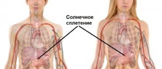
Syntopy of the pharynx. Behind the pharynx are the long muscles of the neck (mm. longus capitis and longus colli) and the bodies of the first cervical vertebrae. Here, between the buccal-pharyngeal fascia, which covers the outside of the pharynx, and the parietal leaf of the fasciae endocervicalis, there is an unpaired retropharyngeal cellular space, spatium retropharyngeum, which is important as a possible location of retropharyngeal abscesses. On the sides of the pharynx there is a second, paired, cellular space - the peripharyngeal space, spatium parapharyngeum, limited medially by the lateral wall of the pharynx, laterally by the branch of the mandible, m. pterygoideus medialis and muscles starting on the styloid process behind - the anterior surface of the massa lateralis atlantis and lamina parietalis fasciae endocervicalis. The peripharyngeal space, in which the internal carotid artery and internal jugular vein are located, passes posteriorly into the retropharyngeal space.
The upper poles of the thyroid gland and the common carotid arteries are adjacent to the lateral surfaces of the laryngeal part of the pharynx. In front of it is the larynx.
The blood supply to the pharynx is carried out from the external carotid artery system: the ascending pharyngeal (from a. carotis ext), the ascending palatine (from a. facialis) and the descending palatine (from a. maxillaris). The laryngeal part of the pharynx, in addition, receives branches from the superior thyroid artery: The intraorgan veins of the pharynx form venous plexuses in the submucosa and on the outer surface of the muscular layer, from where blood flows through the pharyngeal veins into the internal jugular vein or its tributaries.
Lymphatic vessels of the pharynx are formed from capillary networks lying in all layers of the pharyngeal wall. The drainage collectors go to the retropharyngeal (partially to the facial) and mainly to the deep cervical lymph nodes.
The pharynx is innervated by the branches of the vagus, glossopharyngeal and cervical sympathetic nerves, forming the pharyngeal nerve plexus on the posterior and lateral walls of the pharynx.
content .. 110 111 112 113 114 115 116 117 ..
The middle part of the pharynx: its structure and functions
The next part of the system can be considered the oropharynx: this area that extends from the root of the tongue to the esophagus. The entire surface of this tube is covered with mucous membrane, under which muscles are located. They are the ones who compress the pharynx and help push food into the esophagus. It’s hard to believe, but all muscles are in constant motion, thereby ensuring the vital activity of the pharyngeal cavity.
The largest muscles of the oropharynx are called constrictors; they bear a large load during contraction of the muscular system. They are usually located in the posterior part of the pterygoid process (the area of the root of the tongue) and perform the most important functions of the human pharynx in digestion. In addition to swallowing food and mucus, they are involved in the processes of opening and closing the pharynx. Depending on their location, they are divided into the upper constrictor, the middle and two lateral ones.
Innervation and blood supply
The pharynx is innervated through four pathways - afferent, motor, sympathetic and parasympathetic. The activity of the department depends on the person, but the body itself performs some functions independently, for example, the swallowing reflex, increasing or decreasing the secretion of the pharyngeal mucosa, and the gag reflex.
The glossopharyngeal and vagus nerves pass through the pharynx. The superior cervical ganglion has a laryngopharyngeal branch. Nerve fibers control the processes of swallowing, sucking, and protective vomiting mechanisms automatically.
In cases of damage to the vagus or glossopharyngeal nerves, paresis or paralysis of the larynx occurs.
The blood supply to the pharynx involves the facial, superior thyroid, maxillary, and pharyngeal arteries, which arise from the external carotid. The venous plexus is located on the posterior and lateral surface of the pharynx. The pharyngeal vein drains blood rich in carbon dioxide.
The pharynx contains efferent lymphatic vessels and retropharyngeal nodes. Lymph flows to the deep lateral cervical lymph nodes.
Anatomical structure of the pharynx
The anatomical structure of the human pharynx is characterized by the presence of three main sections:
- nasopharynx (from the base of the skull to CI-CII);
- oropharynx (from CIII-IV);
- laryngopharynx (located at the level of CIV, CV, CVI).
There are also conditional lines that allow you to visually separate these departments from each other:
- between the nasopharynx and oropharynx is the level of the hard palate;
- between the oropharynx and hypopharynx this is the upper edge of the epiglottis.
The structure of the pharyngeal wall is characterized by the presence of layers:
- mucous;
- fibrous;
- muscular;
- adventitia.
The structure of the pharyngeal wall is unchanged throughout.
Which is more correct to say - pharynx or throat?
In most cases, people say throat, not pharynx, when they mean the area of the neck between the soft palate and the sternum. Also, the word “throat” is not in modern official anatomical terminology, which can cause confusion.
Thus, pharynx and throat are not synonymous, since one of the concepts implies a region, and the other an organ.
Oral part
Pars oralis is the middle section in the pharynx, communicating in front through the pharynx with the oral cavity, and its posterior part is located at the level of the third cervical vertebra. The functions of the mouth are mixed, due to the fact that the digestive and respiratory systems intersect here.
This crossover is a feature of the human respiratory system and was formed during periods of development of the respiratory organs from the primary intestine (its wall). From the nasal primary bay the oral and nasal cavities were formed, the latter being located above and slightly dorsally relative to the oral cavity. The trachea, larynx and lungs developed from the wall of the (ventral) foregut. That is why the head section of the gastrointestinal tract is located between the nasal cavity (above and dorsally) and the respiratory tract (ventrally), which explains the intersection of the respiratory and digestive systems in the pharynx area.
Possible diseases
Diseases of the nasal cavity are divided into four categories.
Allergic. Symptoms of such diseases manifest themselves through redness and sore throat, lacrimation, itching, and nasal discharge.
Inflammatory. With such diseases of the nasopharynx, general intoxication of the body is most often observed:
- chills,
- apathy,
- febrility,
- appetite and sleep disturbances.
And with tonsillitis - an increase in the size of the nasopharyngeal tonsils.
Traumatic. This category includes diseases characterized by bleeding, bone crepitus, sharp pain, redness and swelling of the affected area.
Oncological. Symptoms characteristic of this group of diseases include the presence of a malignant neoplasm, difficulty swallowing or breathing, a decrease in body weight by 7–10 kg over a month, general weakness of the body, an increase in the size of lymphatic formations, persistent low-grade fever for more than half a month.
Most of the causes of nasopharyngeal diseases can be corrected with medication or by leading a healthy lifestyle. However, a predisposing factor in the occurrence of oncological and allergic pathologies of this organ is burdened heredity, which in no way can be neutralized.
More dangerous pathologies
Any diseases of the nasopharynx are under the supervision of an otolaryngologist. The most common and dangerous pathologies are:
- Sore throat and complications caused by it (inflammation of the tonsils).
- Abscess is a purulent inflammation of the tonsils (a complication of tonsillitis).
- Pharyngitis is an inflammation of the mucous membrane of the pharynx.
- Adenoid vegetation - an increase in the size of the nasopharyngeal tonsils. With this pathology, breathing through the nose is completely impaired.
- Laryngitis is an acute inflammation of the mucous membrane of the larynx.
You can protect yourself from diseases of this organ by taking the following preventive measures:
- Rational and proper nutrition.
- Consumption of mineral and vitamin complexes.
- A healthy lifestyle is partly sports and physical exercise.
- Daily ventilation of living spaces.
Drink more fluids
Drink plenty of cool or warm liquids to stay hydrated. Avoid very hot drinks. If you're tired of all that water, you can also drink tea. Herbal tea may provide relief, but black, green or white tea leaves contain antioxidants that are thought to strengthen the immune system and prevent infections. You can also add some honey.
Try drinking plenty of warm liquids other than caffeine. You can look to drinks like broth, decaffeinated tea, and warm water with honey to soothe your throat. The liquid allows you to keep your throat moist.
The main functions of the pharynx in digestion
The pharynx is the organ through which consumed food enters the esophagus and then into the stomach. The most important processes occur in the pharynx that affect all further digestion. It is here that food is first assessed by its taste: in the oropharynx and on the surface of the tongue there are receptors that form the taste sensations of food and largely contribute to appetite.
Another function of the pharynx is the initial mechanical processing of food: with the help of teeth we bite off food, chew and grind it. An active salivary process occurs in the pharynx, thanks to which food is moistened and easily passes throughout the larynx to the esophagus.
Interesting fact: contraction of the muscles that facilitate swallowing food occurs reflexively; impulses come from the central nervous system that force the muscles to move voluntarily, i.e. the person does not control this process. This feature of the pharynx was discovered when the person was under anesthesia.
How does swallowing occur?
Swallowing is a reflex act, as a result of which a bolus of food is pushed from the mouth into the pharynx and then moves into the esophagus. Swallowing begins with food irritating the receptors in the oral cavity and the back wall of the pharynx. The signal from the receptors enters the swallowing center located in the medulla oblongata (brain section). Commands from the center are sent through the corresponding nerves to the muscles involved in swallowing. The bolus of food, formed by the movements of the cheeks and tongue, is pressed against the palate and pushed towards the pharynx. This part of the act of swallowing is voluntary, that is, it can be suspended at the request of the swallower. When a bolus of food reaches the level of the pharynx (at the root of the tongue), swallowing movements become involuntary.
Swallowing involves the muscles of the tongue, soft palate and pharynx. The tongue moves the food bolus, while the velum palatine rises and approaches the back wall of the pharynx. As a result, the nasal part of the pharynx (respiratory) is completely separated from the rest of the pharynx by means of the velum palatine. At the same time, the neck muscles lift the larynx (this is noticeable by the movements of the protrusion of the larynx - the so-called Adam's apple), and the root of the tongue presses on the epiglottis, which descends and closes the entrance to the larynx. Thus, when swallowing, the airways are closed. Next, the muscles of the pharynx itself contract, causing the bolus of food to move into the esophagus.
Anatomy of cartilage
When studying the structure of the larynx, special attention should be paid to the cartilage present.
They are presented as:
- Cricoid cartilage. This is a wide plate in the form of a ring, covering the back, front and sides. On the sides and edges, the cartilage has articular areas for connection with the thyroid and arytenoid cartilages.
- Thyroid cartilage, consisting of 2 plates that fuse in front at an angle. When studying the structure of a child’s larynx, these plates can be seen to converge in a rounded manner. This happens in women too, but in men it usually develops an angular protrusion.
- Arytenoid cartilages. They have the shape of pyramids, at the base of which there are 2 processes. The first, the anterior one, is the place for fastening the vocal cord, and the second, the lateral cartilage, is where the muscles are attached.
- Horn-shaped cartilages, which are located on the tops of the arytenoids.
- Epiglottic cartilage. It has a leaf-shaped form. The convex - concave surface is lined with mucous membrane, and it faces the larynx. The lower part of the cartilage extends into the laryngeal cavity. The front side faces the tongue.
What is the throat, larynx and pharynx
A common misconception is to call the throat only the small area behind the tongue that turns red and hurts with sore throats. In this case, what is located below is often excluded. For example, there is a difference between the throat and larynx because it is part of this system below and is connected to the pharynx and trachea.
- Sore throat and fever, weakness in the body - what could it be? What to drink for these symptoms
The throat is not an anatomy term; it is the common name for the part of the upper respiratory tract from the hyoid bone to the level of the clavicle or manubrium of the sternum. The throat contains:
- pharynx or oropharynx - begins in the visible part of the mouth, the entrance gates are the tonsils, they are also tonsils that do not allow infections below;
- nasopharynx - cavities located above the palate;
- swallowing department - a small area behind the epiglottis that pushes food and liquids into the esophagus;
- The larynx is a cartilaginous tube for air, lined with mucous membrane and blood vessels.
Important to know: How to treat laryngeal paresis? We understand the aspects of the disease.
The epiglottis serves as a valve that prevents food and water from entering the larynx, at the top of which are the vocal folds (cords). Their closing and opening gives us the opportunity to make sounds. The larynx is protected in front by the thyroid cartilage, and behind it is the esophagus.
The structure and significance of the tonsils
In the nasal part of the pharynx there are such important formations as the tonsils, which belong to the lymphoid (immune) system. They are located on the path of possible introduction of foreign substances or microbes into the body and create a kind of “security posts” on the border of the internal and external environment for the body.
The unpaired pharyngeal tonsil is located in the area of the fornix and posterior wall of the pharynx, and the paired tubal tonsils are located near the pharyngeal openings of the auditory tube, i.e., in the place where microbes, along with the inhaled air, can enter the respiratory tract and tympanic cavity. Enlargement of the pharyngeal tonsil (adenoid) and its chronic inflammation can lead to difficulty in normal breathing in children, so it is removed.
In the area of the pharynx, at the border of the oral cavity and pharynx, there are also paired palatine tonsils - on the side walls of the pharynx (sometimes in everyday life they are called tonsils) - and the lingual tonsil - at the root of the tongue. These tonsils play a significant role in protecting the body from pathogens that enter through the mouth. With inflammation of the palatine tonsils - acute or chronic tonsillitis (from the Latin tonsilla - tonsil) - there may be a narrowing of the passage to the pharynx and difficulty swallowing and speech.
Thus, in the pharynx area, a kind of ring of tonsils is formed, which participate in the body’s defense reactions. The tonsils are significantly developed in childhood and adolescence, when the body grows and matures.
Interaction of the pharynx with the tympanic cavity
On the side walls of the nasal part of the pharynx, on each side there is an opening of the auditory tube, which connects the pharynx with the tympanic cavity. The latter belongs to the organ of hearing and is involved in sound transmission. Due to the connection between the tympanic cavity and the pharynx, the air pressure in the tympanic cavity is always equal to atmospheric pressure, which creates the necessary conditions for the transmission of sound vibrations. Any person has probably encountered the effect of stuffy ears when taking off from an airplane or going up in a high-speed elevator: the ambient air pressure changes quickly, but the pressure in the tympanic cavity does not have time to adjust. The ears become blocked, the perception of sounds is impaired. After some time, hearing is restored, which is facilitated by swallowing movements (yawning or sucking on a lollipop). With each swallow or yawn, the pharyngeal opening of the auditory tube opens and a portion of air enters the tympanic cavity.
Treatment of pharynx diseases
Treatment of pharynx diseases is divided into:
- pathogenetic therapy (aimed at interrupting pathogenetic changes);
- etiological therapy (aimed at eliminating the cause of the disease);
- symptomatic therapy (aimed at eliminating symptoms).
How to treat pain in the throat due to inflammatory processes
In order to eliminate pain in the pharynx, symptomatic treatment aimed at eliminating or reducing the inflammatory process is sufficient. However, if the main cause of the development of the pathological process is not eliminated, then treatment of the pharynx may have low effectiveness and be accompanied by frequent relapses.
As a rule, most often, if the throat hurts, use:
- rinsing the oropharynx with solutions of table salt and chamomile;
- the use of sprays that relieve swelling and reduce inflammation;
- taking drugs that have a systemic effect aimed at the main process (antibiotics, antiviral drugs).
Treatment of pharyngeal tumors
Regardless of whether the pharyngeal tumor is large or small, the degree of malignancy of the process plays a decisive role in the choice of treatment tactics. So, if the neoplasm is benign or malignant, but without signs of metastasis to regional and distant lymph nodes, then radical surgical treatment of the pharyngeal tumor is advisable.
If the tumor has distant metastases, then palliative treatment is carried out aimed at alleviating the symptoms of the disease. Radiation and chemotherapy are carried out.
What to do if your throat hurts for a long time
If the throat hurts, treatment should be carried out by a specialist, in accordance with the existing pathology. As a rule, the most common cause of this unpleasant symptom is an inflammatory process in the tonsils and pharyngeal mucosa. However, the cause of pain may be an oncological process or damage to the peripharyngeal tissue (retropharyngeal or parapharyngeal abscess), which significantly increases the likelihood of death.
In this regard, if your throat hurts for a long time, you should be wary of this and seek qualified help.
Chronic processes
Among chronic lesions of the pharynx, the following are often diagnosed:
- Chronic pharyngitis is a disease characterized by lesions of the mucous membrane of the pharyngeal posterior wall and lymphoid tissue as a result of acute or chronic damage to the tonsils, paranasal sinuses, and so on.
- Pharyngomycosis is damage to the tissues of the pharynx caused by yeast-like fungi and developing against the background of immunodeficiency.
- Chronic tonsillitis is an autoimmune pathology of the palatine tonsils. In addition, the disease is allergic-infectious and is accompanied by a persistent inflammatory process in the tissues of the palatine tonsils.