Polycystic kidney disease is a genetic pathology characterized by the degeneration of the renal parenchyma (functional filling of the organ) into cystic formations. With polycystic disease, instead of healthy functioning kidney tubules, blisters filled with fluid form. Due to the growth of cysts, the kidney gradually loses its ability to filter.
The kidney is an organ that filters the blood and removes toxins. Losing functionality, it cannot ensure the removal of toxic substances, which leads to serious pathological changes in the body, gradually leading to death. Therefore, polycystic disease requires competent and effective treatment, which only a modern clinic can provide.
For the diagnosis and treatment of polycystic kidney disease, we invite you to the Diana private clinic. You can make an appointment with a urologist at any time convenient for you.
What is polycystic disease
The term polycystic kidney disease or polycystic disease refers to a serious defect in the development of organs in which the correct formation of the renal tubules that collect urine and direct it to the pelvis is disrupted.
Small cysts appear in the area of the tubules - vesicles filled with liquid. Kidney damage in polycystic disease always occurs on both sides; the size of the cysts can be from a match head to a large cherry and even more. They contain a light or brown jelly-like mass inside. The growth of cysts with age leads to the fact that the number of functioning cells (nephrons) is reduced, and this leads to the development of renal failure. Often, with polycystic kidney disease, the spleen, pancreas, seminal vesicles, liver, lungs or other organs are also affected.
Classification
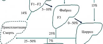
Depending on the time of onset of the first manifestations and the degree of liver damage, the following types of disease are distinguished:
1. Perinatal type - the most common:
- accompanied by oligohydramnios and pulmonary hypoplasia;
- 75% die within 24 hours of birth;
- minimal degree of liver fibrosis;
2. Neonatal type - minimal degree of periportal liver fibrosis;
3. Infantile type - moderate periportal fibrosis;
4. Juvenile type - pronounced periportal fibrosis in combination with portal hypertension, splenomegaly and portosystemic varicose veins.
Causes of polycystic kidney disease in adults
“Polycystic disease is a genetic disease caused by the degeneration of the kidney parenchyma with the formation of multiple cysts of various sizes,” says urologist-andrologist Alexander Danilov.
– This occurs as a result of a disruption in the formation of the functional units of the kidneys - nephrons - in the prenatal period. Some nephrons in polycystic disease do not connect to the collecting system of the kidney. This causes liquid to accumulate in them. They turn into cavities with fluid - cysts. There are from 20 to 50 such cysts. Externally, the affected kidney resembles a bunch of grapes. Cysts can be of different sizes and shapes, and the affected organ can weigh up to 2 kilograms. However, in adults with polycystic disease, the kidney retains a sufficient amount of fully functioning kidney tissue. Therefore, clinical manifestations of the disease may appear late or be completely absent.
Pathological anatomy
Macroscopic structure
The fetus has a characteristic appearance (Potter's face): narrow eyelid slits, a characteristic groove under the eyelid line, micrognathia, a flattened nose, soft large ears of an abnormal shape. The kidneys are increased in size, while maintaining their bean-shaped shape, but cannot perform their function. Poorly functioning kidneys do not produce enough fetal urine, leading to oligohydramnios and pulmonary hypoplasia. The hydrostatic pressure of the amniotic fluid ensures the normal development of the respiratory system.
Figure 1 | Fetus, 23 weeks. Fetal death after premature birth was due to pulmonary hypoplasia, caused by oligohydramnios due to PBP. Enlargement of both kidneys with displacement of the abdominal organs in combination with lung hypoplasia.
Figure 2 | Longitudinal section of a kidney with PCB.
Microscopy
Kidneys Radially oriented dilated collecting ducts form renal cysts with a diameter of 1–2 mm, between which normal glomeruli and tubules are visible.
The size of cysts can vary depending on age. In the early stages, the disease begins with microcysts, which then grow and turn into macrocysts. Cystic kidney disease is accompanied by slight interstitial fibrosis of the renal parenchyma.
Figure 3 | Microscopic specimen of a kidney from a patient with PBC-AR. Twenty-fold magnification, hematoxylin-eosin staining. ✱ — radial renal cysts; ▼—renal capsule. Arrows indicate normal glomeruli between dilated collecting ducts.
Liver Histological changes in the liver include the following malformations of the ductal plate: biliary hyperplasia, biliary ectasia and periportal fibrosis.
Disturbances in the morphogenesis of the development of the bile ducts lead to their dilatation. With subsequent progression of the disease, the dilated ducts turn into macrocysts connected by normal ducts, which allows them to be verified quite well using MRCP.
Figure 4 | Pathological changes in the liver. a) The structure of the normally branched portovenous and ethmoidal systems of the biliary tract (left) is disrupted due to a defect in the development of the ductal plate and errors in the terminal differentiation of cholangiocytes (right); b) Coronal T2-weighted image of the abdominal cavity: - the arrow indicates a cystic fusiform dilatation of the bile ducts; — arrow like an arrowhead — nephromegaly with small cysts; ✱ — splenomegaly. c) Preparation of a liver fragment, hematoxylin-eosin staining, 40x magnification:
✱ — extensive fibrosis of the portal area;
arrow - dilated tortuous bile ducts; arrowhead type arrow —hypoplasia of tributaries of the portal vein.
Symptoms of polycystic kidney disease in adults
In many ways, the symptoms depend on the age at which the first signs of pathology appear.
If they occur in the newborn period, the pathology has an unfavorable course and quickly leads to death due to the development of uremia (poisoning with metabolic products that should be excreted by the kidneys). If it is adult polycystic disease, it typically develops slowly and progressively. The disease occurs in several stages - compensated, subcompensated and then transition to decompensation.
In the compensation stage, there are initially no symptoms. Over time, you may experience:
- feeling of pressure in the lumbar region;
- abdominal pain of varying nature;
- urinary disorders due to stretching of kidney tissue;
- frequent fatigue;
- headache;
- blood in urine.
Basic kidney functions are not impaired at this stage.
At the stage of subcompensation, the first signs of kidney failure appear:
- nausea and dry mouth, thirst;
- attacks of headache or migraine;
- very persistent increase in blood pressure;
- polyuria (excessive urine output) with low density;
- blood in the urine, casts, leukocytes (if pyelonephritis is associated);
- fever, toxicosis, chills, if the cysts suppurate;
- attacks of renal colic if urolithiasis is associated.
In the stage of decompensation, constant uremia (urinary bleeding) is formed. Without hemodialysis, the patient feels extremely unwell and serious complications arise.
Compensation stage
In the compensatory stage, manifestations of polycystic disease are most often characterized by general symptoms: fatigue, general weakness, dysuric phenomena. The patient may also complain of pain in the lumbar region (aching, pressing, prolonged, not relieved by taking antispasmodic drugs). Pain may radiate to the lateral abdominal wall. The occurrence of painful sensations is associated with stretching of the renal capsule with an increase in the volume of the kidney affected by cysts of various sizes. Hematuria (blood in the urine) may occur. The exact cause of bleeding inside cavity formations has not been established. Hematuria can be intermittent and self-limiting. In rare cases, bleeding can be quite massive, requiring surgical intervention.
Diagnostics
In order to make a diagnosis, you need to carefully study the family history - most often familial cases of polycystic disease are identified. The doctor also conducts a full examination, palpating the abdomen - sometimes he can identify enlarged, lumpy kidneys. But the diagnosis can be confirmed only after tests and additional examinations:
- Ultrasound of the kidneys;
- nephroscintigraphy (using radioisotope substances);
- excretory urography (with the introduction of a contrast agent and x-ray of the kidneys);
- CT or MRI of the kidneys;
- selective angiography (to determine how the kidney is fed with blood);
- intravenous urography - it helps to determine significant enlargement of the kidneys on both sides, such as deformation of the calyces and pelvis.
Additionally, urine samples according to Reberg, Zimnitsky, biochemistry of blood, urine, and urine culture for sterility may be prescribed to exclude infection. Genetic research is also carried out.
Additional examination methods
Laboratory:
- clinical urine analysis;
- urine analysis according to Nechiporenko (the number of formed elements in the urine);
- urine analysis according to Zimnitsky (allows you to determine the excretory and concentration function of the kidneys);
- clinical blood test;
- biochemical blood test (level of creatinine, urea - end products of protein metabolism, Ca, K, P);
- bacteriological culture of urine.
Instrumental:
- Ultrasound of the kidneys. Determines the presence of cysts, expansion of the collecting apparatus;
- excretory urography is an x-ray method that is based on the introduction of contrast into the patient’s blood for the purpose of further assessing the excretory function of the kidneys;
- retrograde pyelography is also an X-ray examination method in which contrast is supplied through a urinary catheter. This method allows you to determine the state of the cavity structures of the kidneys;
- computed tomography (CT), nuclear magnetic resonance imaging: allows you to determine the number of cysts, their shape and size;
- renal angiography (determination of the vascular structures of the kidney);
- ECG (evaluation of heart function).
Differential diagnosis of polycystic kidney disease must be carried out with glomerulonephritis, pyelonephritis, and tumor diseases of the kidneys.
Modern methods of treatment
– Since polycystic disease is a genetic disease, there is no specific treatment.
That is, it is not possible to prevent its development, says Dr. Alexander Danilov. – The main thing is symptomatic treatment. It is divided into conservative and surgical. Conservative treatment. Its goal is to reduce the manifestations of the disease and prevent complications. For example, when blood pressure increases, antihypertensive therapy is carried out. With the development of kidney inflammation (pyelonephritis), antibacterial drugs are used. For protein deficiency - amino acid preparations. If anemia develops (decreased hemoglobin concentration in the blood), use iron supplements and erythropoietin.
If the amount of urine produced decreases (reduced diuresis), diuretics may be prescribed. In case of severe renal dysfunction, hemodialysis or peritoneal dialysis is performed.
Surgery. If the cysts rapidly increase in volume, a puncture or laparoscopic excision is performed. Surgical reduction of the volume of cysts leads to reduced compression of the remaining kidney tissue and improved blood circulation in it. This allows the remaining normal kidney tissue to function properly. In cases of severe renal failure, a kidney transplant is performed.
Pathogenesis
Polycystic kidney disease of the autosomal dominant and autosomal recessive type are classified as ciliopathies. In this condition, the normal functioning of cilia on the surface of cells is disrupted. These cilia ensure the reception of signals from the environment outside the cells. The cilia are located on the side of the lumen of the renal tubules, and this provides the sensory function of the tubules, that is, sensitivity to urine flow. Due to the fact that signals are not perceived correctly, cyclic adenosine monophosphate .
The formation of multiple cysts occurs as a consequence of too high proliferation and differentiation of the epithelium of the nephron tubules. As a result, instead of renal canals, cysts are formed - fluid-filled vesicles. As a result, the kidney grows very much - sometimes its weight is several tens of kilograms.
The occurrence of cysts occurs in only 2-5% of nephrons, but large-volume cysts compress healthy nephrons in the neighborhood, and the kidney does not perform a filtering function.
Since primary cilia are also present in the cells of other organs, polycystic liver and pancreatic diseases often develop in parallel with this disease.
Prevention
If there is a genetic predisposition to the disease, the development of polycystic kidney disease cannot be prevented by preventive methods.
- It is important for patients to adhere to a diet low in salt and fat. It is also necessary to control fluid intake to prevent overload of the kidneys.
- It is important to undergo regular examinations to monitor the condition.
- You should not engage in contact sports (boxing, rugby).
- It is important to give up bad habits.
Diet
Diet for polycystic kidney and liver disease
- Efficacy: therapeutic effect after 2 weeks
- Terms: 1 month or more
- Cost of products: 1200-1400 rubles. in Week
The progression of polycystic disease can be reduced if you strictly adhere to dietary recommendations. Meals should be organized in such a way that they help lower blood pressure levels. To do this you need to adhere to the following rules:
- Limit the amount of salt in your diet.
- Reduce the amount of fatty foods and protein foods. Animal fats should be replaced with vegetable fats.
- Eliminate all products containing caffeine - tea, coffee, chocolate.
- Drink enough liquid.
- You should reduce your intake of foods containing potassium and phosphorus.
- Food is best consumed warm.
- Meals should be fractional.
- Simple carbohydrates (baked goods, confectionery) should be minimized or eliminated.
The diet should contain the following products:
- Apples, grapes, grapefruit, kiwi.
- Cabbage, tomatoes, bell peppers, celery.
- Oatmeal, brown rice, barley.
- Low-fat fermented milk products.
- Vegetable soups.
- Rabbit, turkey, beef.
- Carp, bream, pike perch, hake, pike.
- Sugar-free jelly and compotes, herbal teas.
Completely excluded from the diet:
- Fatty dairy products.
- Soy products.
- Oranges, tangerines, bananas.
- Sausages, fatty meat, lard.
- Nuts.
- Mushrooms.
- Fatty fish.
- Pickles, canned food, smoked meats.
- Fast food.
- Alcohol.
During pregnancy
A woman diagnosed with polycystic kidney disease can become pregnant and carry a child to term. However, before conceiving, she must undergo a full examination and carefully follow all the doctor’s recommendations. It is the doctor who must determine whether pregnancy or whether additional treatment is necessary to improve kidney function.
Women diagnosed with polycystic breast disease should also consult a doctor. In general, any disease not only of the mammary glands, but also of other organs and systems should be a reason for consultation before pregnancy.