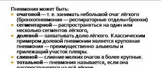
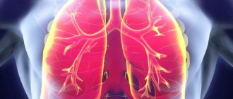
Lobar pneumonia is an acute infectious disease in which one or more lobes of the lung are affected, fibrinous effusion appears in the alveoli, and fibrinous deposits appear on the pleura. Croupous pneumonia affects mainly adults. The disease is characterized by a pronounced clinical picture and symptoms of intoxication. Patients with lobar pneumonia are admitted to the therapy clinic.
To examine patients at the Yusupov Hospital, doctors use modern equipment from leading European, American and Japanese companies. Pulmonologists use European treatment protocols and take an individual approach to the choice of treatment method for each patient. Medicines are administered through the digestive tract, intramuscularly, intravenously and by inhalation. Thanks to complex treatment, the length of stay of patients in hospital is reduced.
Signs of lobar pneumonia in adults
With lobar pneumonia, symptoms appear acutely. Key ones:
- a sharp increase in temperature to 39 - 40 ° C;
- severe chills, weakness;
- chest pain when breathing;
- cough - at first dry, but quickly becomes wet, with rust-colored sputum;
- severe shortness of breath;
- wheezing that may be heard when breathing;
- rapid heartbeat, low blood pressure;
- pale skin, nausea, muscle weakness.
The general condition is serious, the patient needs the help of doctors in the hospital.
Symptoms of lobar pneumonia
Lobar pneumonia has an acute onset. In patients in full health, body temperature rises to 39 ° C, chills and chest pain appear. In the initial stage of the disease, the cough is dry, then it becomes productive, with the release of “rusty” sputum. There is severe shortness of breath, the chest on the affected side lags behind when breathing.
In the initial phase of inflammation, percussion reveals a dull tympanic sound over the lesion. During auscultation, harsh breathing with prolonged exhalation, mild crepitus, and wet and dry rales in a limited area are heard. In the compaction phase of lobar pneumonia, the following symptoms appear:
- a sharp increase in vocal tremors, bronchophony during palpation of the chest;
- with percussion – dull sound;
- vesicular breathing is not audible, crepitus disappears, and pleural friction noise is often heard.
In the resolution phase, vocal tremors gradually normalize, bronchophony disappears, and abundant, sonorous crepitus appears over a long period of time. Loud fine-bubble rales are heard, bronchial breathing gradually gives way to harsh and then vesicular breathing.
When examining the cardiovascular system, a rapid pulse is determined. In the case of severe lobar pneumonia, it is poorly filled, arrhythmic, blood pressure is reduced, and the heart sounds are muffled.
Stages of lobar pneumonia in adults
Lobar pneumonia proceeds according to a special scenario, with a change of stages with typical changes in the lung tissue.
Stage one - high tide. A lot of blood flows to the lung tissue, which is why it turns red, and blood stagnates in small vessels. The stage lasts from 12 - 14 hours to 3 days.
Stage two – red liver. During this period, some of the fluid and red blood cells leak out of the small vessels of the lungs into the alveoli, which is why they work poorly. The affected part of the lung becomes dense, similar in appearance to liver tissue. Fibrin accumulates inside the lung sacs, a sticky substance that gives the tissue its density. This stage lasts up to 3 days.
Stage three – gray hepatization. Red blood cells stop entering the lung tissue, they are replaced by leukocytes and fibrin, epithelium. Therefore, the color of the lungs changes to gray and greenish. The stage lasts from 2 to 6 days.
Stage four – resolution. Gradually, fibrin dissolves, the alveoli are cleared, the lungs are restored and begin to breathe normally. This process is the longest, can last up to 2 - 3 weeks.
How does infection occur?
The causative agents of lobar pneumonia are pneumococci types I-IV. Less commonly, the disease is caused by Friendler's diplobacillus. As a rule, inflammation manifests itself acutely, against the background of absolute health and lack of contact with infected persons. Based on this, we can conclude that the infectious agents were previously in the upper respiratory tract, but their reproduction was restrained due to the work of the immune system. Its weakening is one of the leading factors in the development of lobar pneumonia.
From the point of view of modern medicine, lobar pneumonia is considered an infectious-allergic disease. In the vast majority of cases, the disease develops as a result of infection of the lungs with type I and II pneumococci. The polysaccharide capsule of pneumococci ensures their virulence and also provokes pronounced sensitization of the body.
Conditions that are necessary for the development of lobar pneumonia:
- Primary penetration of pneumococci into the body and development of inflammation. Moreover, its focus may not be located in the lung tissue, but may have a different localization.
- Sensitization of the body to a certain type of pneumococcus and re-entry of infection.
So, lobar pneumonia develops under the condition of secondary infection with pneumococci. An important condition is the fact of re-infection at the peak of sensitization of the body to microbes of a certain type. They can enter the lungs through blood, lymph or airborne droplets.
If all conditions are met, a violent reaction develops in the body, which is similar to what occurs when a foreign protein is introduced. A whole chain of morphological changes is launched in the lungs, which were described by Laennec (flush, red and gray hepatitis, resolution). Inflammatory exudate accumulates in the alveoli, a significant proportion of which is fibrin.
Modern methods of treatment
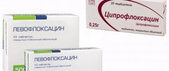
To treat lobar pneumonia, your doctor will prescribe two antibiotics.
One drug is administered intravenously, the second intramuscularly. Usually these are drugs from the group of penicillins and cephalosporins. Stronger medications are required less often. Additionally, complex therapy is required:
- immunocorrection (blood plasma and immunoglobulins are administered);
- correction of blood clotting disorders (intravenous blood thinning solutions, heparin);
- normalization of the protein composition of the blood (intravenous albumin, retabolil);
- oxygen (supplied through a nasal catheter or mask);
- hormonal drugs (for severe cases, prednisone is used for a short course);
- antioxidant therapy (ascorbic acid, rutin);
- bronchodilators (Berodual, Eufillin, Atrovent);
- expectorants (ACC, Bromhexine, Ambrobene, Lazolvan);
- antipyretic drugs (Nurofen, Paracetamol, Ibuklin);
- physiotherapy, massage, exercise therapy.
As the patient’s condition improves, he undergoes a course of rehabilitation to completely eliminate foci of inflammation in the lungs.
Features of the course of the disease in children
Children rarely experience fever and chills, and they do not complain of pain in the side.
An atypical course of lobar pneumonia is observed in young children. At the onset of the disease, there is no cough, but other symptoms occur: dry mouth, bloating, nausea and vomiting, abdominal pain, pale skin, rapid breathing, hyperexcitation or lethargy, enlarged liver. Sometimes there is a stiff neck, headache, convulsions, delirium and hallucinations. The combination of such symptoms can cause an incorrect diagnosis (meningitis). As pneumonia progresses, meningeal signs give way to the classic clinical picture of pneumonia.
Croupous pneumonia rarely develops in children. People aged 18-40 years are most susceptible to it.
In children 7-16 years old, symptoms do not differ from those that occur in adults. Body temperature stabilizes on days 5-9 from the onset of the disease. At the same time, inflammation in the lungs subsides.
Popular questions and answers
Pulmonologist Marina Samoilova told us why lobar pneumonia is so dangerous and how to treat it.
What complications can occur with lobar pneumonia?
If a person has a weakened immune system or treatment is started untimely or incorrectly, complications are possible in the form of:
- abscess (purulent melting of lung tissue); gangrene of the lung;
- purulent pericarditis (inflammation of the lining of the heart);
- purulent brain damage;
- blood poisoning;
- respiratory failure and even death.
Pneumococcal pneumonia, even in our time, is a fairly common cause of death, especially in older people, smokers, people suffering from alcoholism, with cancer and systemic diseases, and HIV-infected people.
Story
Pneumonia was first described by Hippocrates around 460 BC, but until the 19th century it was not known that it was an infection of the lungs and not a symptom of other diseases. The first clinical and pathological manifestations were described by the French physician René Laennec in 1819, and in 1842 the Austrian pathologist Karl Rokitansky was the first to differentiate pneumonia into lobar and bronchopneumonia. The infectious nature was discovered in 1875 by the German pathologist Edwin Klebs, who observed bacteria under a microscope, and in 1884 Karl Friedlander and Frenkel, Albert were able to identify the two most common pathogens of pneumonia at that time - Streptococcus pneumoniae (pneumococcus) and Klebsiella pneumonia. By 1929, the International List of Causes of Death had 94 terms for pneumonia; later in the ICD-10 classifier, many of the descriptive terms that included pneumonia were eventually removed, but it is still listed in descriptive terms for some infectious and noninfectious causes, including complications from certain diseases and procedures. In the 1930s, penicillin developed treatments for bacterial pneumonia, but pneumonia is still the leading cause of death among children under 5 years of age.
23.01.2021 7759
Is it possible to treat lobar pneumonia with folk remedies?
As a rule, patients with lobar pneumonia should be hospitalized for constant monitoring by medical staff and emergency measures at the slightest sign of complications.
Often such patients experience nausea, vomiting, bowel movements, and confusion, so medications and solutions are administered intravenously. Thus, we cannot talk about treatment at home without the participation of qualified specialists. Fortunately, in our time, subject to a healthy lifestyle, such serious diseases as lobar pneumonia are becoming less and less common, and with modern treatment methods, in most cases it is possible to avoid serious complications and save the patient’s life.
Published on the portal kp.ru
Prevention
Prevention of pneumonia is an important factor in reducing child mortality. Vaccination against Haemophilus influenzae type B, pneumococcus, measles and whooping cough is the most effective way to prevent pneumonia. For young children, nutrition plays an important role, particularly exclusive breastfeeding for the first 6 months of life. Reduces the incidence of illness among children and combats air pollution, for example, through the use of environmentally friendly cookstoves in residential buildings. In overcrowded homes, providing hygienic conditions can help reduce disease incidence.
In health care settings, effective measures to reduce the spread of infection include hand washing and the use of gloves and masks, and patient isolation is recommended.
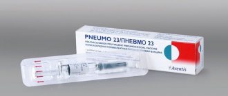
Vaccinal prevention
Vaccinal prevention of pneumococcal infections
According to the position of WHO and the Russian Respiratory Society, “Vaccination is the only way to prevent the development of pneumococcal infection.” In the Russian Federation, 94% of all etiologically deciphered cases of complicated pneumococcal infection in children are caused by pneumococcal community-acquired pneumonia. Pneumococcus is the cause of up to 76% of community-acquired pneumonia in Russian adults. For vaccination against pneumococcal infection of persons over 2 years of age in the USA since 1983, and in the Russian Federation since 1999, polysaccharide polyvalent vaccines containing antigens of 23 serotypes, causing up to 90% of invasive diseases of pneumococcal etiology, have been successfully used. Vaccination is carried out once, followed by revaccination for patients from high-risk groups (over 65 and immunocompromised individuals) after 5 years. The effectiveness of polysaccharide vaccines reaches 80%, but may be lower in elderly people, patients with immunodeficiency conditions, and in children under 2 years of age. These vaccines cause the formation of T-independent B-cell immunity.