Hydatidiform mole (MH) is a fatal disease for the fetus concerning the pathological development of the fetal egg and associated with the transformation of the chorion into cysts (protective membranes formed due to unfavorable conditions in certain life cycles, in the form of bubbles containing fluid) and the proliferation of the villi epithelium outer germinal membrane.
This pathological condition is accompanied by the following negative manifestations:
- bleeding;
- early toxicosis;
- enlargement of the uterus relative to the gestational age of the fetus.
PZ is diagnosed using a vaginal examination, ultrasound, fetal phonocardiography, and determination of the content of human chorionic gonadotropin (β-hCG).
Treatment includes techniques such as vacuum aspiration, curettage of the uterine cavity, and removal of the organ (hysterectomy).
What is a hydatidiform mole?
Hydatidiform mole is a pathology that is characterized by modification of the membrane tissue (chorionic hyperplasia) into peculiar cysts. These are cavities, bubbles filled with liquid contents. Next, the growth of such chorionic villi occurs, which is accompanied by the death of the embryo developing in utero.
This pathology belongs to trophoblastic diseases. The incidence of such a diagnosis is about 0.5%. Characteristic features of the pathological condition are swelling and a sharp proliferation of villi tissue, the formation of bubble-like formations. In its form, this process resembles a bunch of grapes. The size of these bubbles is quite impressive. Can be about 2-2.5 centimeters in diameter. There is practically no blood flow in such bubbles.
Reasons for the development of the anomaly
There are several reasons for the formation of such a pathological condition as hydatidiform mole:
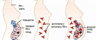
- Chromosomal abnormalities resulting from the prenatal period. With the complete variant of the pathology, there is a complete loss of maternal genetic material. The same situation can arise when two male reproductive cells fertilize an inferior egg without a nucleus.
- Viral or bacterial infection of the chorionic villi, leading to the above changes.
- Multiparous women, as well as young primigravidas, are at risk of developing the process.
- Immunodeficiency states.
- Thyrotoxicosis.
Causes of bladder diarrhea
There are several possible causes of hydatidiform mole. These include:
- A genetic error in which two sperm penetrate into one egg at once. Moreover, if the egg turns out to be nuclear-free, then a complete hydatidiform mole occurs. If the egg has a nucleus, then a partial pregnancy may occur.
- Various infectious diseases. Most often, this disease is caused by Toxoplasma viruses.
- Lack of the hormone estrogen.
Predisposing factors for the development of hydatidiform mole include:
- multiple abortions;
- thyrotoxicosis;
- age (20 - 24 years and 40 - 49 years);
- a large number of births;
- immunodeficiency;
- lack of vitamin A;
- consanguineous marriages.
Types of hydatidiform mole
- Complete hydatidiform mole is a condition in which all chorionic villi are subject to pathological changes in the form of stromal edema, trophoblast hypertrophy and the formation of vesicles, while the signs of a developing embryo are completely absent. All tissue is affected. This pathology is most often detected at 10-12 weeks of gestation. Such a hydatidiform mole contains a diploid number of chromosomes: 46 XX. It is worth saying that all chromosomes are paternal. There may be a karyotype of 46 XY, however, both X and Y are also paternal. The danger of this pathology is that in twenty percent of cases this process can become malignant with the formation of many metastases.
- Partial hydatidiform mole is characterized by the presence of some intact tissue. Intact parts of the embryo and placenta may be identified. The chromosome set is defined as 69 XXX, 69 XXX. The set contains 1 maternal chromosome. The frequency of transition to a malignant form of the process is about five percent.
- Invasive hydatidiform mole. This pathology is a process in which chorion grows into the myometrium of the uterus. This is a severe pathology that can lead to massive bleeding during removal of hydatidiform mole tissue.
According to histological structure, pathology is also divided into:
- Syncytial type;
- Cytotrophoblastic;
- Mixed form of hydatidiform mole.
Malignant gestational trophoblastic disease
Pathogenesis . In 10% of patients with HTC, a persistent (malignant, malignant) form of the disease develops. Malignant HTC is divided into 3 histological types:
1) invasive hydatidiform mole;
2) choriocarcinoma;
3) trophoblastic tumor of the placental plane.
In 50% of cases, malignant GTX develops months and years after hydatidiform mole. The remaining 25% of cases occur after a normal pregnancy, another 25% after a spontaneous miscarriage, ectopic pregnancy or abortion.
Due to different treatment and prognosis, malignant HTC is also divided into non-metastatic (does not spread beyond the uterus) and metastatic (spreads beyond the uterus). Metastatic disease, in turn, may have a favorable or unfavorable prognosis depending on the time interval after the previous pregnancy, the level of hCG, the presence of metastases in the brain and liver, the type of previous pregnancy, and the results of previous chemotherapy.
Clinical manifestation. Although the three forms of malignant HTC are histologically distinct, clinical severity has more prognostic value than histological features. Gestational trophoblastic disease occurring after hydatidiform mole is typically diagnosed by the presence of a plateau or elevated r-hCG level during the monitoring period after evacuation of the hydatidiform mole. Unlike other forms of HTC, trophoblastic tumor of the placental plane is characterized by chronically low levels of beta-hCG. Patients with r-hCG levels >100,000 mIU/ml, enlarged uterus, and the development of a theca-luteal cyst have the highest risk of malignant HTC. Choriocarcinoma tends to manifest as metastatic disease.
Treatment . Gestational trophoblastic disease is extremely sensitive to chemotherapy. Treatment of non-metastatic HTC is with a single agent, usually methotrexate or actinomycin. Methotrexate is an antimetabolite that stops cell development in phase 8 of the cell cycle by binding to dihydrofolareductase, which prevents the reduction of dihydrofolate to tetrahydrophiolic acid. This in turn inhibits thymidylate synthetase and purine production, reducing DNA, RNA and protein synthesis.
Side effects of methotrexate treatment
With a favorable prognosis, metastatic HTC is of course treated with one chemotherapy agent; with an unfavorable prognosis, polychemotherapy is used. The effectiveness of such treatment is 90-100% for GTX with a favorable prognosis and 50-70% for GTX with a poor prognosis.
Surgery does not play a significant role in the treatment of malignant gestational trophoblastic disease, with the exception of placental plane trophoblastic tumor, which is insensitive to chemotherapy and can be treated by hysterectomy. Radiation therapy is usually reserved for the treatment of distant metastases in the brain and liver.
Monitoring of patients . As with other forms of HTC, careful monitoring of r-hCG levels is necessary when managing patients with malignant disease. During this period, patients should be advised to use contraception in order to accurately assess r-hCG levels.
Symptoms
There are several signs for the clinical picture of hydatidiform mole:
- Dark bloody discharge from the genital tract, which contains bubbles rejected from the site of hydatidiform mole. The volume of such bleeding can vary significantly, ranging from spotting to massive bleeding.
- If the bleeding is large or prolonged, the woman may develop anemia. Its symptoms include weakness, fatigue, pale skin, rapid heartbeat, and sometimes episodes of loss of consciousness.
- In the invasive form of this pathology, when chorionic villi grow into the wall of the uterus (myometrium), pain in the lower abdomen may occur. When a perforation of the uterine wall occurs, massive bleeding into the abdominal cavity may occur, which is accompanied by acute abdominal pain and loss of consciousness.
- The size of the uterus is not commensurate with gestational age. When such a pathological condition develops, the uterus is larger than the period calculated from the first day of the last menstruation.
- Toxicosis during hydatidiform mole is often several times more pronounced. Symptoms quickly increase, nausea and vomiting are debilitating, and liver and kidney failure may occur. This quickly leads a woman to a moderate or even severe state.
Benign gestational trophoblastic disease
Benign gestational trophoblastic disease is represented by hydatidiform mole, which accounts for 80% of GTX cases. More than 90% of cases of hydatidiform mole are classified as classic, or complete hydatidiform mole, and are a consequence of molar degeneration in the absence of a fetus. In 10% of cases of molar pregnancy, there is a partial or incomplete hydatidiform mole, which is represented by molar degeneration in combination with the presence of an abnormal fetus. Hydatidiform mole in 90% of cases is a benign disease. The remaining cases are represented by invasive hydatidiform mole, choriocarcinoma, or trophoblastic tumor of the placental plane.
Complete hydatidiform mole
Pathogenesis . Although the cause of this condition remains unknown, most cases of complete hydatidiform mole are believed to result from the fertilization of an anucleate egg (an "empty egg"), in which the nucleus is lost or nonfunctional, with a single normal sperm. So, all chromosomes of the fertilization product in this case are of paternal origin. In 90% of cases, a complete hydatidiform mole has a karyotype of 46, XX, in 10% of cases - 46, XV. In rare cases, a complete hydatidiform mole occurs when two normal sperm fertilize an empty egg. So, in this case too, all chromosomes of the fertilization product are of paternal origin.
Placental anomaly in complete hydatidiform mole is characterized by trophoblast proliferation and hydropic degeneration or absence of fetal parts. Although most cases are benign, complete hydatidiform mole has a greater malignant potential than complete mole.
Epidemiology . The incidence of hydatidiform mole ranges from 1:200 to 1:1000 pregnancies around the world, increasing in Asian women.
Risk factors . Gestational trophoblastic disease occurs during reproductive age (20-40 years). An increase in the incidence of the disease occurs in those geographical areas where the diet is poor in beta-carotene and folic acid. More often, GTX develops in women who have a history of spontaneous miscarriages or hydatidiform mole.
Symptoms
History . The most common symptom of a hydatidiform mole is irregular or significant vaginal bleeding in early pregnancy (97% of cases). Bleeding is usually painless but may be associated with uterine contractions. But thanks to modern early diagnosis of hydatidiform mole, these complications are rarely observed.
Symptoms of hydatidiform mole require differentiation from other conditions associated with increased hCG levels: severe nausea and vomiting - with uncontrollable vomiting of pregnancy; irritability, dizziness, photophobia - with preeclampsia; nervousness, anorexia, tremor - with hyperthyroidism. Preeclampsia before 24 weeks of pregnancy has a strong correlation with hydatidiform mole.
Objective examination . In the case of a complete hydatidiform mole, gynecological examination may reveal expulsion of racemose molar lobes into the vagina or bleeding from the cervical canal. Sometimes bilateral theca-luteal cysts may be found, resulting from stimulation of the ovaries by high levels of hCG. They show inconsistency of the uterus for gestational age, absence of heartbeat and fetal movements. Clinical examination may reveal signs of hyperthyroidism or preeclampsia: tachycardia, tachypnea and hypertension.
Diagnostics . In a hydatidiform mole, the hCG level is significantly higher (>100,000 mIU/ml) than in a normal pregnancy. The diagnosis is confirmed by ultrasound examination, which reveals “blizzard syndrome” due to swelling of the chorionic villi. In the case of complete molarity, the fetal elements are not visualized during ultrasound examination.
Differential diagnosis is performed when there is an increase in hCG levels and / or an enlarged placenta (multiple pregnancy, non-immune hydrops fetalis, intrauterine infection, uterine fibroids, threatened abortion, ectopic and intrauterine pregnancy).
Treatment of hydatidiform mole, regardless of the stage of pregnancy, consists of urgent removal of the contents of the uterus. If the patient wishes to preserve fertility, vacuum aspiration of the contents of the uterus is performed, followed by curettage of its cavity with a sharp curette. After removing the entire mass of the hydatidiform mole, intravenous oxytocin is administered to stimulate uterine contractions and minimize blood loss. If the patient does not wish to preserve her fertility, a hysterectomy may be an alternative treatment option. The risk of recurrence is 3-5%, even with hysterectomy.
Monitoring of patients . The prognosis for hydatidiform mole is favorable (95-100% of cases of effective treatment). Persistence of the disease is observed in 15-25% of patients with complete and in 4% with partial drift. So, careful monitoring of patients is necessary.
After evacuation of the hydatidiform mole, control studies of r-hCG levels are carried out weekly until three normal results are obtained. After this, r-hCG levels are monitored monthly for less than 1 year. During the monitoring period, the patient should abstain from pregnancy.
Patients after successful treatment of hydatidiform mole do not have an increased risk of spontaneous abortions and congenital anomalies. The risk of developing HTC in a subsequent pregnancy is 1-5%.
Incomplete hydatidiform mole
Pathogenesis . Incomplete hydatidiform mole is formed due to the fertilization of a normal egg by two sperm at the same time. This results in the development of a triploid karyotype with 69 chromosomes, of which two chromosome sets are of paternal origin.
Placental anomalies in incomplete hydatidiform mole are accompanied by local hydropic villi and syncytiotrophoblast hyperplasia.
Incomplete hydatidiform mole is often accompanied by the presence of an accompanying fetus with a triploid genotype and numerous anomalies. Most fetuses associated with hydatidiform mole can survive only a few weeks in the uterus before spontaneous abortion occurs in the late first to early second trimester of gestation. Incomplete hydatidiform mole is usually more benign and has less malignant potential than complete mole.
Clinic and diagnostics . Incomplete hydatidiform mole manifests itself as delayed menstruation and symptoms of pregnancy. In 90% of patients with incomplete hydatidiform mole, a miscarriage occurs. Symptoms and signs of incomplete hydatidiform mole are similar to those of complete mole, but are usually less pronounced. The size of the uterus may initially correspond to the gestational age, and fetal cardiac activity may be present. Therefore, incomplete hydatidiform mole can be diagnosed later. The diagnosis is determined by histological examination of the fertilization product.
Treatment for partial molar pregnancy involves urgent evacuation of the contents of the uterus. In less than 4% of patients, incomplete hydatidiform mole can progress to a malignant disease.
Monitoring of patients consists of serial testing of r-hCG levels according to the same principles as in the case of complete hydatidiform mole. To accurately control r-hCG levels, pregnancy prevention is important.
Diagnostics
Gynecological examination.
- When examined in the doctor's chair, you should be alert to the uterus, which is revealed to be much larger in size than the expected gestational age based on the date of the last menstruation.
- Dense consistency of the uterus with areas of strong softening.
- Bloody discharge from the genital tract with blisters.
Ultrasound examination, which is the most informative method for diagnosing this pathological condition. Ultrasound signs of hydatidiform mole include:
- Increased size of the uterus, which does not correspond to the timing of menstruation;
- The presence of a fine-grained tissue structure in the uterine cavity, reminiscent of a “blizzard”. This is a characteristic sign of a hydatidiform mole. This ultrasound picture is also sometimes called a “bunch of grapes”.
- A frequent occurrence during ultrasound diagnosis of the uterine appendages with such pathology is the presence of thecal lutein cysts.
- Blood sampling to determine hCG levels. Indeed, with hydatidiform mole, its level may exceed the borderline norms.
- If there is a suspicion of metastasis of a hydatidiform mole, a computed tomography or MRI is prescribed.
Treatment
The treatment tactics for hydatidiform mole are based on the evacuation of pathological tissue from the uterus (removal of hydatidiform mole). This can be done using the method of vacuum aspiration of the contents of the female reproductive organ. And then curettage of the uterine cavity is performed. If there is a condition that threatens the patient's life, for example, bleeding from an invasive mole, then doctors may decide to remove the uterus.
After evacuation of the contents from the uterine cavity, mandatory monitoring of the level of hCG in the blood is carried out weekly. If the hCG level does not fall over time, then chemotherapy treatment is necessary. Once every two weeks, ultrasound diagnostics of the uterine cavity and appendages is performed.
If no signs of chorionepithelioma are found, then chemotherapy is not performed, and the woman is prescribed clinical observation for a period of two years.
Indications for urgent medical attention
- High blood pressure.
- Heavy bleeding.
- Severe nausea and vomiting.
- Nagging pain or various pain symptoms in the uterus and appendages.
Pregnancy is a very difficult and at the same time responsible period for every woman. She must listen to what signals her body gives both for her own sake and for the future baby. Only a timely visit to a doctor is often the only opportunity to give birth to a child and maintain your own health.
New pregnancy after hydatidiform mole
The most pressing question after a case of hydatidiform mole is whether pregnancy can occur after a hydatidiform mole.
The diagnosis of hydatidiform mole is not a death sentence. With proper and timely diagnosis and proper treatment, pregnancy is possible. An important point is the timely administration of chemotherapy after a hydatidiform mole in the absence of a drop in hCG levels.
Pregnancy after a hydatidiform mole simply needs to be planned. If chemotherapy was not prescribed after such a pathological condition, then the minimum period of absence of pregnancy is 6-12 months. If chemotherapy was nevertheless prescribed, then the recommended break is 12-24 months. The main point in planning such a pregnancy is the appointment of competent contraception by the obstetrician-gynecologist who is caring for you.
Trophoblastic tumor of the placental plane
Pathogenesis . Trophoblastic tumor of the placental plane is very rare and occurs at the site of placental implantation. The cells of this tumor infiltrate the muscular layer of the uterus, grow between smooth muscle fibers and gradually invade the myometrium and blood vessels. Histologically, the typical feature of this tumor is the absence of villous tissue and cytotrophoblast proliferation.
Symptoms and diagnosis . The most common symptom of a trophoblastic tumor of the placental plane is uterine bleeding, which can occur several months or years after a previous pregnancy. Unlike other forms of HTC, this tumor produces chronically low levels of r-hCG and human placental lactogen.
Treatment . Another distinguishing feature of placental plane trophoblastic tumor is its insensitivity to chemotherapy. But this tumor rarely metastasizes beyond the uterus, so hysterectomy is the treatment of choice for this disease.