Cataracts are a serious pathology that, without proper treatment, can lead to complete loss of vision over time. Most often it appears in both eyes.
This disease of the visual system is characterized by clouding of the lens of the eye. The problem can be either congenital - diagnosed immediately after birth, or acquired - can appear at any age.
Congenital cataracts usually occur due to health problems in the pregnant woman and genetic abnormalities. Taking medications, bad habits, and prolonged exposure to a toxic environment also have an impact.
Acquired cataracts can appear at any age, but problems most often occur after 55 years of age. At this age, natural biological processes begin, the lens loses its elasticity, becomes cloudy and dense. In addition to age-related changes, other factors may influence the early appearance of cataracts:
- long-term use of medications, such as steroid therapy;
- other diseases of the visual system;
- eye and head injuries;
- X-ray irradiation;
- negative effects of ultraviolet and infrared rays;
- poor lifestyle, vitamin deficiency.
If we talk about natural processes, cataracts develop in every person and it is only a matter of time. Some people may experience significant changes in just a few months, while others may not be aware of the presence of the disease for many years.
Treatment with conservative methods is ineffective and makes no sense. The only way to get rid of the problem is through cataract surgery.
Primary cataract – what is it?
Cataract is an age-related disease that manifests itself under the influence of negative factors: genetic disruptions, endocrine disorders, eye injuries, taking potent medications, concomitant ophthalmological diseases, etc. Hereditary predisposition also plays a significant role in the development of the disease.
There are 4 stages of cataract development:
- Initial, in which only peripheral damage to the lens is diagnosed.
- Immature, in which the clouding gradually spreads to the optical zone. At this stage, the patient notices a decrease in visual acuity. Surrounding objects are visible as if through a foggy haze.
- Mature. The area of opacity covers the entire lens. At this stage, the patient no longer sees anything, but only feels light.
- Overripe. Complete blindness and disintegration of the lens occurs. At this stage, the damaged fibers disintegrate.
Cataract ranks first in the world among diseases leading to vision loss.
Cost of services for cataract treatment
| № | Service name | Price in rubles | Make an appointment |
| 2009003 | Optical-reconstructive intervention in the anterior segment of the eye for cataracts and post-traumatic and post-traumatic changes | 90000 | Sign up |
| 2008047 | Phacoemulsification for complicated, mature and overmature cataracts, category 3 of complexity | 86880 | Sign up |
| 2008046 | Phacoemulsification for complicated, mature and overmature cataracts, category 2 of complexity | 79650 | Sign up |
| 2008045 | Phacoemulsification for complicated, mature and overmature cataracts, category 1 of complexity | 77400 | Sign up |
| 2008044 | Phacoemulsification for initial and immature cataracts, category 3 of complexity | 71220 | Sign up |
| 2008043 | Phacoemulsification for initial and immature cataracts, category 2 of complexity | 67080 | Sign up |
| 2014001 | Penetrating keratoplasty + phacoemulsification or cataract extraction with IOL implantation (complexity category 2) | 96000 | Sign up |
| 2014003 | Penetrating keratoplasty + reconstruction of the anterior chamber with iris plastic surgery, phacoemulsification or cataract extraction with IOL implantation | 120000 | Sign up |
| 2014005 | Deep anterior lamellar keratoplasty + phacoemulsification or cataract extraction with IOL implantation (complexity category 2) | 108000 | Sign up |
| 2014007 | Posterior layered endothelial keratoplasty + phacoemulsification or cataract extraction with IOL implantation | 84000 | Sign up |
| 2008041 | Discision of secondary cataract | 9000 | Sign up |
| 2008053 | A set of consumables and an imported intraocular lens for phacoemulsification of cataracts with cataract removal. | 42000 | Sign up |
| 2008005 | Ultrasound phacoemulsification with IOL implantation for initial and immature age-related cataracts | 79650 | Sign up |
| 2008007 | Ultrasound phacoemulsification with IOL implantation for complicated, mature and overmature age-related cataracts | 84440 | Sign up |
| 2008012 | Cataract removal without phacoemulsification + IOL | 40200 | Sign up |
| 2008021 | Cataract extraction with implantation of an artificial lens of the first category of complexity | 40500 | Sign up |
| 2008022 | Cataract extraction with implantation of an artificial lens of the second category of complexity | 45600 | Sign up |
| 2008023 | Cataract extraction with implantation of an artificial lens of the third category of complexity | 50400 | Sign up |
Making an appointment Today registered: 21
What to do when diagnosing cataracts at an early stage
The only effective and sufficient method of treating cataracts is surgery, during which the clouded lens is removed and an implant is installed in its place. Modern implants can restore vision. The patient notices the result almost immediately after the operation, which lasts no more than 15 minutes. Within an hour, vision returns, but not yet in full; the maximum effect will occur after the prosthesis has completely engrafted and the wound has healed.
Does this mean that initial cataracts should not be treated, since the patient is not in danger of going blind? Not really. Symptomatic treatment can be prescribed, as well as eye drops that prevent the development of the disease. Most often at this stage, general strengthening drugs, vitamins, minerals, antioxidants, and catalin are prescribed. However, these remedies can only slightly slow down the regression, but not stop it completely. Cataracts will inevitably progress and, if the patient refuses surgery, eventually complete blindness will occur.
It is also not recommended to perform surgery at the initial stage. Since the damage to the lens is still small, it does not cause physical discomfort and does not cause an aesthetic defect. Surgical intervention is indicated at the second stage, when the lesion is already noticeable and interferes with normal life activities. The doctor will recommend surgery if there is 50% damage to the lens. If the patient wishes, the implant can be installed at the first external signs of the disease.
Important : after the initial diagnosis of cataracts, it is necessary to undergo regular examinations by an ophthalmologist.
Since in the later stages the cloudy lens becomes deformed, which leads to an increase in IOP and the development of glaucoma. It is also possible that other concomitant diseases will appear that will not be noticed by the patient himself against the background of cataract symptoms. The Clean View clinic provides a full range of procedures for successful cataract removal. More details by phone +7 (499) 141-13-75.
SYMPTOMS
Cataracts significantly impair a person's quality of life. At the initial stage, patients may not be aware of the presence of a problem.
Over time, warning signs appear:
- the perception of colors changes. Patients feel that the color rendering of the TV has become worse and want to increase the brightness;
- photosensitivity appears;
- in twilight and in poor lighting conditions, vision deteriorates;
- tearing appears;
- spots, stripes, circles appear before the eyes;
- double vision is observed, objects become blurry;
- There is difficulty in selecting points.
Features of complicated cataracts
Complicated cataract has its own differences:
- Localization. Cloudiness of the lens occurs in the cortical zone, adjacent to the anterior or posterior capsule.
- Morphology. A cataract is an irregular, cloudy area that includes air bubbles and resembles a “pumice stone.” The color of the pathological area has a yellowish, brownish or grayish tint. As the process progresses, this opacification spreads to the middle lens layers.
- Symmetry. Complicated cataracts can be unilateral (if one eye is affected by an ophthalmological disease) or bilateral (in the case of general diseases).
- Progression. In the absence of adequate treatment of the primary disease, complicated cataracts can progress.
Treatment
In cases where a complicated cataract leads to a significant decrease in vision that interferes with the patient’s usual activities, surgery to remove it is recommended.
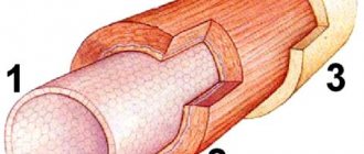
The essence of the operation is the surgical removal of the cloudy lens, replacing it with an intraocular lens (IOL). An intraocular lens is an optical system that will subsequently assume the function of refraction. During the operation, a micro-incision is made on the capsule, then the lens substance is crushed with ultrasound and removed from the eye. An intraocular lens is implanted into the remaining capsular bag.
The right moment for surgery is the time of stabilization of complicated cataracts, with simultaneous compensation of the underlying disease (ophthalmological or general). For this purpose, they relieve the acute inflammatory process in the tissues of the eye, achieve remission of rheumatoid arthritis, compensate for the level of glycemia in the case of diabetes, etc. Contraindications to surgical intervention for complicated cataracts may include:
- Blindness. A complete lack of light perception gives an unfavorable prognosis for vision, even if the operation is successfully performed.
- IOP instability. Before performing phacoemulsification, it is necessary to achieve stabilization of IOP figures.
When contacting the Moscow Eye Clinic with complicated cataracts, the patient is guaranteed a comprehensive diagnosis with determination of the root causes of the disease and the development of an individual treatment plan, which will include the participation of doctors of other specialties.
Publications in the media
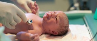
Congenital cataracts are observed in 5 cases out of 100,000 newborns; it accounts for 10–38% of cases of childhood blindness.
Classification • Coronal (coronary) - opacities are located radially in the deep layers of the cortex of the peripheral parts of the lens and are club-shaped, light gray or greenish-blue in color • Fusiform - fusiform opacities are located along the axis of the lens • Discoid - disc-shaped opacities are located between the nucleus and posterior pole of the lens • Zonular (layered) - lens opacities are located in the form of 1-3 concentric layers around its core • Stellate cataract (lens suture) is characterized by the presence of point opacities in the area of the embryonic sutures of the lens, forming a star figure • Cataract of the anterior embryonic suture - stationary cataract , characterized by multiple point white opacities in the area of the anterior embryonic suture of the lens • Polar cataract - a small round grayish-white opacification, often of a layered structure, located at one of the poles of the lens • Pyramidal cataract - polar cataract, opacification in the form of a cone, the apex is directed towards the pole lens
Genetic aspects. Types and genes: • 182500, SORD gene, 15q15; • blue, type 1, CCA1, 115660, 17q24; • blue, type 2, 601547, CRYB2 (123620), 22q11.2 q12.2; • zonular Marner, CTM, 116800, 16q22.1; • zonular powder, type 1, 116200, GJA8, CX50, CAE1, 600897, 1q12 q21; • zonular powder, type 2, CAE2, CZP2, 601885, 13q11 q12; • zonular, CCZS, 600881, 17q11 q12; • with hyperferritinemia 600886, FTL, 134790, 19q13.3 q13.4; • with microphthalmia, CATM, 156850, 16p13.3; • full, X-linked (?), CCT, 302200; • Volkmann, CCV, 115665, 1pter p36.13; • with late corneal dystrophy, PAX6, AN2, 106210, 11p13; • Coppock similar, CRYGA, CRYG1, 123660, 2q33 q35; • polar posterior, CPP, 116600, 1pter p36.1; • polar anterior, type 1, CTAA1, 115650, 14q24 qter.
Risk factors • Metabolic disorders: diabetes in the mother, galactosemia in the fetus • Infectious diseases of the mother in the first trimester of pregnancy: rubella, herpes, mumps, toxoplasmosis, etc.
Clinical picture • The disease is often asymptomatic • Sometimes parents notice disturbances in gaze fixation or strabismus in the child due to the lack of cortical control over the friendly contractions of the external muscles of the eye • Leukocoria (light reflection from the pupil - a visible manifestation of clouding of the lens), nystagmus, signs of the underlying disease ( eg, Down syndrome, rubella) • Lens opacities are detected immediately after birth or within 3 months.
Differential diagnosis • Tumors of the external part of the visual analyzer, pathways • Retinopathy.
Treatment • The most optimal surgical treatment is in the first months of life (cataract extraction) under anesthesia, which allows for the correct formation of the visual cortex and the retina itself, as well as adequate development of muscle coordination necessary for binocular vision •• Implantation of an intraocular lens is not indicated, since the eye is changes the refraction during the growth process •• For bilateral cataracts with visual acuity less than 0.005, not corrected with lenses, surgical intervention is indicated at the age of 2 to 6 months •• With less severe cataracts (visual acuity - below 0.3), object vision is possible, therefore, the operation is performed at the age of 2–5 years •• Postoperative care: to combat amblyopia, it is necessary to wear a patch on the healthy eye for a long time. Correction of the refraction of the operated eye is indicated, frequent repeated examinations of visual acuity •• If visual acuity is above 0.3 (improvement to 0.4 with pupil dilation), surgical extraction is not performed • With delayed surgical treatment, instillation of mydriatic agents into the eye is necessary 1-2 r/ weeks until surgery to ensure the full development of the retina, which often suffers from a lack of light reaching it, to prevent nystagmus, strabismus, and amblyopia.
Forecast. If congenital cataracts are not detected in a timely manner, the prognosis is often unfavorable due to the high risk of developing amblyopia.
ICD-10 • Q12.0 Congenital cataract.
Application. AMC syndrome (from: A taxia- M icrocephaly- C ataract, ataxia-microcephaly-cataract syndrome, 208870, r). Clinically: ataxia, mental retardation, microcephaly, congenital cataract, nystagmus.
Clinical picture of complicated cataract
The course of complicated cataracts has its own characteristics, which are determined by the clinical picture of the underlying disease, for example:
- With tuberculosis, pupil occlusion, iris atrophy, and adhesions may develop. The inflammatory process can involve the entire eyeball.
- With rheumatoid arthritis, the substance of the lens may harden (calcification process) or turn into a liquid mass with heterogeneous inclusions. The lens capsule becomes thinner.
- In glaucoma, the development of cataracts occurs with long-term elevated IOP values. The opacities are located close to the posterior capsule.
Secondary and membranous forms
- The secondary type occurs in an eye without a lens (aphakic) after extracapsular extraction. It is nothing more than the growth of the subcapsular epithelium, which remains in the equator zone of the lens bag. The absence of a nucleus provides special spatial space, so the epithelial cells do not stretch and grow freely, while they swell and take the form of small transparent balls of different sizes lining the posterior capsule. Biomicroscopy reveals their similarity to caviar grains or soap bubbles. After the names of the researchers who described secondary cataracts for the first time, they are called Adamyuk-Elschnig balls.
The initial stage in the development of the disease occurs without subjective symptoms. A decrease in visual acuity occurs when epithelial growths reach the central region.
Treatment of cataracts is exclusively surgical. To do this, an operation called discision of the posterior lens capsule is performed. This is a linear dissection within the pupillary area on which the Adamyuk-Elschnig balls are localized. As a rule, such procedures are performed using a laser. The resulting secondary cataract is also destroyed in the pupil area. During the operation, a round hole measuring 2-2.5 mm is formed with a laser beam. If this is not enough to restore high visual acuity, the diameter of the puncture can be increased. Pathology can also develop in pseudophakic eyes, however, this happens much less frequently than in aphakic eyes.
- Membranous cataracts are formed due to spontaneous resorption of an injured lens. In this case, after the injury, only the anterior and posterior fused lens capsules remain, which are a thick cloudy film.
Surgical treatment consists of cutting the central zone with a special knife or laser beam. Then, if indicated, an artificial lens with a special design can be implanted in the resulting hole.
Reviews
All reviews
Successful surgery to correct strabismus
5 ★
24.03.2016
Dear Natalia Ivanovna! I accidentally visited the website of this wonderful clinic again, due to the fact that I advised my friend to contact it, since they work here with great professionals. I would like to once again express my gratitude to you for your attentive and kind attitude and for correcting my strabismus two years ago. Thanks a lot!
This is the second time our family has contacted you.
5 ★
05.03.2016
Natalya Ivanovna. This is the second time our family has contacted you. Thank you for your professional work, attentiveness and sensitive attitude.
Review from Vera
5 ★
08.05.2020
Many thanks to doctor Natalia Ivanovna Fomenko for her quick, clear answer to my question. She explained everything in detail and gave recommendations. She advised me to carry out the necessary examination and take the necessary tests. Together with my 90-year-old mother, we thank you for your attentive and responsible attitude.
Cataract surgery was performed, everything went well
5 ★
10.04.2017
Dear Natalya Ivanovna, we express our gratitude for your experience and attitude towards our family! We have turned to the Moscow Eye Office for help more than once. The first time we came was on the advice of a friend. The cataract was operated on, everything was successful. Thanks to all the doctors with whom we communicated during all this time.
Author:
Dagaev Adam Huseinovich 5/5 (1 rating)
Honey. portal:
Fibrosis of the posterior capsule of the lens
The pathology is clouding of the capsular membrane, with its compaction. The condition occurs after extracapsular extraction.
It is very rarely found after removal of the lens nucleus on the operating table during surgery. As a rule, it develops 1-2 months after the procedure. This happens because the capsular bag was not sufficiently cleaned, that is, very thin areas of lens masses invisible to the eye were left, which subsequently became cloudy. After cataract surgery, compaction and contraction of the posterior capsule always occurs; this is a common phenomenon of physiological fibrosis, but normally it should always be transparent.
Fibrosis does not always lead to decreased visual acuity. Sometimes even with significant opacities it persists. That is, it all depends on the location. When even a small gap remains in the center, in most cases this is enough to allow rays of light to penetrate, providing normal visual perception. However, if visual function is sharply reduced, surgical restoration is required. To do this, the clouded lens capsule is dissected with a laser. However, the question of the need for such manipulations is decided by the surgeon after diagnosis.
Short description
Congenital cataracts are observed in 5 cases out of 100,000 newborns; it accounts for 10–38% of cases of childhood blindness.
Code according to the international classification of diseases ICD-10:
- Q12.0 Congenital cataract
Classification • Coronal (coronary) - opacities are located radially in the deep layers of the cortex of the peripheral parts of the lens and are club-shaped, light gray or greenish-blue in color • Fusiform - fusiform opacities are located along the axis of the lens • Discoid - disc-shaped opacities are located between the nucleus and posterior pole of the lens • Zonular (layered) - lens opacities are located in the form of 1-3 concentric layers around its core • Stellate cataract (lens suture) is characterized by the presence of point opacities in the area of the embryonic sutures of the lens, forming a star figure • Cataract of the anterior embryonic suture - stationary cataract , characterized by multiple point white opacities in the area of the anterior embryonic suture of the lens • Polar cataract - a small round grayish-white opacification, often of a layered structure, located at one of the poles of the lens • Pyramidal cataract - polar cataract, opacification in the form of a cone, the apex is directed towards the pole lens
Causes
Among the particularly common common diseases that lead to the development of cataracts are usually noted:
- Diabetes;
- Skin diseases (eczema, psoriasis);
- Rheumatoid arthritis;
- Tuberculosis and other infectious diseases;
- Intoxication.
Diseases accompanied by damage to the choroid, retina, iris or lens itself can also lead to clouding. Very often, cataracts occur against the background of glaucoma.
Treatment prices
Prices for cataract treatment are set based on the required volume of treatment and are calculated individually.
The cost of phacoemulsification surgery in our ophthalmology clinic starts from 45,000 rubles (complexity category I, per eye). You can find out more information in the section Cataract surgical treatment or from the clinic administrators.
You can find out the cost of the procedure and make an appointment at the Moscow Eye Clinic by calling the multi-line phone number 8 (800) 777-38-81 (daily from 9:00 to 21:00, free for mobile phones and regions of the Russian Federation) or through the online registration form .
Make an appointment
Disease prevention
The initial stage of cataracts is not a death sentence; modern cataract removal techniques can restore your vision within a few seconds. Cataracts are associated with age-related changes, so no one can avoid them. But you should still adhere to medical recommendations for a healthy lifestyle:
- protect your eyes from strong and prolonged exposure to ultraviolet radiation by wearing sunglasses with appropriate filters outdoors;
- do not drink alcohol;
- no smoking;
- do not eat harmful foods;
- maintain weight within normal limits, if it is higher, then take measures to lose weight;
- treat hypertension with antihypertensive medications;
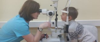
- Visit an ophthalmologist regularly.
It is better not to neglect the last point. Only an ophthalmologist will be able to recognize the onset of the disease and prescribe effective treatment. A visit to the doctor is necessary for inflammation of the eye, as well as for regular monitoring of myopia.