It branches like a tree, first into large branches (trunks), then into smaller branches and twigs, and is conventionally divided into several parts or sections:
- 1. The ascending aorta is the area from the aortic valve to the brachiocephalic trunk.
- 2. The aortic arch is a short section from which all the vessels supplying the arms and head (brachiocephalic arteries) depart. They anatomically form an arch connecting the ascending and descending aorta.
- 3. The descending (thoracic) aorta begins from the mouth of the left subclavian artery and continues to the diaphragm.
- 4. Below the diaphragm and before the bifurcation of the aorta (bifurcation) is the abdominal aorta.
Dividing the aorta into sections is very important for assessing risk and choosing optimal treatment tactics in patients with aortic aneurysms.
An aortic aneurysm is an area of local expansion.
What is the aorta
The aorta is the largest vessel in the human body, which carries blood from the heart to the organs and limbs. The upper section of the aorta runs inside the chest, this section is called the thoracic aorta . The lower part is in the abdominal cavity and is called the abdominal aorta . It delivers blood to the lower part of the body. In the lower abdomen, the abdominal aorta divides into two large vessels - the iliac arteries , which carry blood to the lower extremities.
The aortic wall consists of three layers: internal (intima), middle (media), external (adventitia).
Anatomy and topography of the aorta
The aorta is the largest arterial trunk of the systemic (main) circulation.
It starts in the left ventricle of the heart and continues to the level of the body of the fourth lumbar vertebra. The structure of the aortic wall is divided into:
- endothelium - the inner lining that ensures selective permeability of substances from the blood into the vascular wall;
- muscle layer consisting of smooth muscle cells;
- the outer layer, which contains large quantities of elastic fibers, nerve endings and feeding vessels.
The aortic wall has a three-layer structure.
Due to the control of the central nervous system, regular contraction and relaxation of smooth muscle fibers occurs. This promotes the physiological flow of blood and its active distribution from the center to the peripheral tissues. You can find out in more detail which vessels depart from the aortic arch only by reading this article in full.
This is interesting. The speed of blood movement through the aorta is on average 0.5-1.3 m/s.
In anatomy and clinical medicine, it is customary to distinguish the following parts in the structure of the aorta:
- ascending part about 6 cm long;
- arc;
- descending part:
- thoracic region, which lasts 16-17 cm;
- abdominal section
The sections of the great vessel are divided according to topographical characteristics.
The ascending part of the aorta emerges from the left ventricle of the heart, continuing its arterial cone. At its very beginning, the vessel forms an extension - a bulb (bulbus) with a diameter of 25-30 mm. Behind the sternum it goes up and gradually turns into an arch.
The aortic arch is characterized by deviation to the left and posteriorly. At the level of the fourth thoracic vertebra, it narrows slightly, forming an isthmus, and passes into the thoracic region.
The descending aorta is the longest section of the arterial vessel. It continues from the thoracic (IV vertebra) to the lumbar (IV vertebra) spine and, when passing the diaphragm, is divided into two parts - the thoracic and abdominal.
All vessels leaving the aorta are presented in the table below.
- Internal carotid artery (segments according to Bouthillier)
| Part of the aorta (see photo) | Outgoing vessels | |
| Rising | Coronary arteries (right, left) | |
| Arc | Brachiocephalic trunk | |
| General a. carotis (left) | ||
| Subclavian a. (left) | ||
| Descending | Thoracic region | Intercostal branches |
| Esophageal branches | ||
| Tracheal branches | ||
| Pleural branches | ||
| Pericardial branches, etc. | ||
| Abdominal | Celiac trunk | |
| Mesenteric a. | ||
| Renal a. and etc. |
Note! The vessels of the ascending aorta nourish the heart muscle itself, ensuring its stable functioning. Narrowing of the coronary arteries leads to such serious pathologies as coronary artery disease, angina pectoris, acute coronary syndrome (heart attack).
Abdominal aortic aneurysm
Abdominal aortic aneurysm is a chronic degenerative disease with life-threatening complications. An abdominal aortic aneurysm is defined as an increase in its diameter by more than 50% compared to the norm or a local bulging of its wall. Under the pressure of blood flowing through this vessel, the dilatation or bulging of the aorta may progress. The diameter of a normal aorta in the abdominal region is approximately 2 cm. However, at the site of an aneurysm, the aorta can be dilated to 7 cm or more.
Why is an aortic aneurysm dangerous?
An aortic aneurysm poses a major health risk as it can rupture. A ruptured aneurysm can cause massive internal bleeding, which in turn leads to shock or death.
An abdominal aortic aneurysm can cause other serious health problems. Blood clots (thrombi) often form in the aneurysm sac or parts of the aneurysm tear off, which move with the blood flow along the branches of the aorta to the internal organs and limbs. If one of the blood vessels becomes blocked, it can cause severe pain and lead to organ death or loss of a lower limb. Fortunately, if an aortic aneurysm is diagnosed early, treatment can be timely, safe and effective.
VARIANT ANATOMY OF THE AORTIC ARCH:
ARTERIA LUSORIA
Chernykh A.V.
Doctor of Medical Sciences, Professor, Head of the Department of Operative Surgery with Topographic Anatomy, Voronezh State Medical University named after. N.N. Burdenko of the Russian Ministry of Health
Zakurdaev E.I.
Candidate of Medical Sciences, Assistant at the Department of Operative Surgery with Topographic Anatomy of the Voronezh State Medical University named after V. N.N. Burdenko of the Russian Ministry of Health
Yakusheva N.V.
Candidate of Medical Sciences, Associate Professor of the Department of Operative Surgery with Topographic Anatomy of the VSMU (Voronezh State Medical University) named after. N.N. Burdenko of the Russian Ministry of Health
Sudakov D.V.
Candidate of Medical Sciences, Assistant at the Department of Operative Surgery with Topographic Anatomy of the Voronezh State Medical University named after V. N.N. Burdenko of the Ministry of Health of Russia, surgeon of the BUZ VOKB No. 1 (Voronezh Regional Clinical Hospital No. 1).
annotation
During topographic-anatomical dissection of the fixed corpse of a man who died at the age of 62 years, an aberrant right subclavian artery was discovered. It was located behind the branches of the aortic arch and the organs of the neck. The close location of the right aberrant subclavian artery with numerous organs, blood vessels and nerve trunks can cause various complications during surgical interventions.
Key words:
aortic arch, aberrant right subclavian artery, vascular anomalies.
Relevance of
the problem
Interest in studying the variant anatomy of blood vessels is determined by the need to minimize the risk of intraoperative complications. The most common anomaly of the aortic arch is the aberrant right subclavian artery - arteria
lusoria
. This anomaly occurs in 0.4-2.0% of cases[1][3]. Typically, the aberrant right subclavian artery arises from the aortic arch distal to the orifice of the left subclavian artery and is located behind the great vessels and the esophagus[9]. Rarely are cases of an aberrant vessel located between the esophagus and trachea. The aberrant right subclavian artery can also be located anterior to the trachea[10].
Materials and methods
The fixed corpse of a man who died at the age of 62 was examined. A topographic-anatomical preparation of the anterior neck and mediastinum was performed. Access to the organs and vessels of the neck was performed through two horizontal and one vertical incisions[7][8]. The upper horizontal incision connected the apices of the mastoid processes of the temporal bone and was carried out along the lower edge of the lower jaw. The lower horizontal incision connected the bases of the acromial processes of the scapula and was carried out along the upper edge of the clavicles and the manubrium of the sternum [2] [4]. The vertical incision connected the horizontal ones and corresponded to the midline. A median sternotomy was performed to examine the anterior mediastinum[7].
Research results and discussion
In the case studied, the aortic arch gave off four branches, whereas in typical situations there are three. The first branch of the aortic arch, counting from right to left, was the right common carotid artery. The second and third branches of the aortic arch were the left common carotid and subclavian arteries. The fourth branch of the aortic arch was the aberrant right subclavian artery – arteria
lusoria
(Fig. 1).
Rice. 1. Aberrant topography of the right subclavian artery.
Designations:
1 - aortic arch, 2 - right common carotid artery, 3 - trachea, 4 - esophagus, 5 - left common carotid artery, 6 - left subclavian artery, 7 - aberrant right subclavian artery (
arteria lusoria
).
The abnormal vessel originated from the posterior surface of the aortic arch 0.6 cm distal to the orifice of the left subclavian artery[6]. From left to right, the aberrant right subclavian artery crossed the remaining branches of the aortic arch and the esophagus along the posterior surface. The initial segment of the aberrant blood vessel was located in the left scalenovertebral space, the main trunk was in the retrovisceral cellular space of the neck[5][14]. Next, the aberrant artery penetrated into the right scalenovertebral space, where it gave off the vertebral and internal thoracic arteries.
The data obtained in the study are somewhat at odds with the literature. Thus, on the studied sectional material, the diameter of the superficial epigastric artery at the level of the inguinal ligament was 1.7±0.2 mm, and the vein – 2.1±0.4 mm. It follows from this that the diameter of the studied blood vessels meets the requirements of plastic surgeons for performing microvascular anastomoses.
Conclusion
1. A rather rare variant of the aortic arch, the aberrant right subclavian artery, has been identified and described. In this case, the anomalous vessel originated from the aortic arch distal to the origin of the left subclavian artery and was directed from left to right through the retrovisceral cellular space of the neck.
Literature:
1. Treatment of purulent wounds in patients with diabetes mellitus using electrically activated aqueous solutions / D.V. Sudakov, E.A. Fursova, M.V. Frolov, O.V. Sudakov // System analysis and management in biomedical systems. 2011. T. 10. No. 2. P. 427-429.
2. New data in the treatment of purulent wounds in microsurgical patients // D.V. Sudakov, A.V. Chernykh, E.I. Zakurdaev, A.N. Tikhonov // Central Scientific Bulletin. 2022. T. 2. No. 1 (18). pp. 9-11.
3. Assessing the information content of clinical examination indicators in patients with type 2 diabetes mellitus and purulent wounds / D.V. Sudakov, E.V. Starodubtseva, O.V. Sudakov, V.N. Snopkov // System analysis and management in biomedical systems. 2013. T. 12. No. 4. P. 1163-1165.
4. Experience of autotransplantation of the main nerve trunks of the forearm with the sural nerve against the background of bite wounds of the upper extremities / D.V. Sudakov, A.V. Chernykh, N.V. Yakusheva, A.N. Tikhonov // Central Scientific Bulletin. 2022. T. 2. No. 1 (18). pp. 12-14.
5. Constructing a forecast for the effectiveness of autotransplantation with the sural nerve in microsurgery of the upper extremities in patients with type 2 diabetes mellitus / D.V. Sudakov, A.V. Chernykh, N.V. Yakusheva, N.O. Vasiliev // In the collection: Current issues of modern medicine. Collection of scientific papers based on the results of the III international scientific and practical conference. 2016. pp. 122-124.
6. The problem of antibiotic resistance in microsurgery / D.V. Sudakov, A.V. Chernykh, E.I. Zakurdaev, A.N. Tikhonov // Central Scientific Bulletin. 2022. T. 2. No. 1 (18). pp. 15-17.
7. A rare case of a giant sliding inguinal-scrotal hernia of the bladder / A.V. Chernykh, E.N. Lyubykh, Yu.V. Maleev, E.I. Zakurdaev, A.N. Shevtsov, V.V. Spitsin // Journal of Anatomy and Histopathology. 2013. T. 2. No. 3 (7). pp. 67-69.
8. Sudakov D.V. Analysis of the composition of the microbial flora in patients with type 2 diabetes mellitus after autotransplantation of damaged main nerves of the forearm with the sural nerve / D.V. Sudakov, A.V. Chernykh, N.V. Yakusheva // In the collection: Modern medicine: current issues and development prospects. Collection of scientific papers based on the results of the international scientific and practical conference. Innovation Center for the Development of Education and Science. 2016. pp. 134-138.
9. Sudakov D.V. Modeling the process of treating purulent wounds in patients with diabetes mellitus / D.V. Sudakov // System analysis and management in biomedical systems. 2013. T. 12. No. 1. P. 327-330.
10. Sudakov D.V. Constructing a forecast for the effectiveness of using the Ilizarov wrist extrafocal compression-distraction apparatus in microsurgery / D.V. Sudakov, A.V. Chernykh, N.V. Yakusheva // In the collection: Modern medicine: current issues and development prospects. Collection of scientific papers based on the results of the international scientific and practical conference. Innovation Center for the Development of Education and Science. 2016. pp. 138-143.
11. Chernykh A.V., Vitchinkin V.G., Yakusheva N.V., Maleev Yu.V., Zakurdaev E.I., Bolotskikh V.A., Spitsyn V.V. High origin of the radial and ulnar arteries // Journal of Anatomy and Histopathology. – 2014. – T. 3, No. 3. – P. 63-65.
12. Chernykh A.V., Zakurdaev E.I., Yakusheva N.V., Vitchinkin V.G., Maleev Yu.V., Zakurdaeva M.P., Andrianova K.A., Lazareva O.A. Applied aspects of variant anatomy of the inferior epigastric artery // Journal of Anatomy and Histopathology. – 2016. – T. 5, No. 4. – P. 74-78.
13. Chernykh A.V. Autotransplantation of the sural nerve in microsurgery of the upper extremities in patients with type 2 diabetes mellitus / A.V. Chernykh, D.V. Sudakov, N.V. Yakusheva // Applied information aspects of medicine. 2016. T. 19. No. 3. pp. 107-112.
14. Chernykh A.V. Problems and prospects for studying the topographic anatomy of the parathyroid glands / A.V. Chernykh, Yu.V. Maleev, A.N. Shevtsov // Journal of Anatomy and Histopathology. 2013. T. 2. No. 2 (6). pp. 15-23.
Information about authors:
Chernykh Alexander Vasilievich
– Doctor of Medical Sciences, Professor, Head of the Department of Operative Surgery with Topographic Anatomy of the Federal State Budgetary Educational Institution of Higher Education VSMU named after N.N. Burdenko of the Russian Ministry of Health.
Zakurdaev Evgeniy Ivanovich
– Candidate of Medical Sciences, assistant at the Department of Operative Surgery with Topographic Anatomy of the Federal State Budgetary Educational Institution of Higher Education VSMU named after N.N. Burdenko of the Russian Ministry of Health.
Yakusheva Natalya Vladimirovna
– Candidate of Medical Sciences, Associate Professor of the Department of Operative Surgery with Topographic Anatomy of the Federal State Budgetary Educational Institution of Higher Education VSMU named after. N.N. Burdenko of the Russian Ministry of Health.
Sudakov Dmitry Valerievich –
Candidate of Medical Sciences, Assistant of the Department of Operative Surgery with Topographic Anatomy of the Federal State Budgetary Educational Institution of Higher Education VSMU named after N.N. Burdenko of the Ministry of Health of Russia, doctor-surgeon of the BUZ VOKB No. 1.
VARIANT ANATOMY OF THE AORTIC ARCH: ARTERIA LUSORIA
Chernyh AV, Zakurdaev EI, Yakusheva NV, Sudakov DV
Summary
When topographic anatomical dissection of a fixed cadaver of a man, who died at the age of 62 years, found an aberrant right subclavian artery. It was located behind the branches of the aortic arch and organs of neck. The proximity of the aberrant right subclavian artery with multiple organs, blood vessels and nerve trunks can cause a variety of complications during surgery.
Keywords:
aortic arch, aberrant right subclavian artery, vascular abnormalities.
References:
1. Treatment of purulent wounds in patients with diabetes mellitus using electro-aqueous solutions / DV Sudakov, EA Fursova, MV Frolov, OV Sudakov // System analysis and control in biomedical systems. 2011. V. 10. No. 2. P. 427-429.
2. New data in the treatment of purulent wounds in patients with microsurgical Profile / DV Sudakov, AV Chernyh, EI Zakurdaev, AN Tikhonov // Central Scientific Gazette. 2022. V. 2. No. 1 (18). P. 9-11.
3. Evaluation of informative indicators of clinical examination of patients with type 2 diabetes and purulent wounds / DV Sudakov, EV Starodubceva, OV Sudakov, VN Snopkov // System analysis and control in biomedical systems. 2013. V. 12. No. 4. P. 1163-1165.
4. Experience sural nerve autografting main nerve trunks of the forearm against the backdrop of bite wounds upper limb / DV Sudakov, AV Chernyh, NV Yakusheva, AN Tikhonov // Central Scientific Gazette. 2022. V. 2. No. 1 (18). P. 12-14.
5. Building efficiency forecast sural nerve autografting in microsurgery of the upper extremities in patients with type 2 diabetes / DV Sudakov, AV Chernyh, NV Yakusheva, NO Vasiliev // In: Actual problems of modern medicine. Collection of scientific papers on the results of the III international scientific - practical conference. 2016. P. 122-124.
6. The problem of antibiotic resistance in microsurgery / DV Sudakov, AV Chernyh, EI Zakurdaev, AN Tikhonov // Central Scientific Gazette. 2022. V. 2. No. 1 (18). P. 15-17.
7. A rare case of giant sliding inguinal-scrotal hernia bladder / AV Chernykh, EN Lubih, YV Maleev, EI Zakurdaev, AN Shevtsov, VV Spitsin // Journal of anatomy and histopathology. V. 2. No. 3. 2013. 3 (7). P. 67-69.
8. Sudakov DV Analysis of the composition of the microbial flora in patients with type 2 diabetes after autologous transplantation sural nerve trunk damaged forearm nerves / DV Sudakov, AV Chernyh, NV Yakusheva // In: Modern medicine: current issues and prospects. Collection of scientific papers on the results of the international scientific-practical conference. Innovation Center for Development of Education and Science. 2016. P.134-138.
9. Sudakov DV Simulation of the process of treatment of purulent wounds in patients with diabetes / DV Sudakov // System analysis and control in biomedical systems. 2013. V.12. No. 1. P. 327-330.
10. Sudakov DV Construction of the forecast of effectiveness of use of carpal extrafocal compression – distraction apparatus Ilizarov in microsurgery / DV Sudakov, AV Chernyh, NV Yakusheva // In: Modern medicine: current issues and prospects. Collection of scientific papers on the results of the international scientific-practical conference. Innovation Center for Development of Education and Science. 2016. P. 138-143.
11. Chernyh AV, Vitchinkin VG, Yakusheva NV, Maleev Yu.V., Zakurdaev EI, Bolotskikh VA, Spitsin VV Case report of high origin of radial and ulnar arteries // Journal of anatomy and histopathology. 2014. V. 3. No. 3. P. 63-65.
12. Chernykh AV, Zakurdaev EI, Yakusheva NV, Vitchinkin VG, Maleev Yu.V., Zakurdaeva MP, Andrianova KA, Lazareva OA // Journal of anatomy and histopathology. 2014. V. 5. No. 4. P. 74-78.
13. Chernyh AV Autotransplantation microsurgery sural nerve in the upper limbs in patients with type 2 diabetes / AV Chernyh, DV Sudakov, NV Yakusheva // Applied information aspects of medicine. 2016. V. 19. No. 3. P. 107-112.
14. Chernyh AV Problems and prospects of the study of topographic anatomy of the parathyroid glands / AV Chernyh, YV Maleev, AN Shevtsov // Journal of anatomy and histopathology. 2013. V. 1. no. 2 (6). P. 15-23.
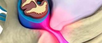
Types of aortic aneurysms
There are “true” and “false” aortic aneurysms. A true aneurysm develops due to the gradual weakening of all layers of the aortic wall. A false aneurysm is usually the result of trauma. It is formed from the connective tissue surrounding the aorta. The cavity of the false aneurysm is filled with blood through a crack in the aortic wall. The aortic walls themselves do not participate in the formation of an aneurysm.
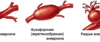
Depending on the form there are:
- saccular aneurysm - expansion of the aortic cavity on only one side;
- spindle-shaped (fusiform) aneurysm - expansion of the aneurysm cavity on all sides;
- mixed aneurysm - a combination of saccular and fusiform forms.
Causes and risk factors for the development of abdominal aortic aneurysm
The causes of the development of abdominal aortic aneurysms are very diverse. The most common cause of aneurysm development is atherosclerosis. Atherosclerotic aneurysms account for 96% of the total number of all aneurysms. In addition, the disease can be either congenital (fibromuscular dysplasia, Erdheim's cystic medianecrosis, Marfan syndrome, etc.) or acquired (inflammatory and non-inflammatory). Inflammation of the aorta occurs when various microorganisms invade (syphilis, tuberculosis, salmonellosis, etc.) or as a result of an allergic-inflammatory process (nonspecific aortoarteritis). Non-inflammatory aneurysms most often develop with atherosclerotic lesions of the aorta. Less commonly, they are the result of injury to its wall.
Risk factors for developing an aneurysm
- Arterial hypertension;
- Smoking;
- The presence of aneurysms in other family members. Which indicates the role of hereditary factors in the development of this disease;
- Gender: men over 60 years of age (abdominal aortic aneurysms occur less frequently in women).
What it is
We should start by defining the aorta. The aorta is the largest vessel in our body, and accordingly its importance is high. An aneurysm is an enlargement of a blood vessel. An aortic aneurysm can form in the thoracic or abdominal regions. Moreover, aneurysm of the abdominal aorta is much more common. This condition of the vessel may not cause any harm for a long time, however, an aneurysm is very dangerous due to the unpredictable course of expansion and the threat of rupture of the vessel walls when they become thinner - and this condition already poses a threat to life due to severe internal bleeding. In addition, blood clots can form at the site of vessel expansion due to changes in blood flow - the danger of this condition is the risk of the blood clot breaking off and clogging a smaller vessel.
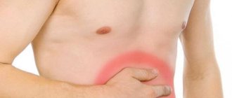
Symptoms and signs of abdominal aortic aneurysm
In most patients, abdominal aortic aneurysms occur without any symptoms and are an incidental finding during examinations and operations for other reasons.
When signs of an aneurysm develop, the patient experiences one or more of the following symptoms:
- A feeling of pulsation in the abdomen, similar to a heartbeat, an unpleasant feeling of heaviness or fullness.
- Dull, aching pain in the abdomen, in the navel area, usually on the left.
Indirect signs of abdominal aortic aneurysm are important :
- Abdominal syndrome. Manifested by the appearance of belching, vomiting, unstable stool or constipation, lack of appetite and weight loss;
- Ischioradic syndrome. Manifested by lower back pain, sensory disturbances and movement disorders in the lower extremities;
- Syndrome of chronic ischemia of the lower extremities. Manifests itself in the appearance of pain in the muscles of the lower extremities when walking, sometimes at rest, coldness of the skin of the lower extremities;
- Urological syndrome. It manifests itself as pain and heaviness in the lower back, difficulty urinating, and the appearance of blood in the urine.
Harbingers of rupture may be increased abdominal pain.
When an aneurysm ruptures, the patient suddenly feels an increase or appearance of abdominal pain, sometimes “radiating” to the lower back, groin area and perineum, as well as severe weakness and dizziness. These are symptoms of massive internal bleeding. The development of such a situation is life-threatening! The patient needs emergency medical care!
Treatment
As already noted, the most dangerous complication of an aortic aneurysm is the threat of its rupture and internal bleeding. If the patient did not consult a doctor and the aneurysm was not detected in a timely manner, then in the event of a rupture, the only option would be open surgery: the surgeon will remove the affected area of the aorta and install a prosthesis in its place. This method of emergency treatment of an aneurysm has contraindications; in addition, the operation is a serious intervention with associated risks and a long rehabilitation period (up to three months). Therefore, it is so important to contact a specialist if you are at risk and feel periodic pain and/or throbbing in the abdominal area.
Once an aneurysm is diagnosed, its further expansion is difficult to predict; regular monitoring by an experienced specialist, lifestyle adjustments, and control of risk factors are required. When the attending physician realizes that there is a risk of rupture, an alternative to emergency open surgery will be the planned installation of a stent graft. The installation is minimally invasive, i.e. the stent is inserted through the vessel, advanced to the site of the aneurysm and secured there. The structure of the stent resembles a vessel; it is fixed with one edge above the expansion of the aorta, and with the other below. After the operation, blood will flow through the stent, and the resulting cavity between it and the aortic wall will decrease over time. The stent graft is inserted under local anesthesia and requires only a couple of days of recovery.
Of course, any surgical intervention carries its own risks, which is why it is so important to choose a good medical center with experienced cardiovascular surgeons. Understanding the danger of aortic aneurysm as a disease, not only doctors in the cardiology department, but also in other departments of our center, when conducting routine examinations or examinations due to other diseases, monitor the patient’s condition and analyze the entire volume of data received: if a problem with the aorta is suspected, the patient within one center will be transferred for consultation with a cardiologist. For our patients, we try to organize the most convenient logic for staying in our center and consulting with specialists in order to avoid double examinations and unnecessary manipulations. Comprehensive patient management is one of the main principles of our work. And the quality of surgical and conservative care is maintained thanks to a team of experienced specialists, provided with the necessary diagnostic and treatment facilities.
You can sign up for a consultation using a special form on the website or by phone.
Diagnosis of abdominal aortic aneurysms
Most often, abdominal aortic aneurysms are detected by ultrasound examination of the abdominal organs. Typically, the discovery of an aneurysm is an incidental finding. If a doctor suspects a patient has an aortic aneurysm, modern diagnostic methods are used to clarify the diagnosis.
Methods for diagnosing abdominal aortic aneurysm
- Computed tomography in angio mode;
- Magnetic resonance imaging in angio mode;
- X-ray contrast aorto- and angiography;
- Ultrasound duplex or triplex angioscanning of the abdominal aorta.
If necessary, the abdominal and thoracic aorta is examined.
Symptoms of the disease
As a rule, the disease occurs without symptoms. In most cases, an aneurysm is discovered accidentally - during a routine examination. If symptoms appear, they are expressed in the area of the aortic arch. Some of the most striking signs of the development of the disease include:
- heavy snoring;
- shortness of breath, cough;
- sharp chest pain;
- discomfort during swallowing.
In some cases, individual signs of a cardiac aortic aneurysm may appear.
Diagnosis of the disease
Diagnosis of the disease begins with the patient visiting a therapist. In this case, the specialist collects anamnesis, analyzes the existing symptoms, and then refers the patient to a highly specialized specialist. To confirm the diagnosis, additional laboratory and hardware tests are prescribed:
- clinical, general urine and blood tests: allow you to determine the presence of pathologies that affect the further development of the disease;
- echocardiography: helps determine the type, shape, size of the aneurysm;
- X-ray: shows enlarged heart, pulmonary edema.
In some cases, MRI of the heart vessels is prescribed to obtain important data.
Make an appointment Make an appointment and get a high-quality examination of the heart and blood vessels in our center
Prevention
When diagnosing this disease, it is important to resort to effective preventive measures. These include the following activities:
- maintaining a healthy lifestyle;
- refusal to drink alcohol, smoke;
- timely examination by a doctor.
In addition, the patient must form the correct diet. Meals for aortic aneurysm should be nutritious. At the same time, the menu should not include fatty foods. Sufficient physical activity is another component of prevention. If a patient has acute chest pain for more than 6 minutes, the patient should seek immediate medical attention.
Treatment methods for aortic aneurysm
There are several methods for treating aortic aneurysm. It is important to know the advantages and disadvantages of each of these techniques. Approaches to the treatment of abdominal aortic aneurysms:
Monitoring the patient over time
If the aneurysm is less than 4.5 cm in diameter, the patient is recommended to be monitored by a vascular surgeon, since the risk of surgery exceeds the risk of rupture of the aortic aneurysm. Such patients should undergo repeated ultrasound examinations and/or computed tomography at least once every 6 months.
When the aneurysm diameter is more than 5 cm, surgical intervention becomes preferable, since as the size of the aneurysm increases, the risk of aneurysm rupture increases.
If the size of the aneurysm increases by more than 1 cm per year, the risk of rupture increases and surgical treatment also becomes preferable.
Open surgery: aneurysm resection and aortic replacement
Surgical treatment is aimed at preventing life-threatening complications. The risk of surgery is associated with possible complications that include heart attack, stroke, limb loss, acute intestinal ischemia, male sexual dysfunction, embolization, prosthetic infection, and renal failure.
The operation is performed under general anesthesia. The essence of the operation is to remove the aneurysmal expansion and replace it with a synthetic prosthesis. The average mortality rate for open procedures is 3-5%. However, it may be higher if the renal and/or iliac arteries are involved in the aneurysm, as well as due to the patient’s concomitant pathology. Observation in the postoperative period is carried out once a year. Long-term treatment results are good.
Endovascular repair of aortic aneurysm: installation of a stent graft
Endoprosthetics of aortic aneurysm is a modern alternative to open surgery.
The operation is performed under spinal or local anesthesia through small incisions/punctures in the groin areas. Through the above approaches, catheters are inserted into the femoral artery under X-ray control. According to which, in the future, the endoprosthesis will be brought to the aneurysmal extension. An endoprosthesis or stent-graft of the abdominal aorta is a mesh frame made of a special alloy and wrapped in synthetic material. The last stage of the operation is the installation of a stent graft at the site of the aneurysmal expansion of the aorta. Eventually, the aneurysm is “switched off” from the bloodstream and the risk of rupture becomes unlikely. After aortic replacement, the patient is observed in the hospital for 2-4 days and discharged.
This technique allows to reduce the incidence of early complications, shorten the length of patient stay in the hospital and reduce the mortality rate to 1-2%. Observation in the postoperative period is carried out every 4-6 months using ultrasound techniques, CT angiography, X-ray contrast angiography. The endovascular treatment method is certainly less traumatic. About 40,000 such operations are performed annually in the United States alone.
Thus, the choice of treatment method for abdominal aortic aneurysm is based on the individual characteristics of the patient.
Diagnostics
Due to the absence of clear symptoms and gradual expansion of the aorta, most often the suspicion of an aneurysm appears to the doctor during an examination for another reason. The diagnosis is confirmed using ultrasound or CT scan of the abdominal cavity, angiography - which examination is suitable for a particular patient is decided by the attending physician. Examinations will provide information about the size of the aneurysm, the presence of a blood clot in this location, and the condition of the vessel walls.
In many patients, the identified dilatation of the aorta does not require surgical intervention by a specialist for a long time - the main task will be observation. In this case, the patient will be given a list of recommendations so as not to worsen the condition of the blood vessels (first of all, quitting smoking, if such an addiction is present), and will also be offered to undergo periodic examinations to monitor the condition of the aorta. But in any case, the doctor must exclude the risk of aortic rupture at the site of expansion. For this purpose, examination data is used: the width of the aorta and the rate of its expansion over time. If there is a threat of rupture, the doctor may decide to perform an operation, and this will require additional tests of blood, blood vessels, and urine to prepare for the operation.