Amebiasis - symptoms and treatment
Amoebiasis (Amoebic Dysentery) is an acute and chronic disease that is caused by pathogenic strains of dysenteric amoeba. Penetrating into the body, they lead to ulcerative damage to the intestines, which is accompanied by moderate intoxication, stool disorders, weight loss and sometimes the appearance of abscesses in the liver, intestines, lungs and brain. The disease can last a long time, sometimes leading to death, especially in weakened patients and in the absence of medical care.
Pathogen
Taxonomy:
- domain - eukaryotes;
- branch - amoeba-like;
- type - Evosea;
- class - Archamoebae;
- family - Entamoebidae;
- genus - Entamoeba (entamoeba);
- species - dysentery amoeba (Entamoeba histolytica).
The Russian scientist F. A. Lesh was the first to describe the pathogen and prove its pathogenicity in 1875. At least 22 strains have now been isolated, of which 9 are pathogenic, and the remaining 13 are most likely not pathogenic for humans.
Changing during life, amoebas go through two stages of development:
- the vegetative, or active, stage in the form of a trophozoite ;
- a resting stage, or "dormant" stage, in the form of a cyst .
The vegetative stage divides amoebas according to function and structure into large vegetative, luminal and tissue. All of them, suddenly entering the external environment, die within 30 minutes.
Large vegetative trophozoites (forma magna) reach 20-60 microns. This form of amoebas moves due to forward thrusts. Contains the nucleus and cytoplasm - the main part of the cell. The cytoplasm is divided into a glassy transparent mass and an inner liquid layer with digestive vacuoles in which red blood cells captured by the amoeba are digested. In acute amoebiasis, large trophozoites are detected in fresh feces - “warm feces”. Capable of breaking down protein molecules. They have surface pectins, with the help of which amoebas attach to the intestinal mucosa.
The luminal vegetative trophozoites (forma minuta) are much smaller than the large form: they reach 15-20 microns. Such trophozoites are inactive. They have one core, which is not visible without coloring. Their cytoplasm contains small vacuoles, but without erytrophytes. Luminal trophozoites live in the upper part of the large intestine, feed on bacteria, reproduce, and do not cause obvious harm. They are detected in the feces of acute amoebiasis at the beginning of recovery, in chronic amoebiasis, as well as in amoeba carriers, but to identify them it is necessary to carry out deep intestinal lavages or examine the final portions of feces after taking a saline laxative. In the lower part of the large intestine, with a gradual deterioration of conditions, for example, lack of fluid, disruption of the bacterial flora, taking medications or changes in the pH of the environment, they usually transform into a cyst form, which is gradually released into the environment. If the body's defenses are weakened, luminal trophozoites can transform into a larger vegetative form and become aggressive.
Tissue vegetative trophozoites are formed from the luminal form, but are similar in appearance to the vegetative one. They reach 20-25 microns, are mobile, parasitize, penetrating the mucous membrane of the colon, and infect the intestines. They are detected only in acute amoebiasis in the affected organs; they are found extremely rarely in liquid feces - during the disintegration of intestinal ulcers.
Cysts form from the luminal form in the lower parts of the colon. They have a round shape and reach 8-15 microns. They are immobile, covered with a dense shell of chitin, and contain chromatoid bodies (RNA with protein) and glycogen. Depending on the maturity of the cysts, they contain from 1 to 4 nuclei. They are more often detected in patients with chronic amoebiasis and parasite carriers. They are very stable in the external environment: they remain in feces at room temperature for up to 2 weeks or longer, when frozen to −21°C - no more than 3 months, in water - up to 8 months. When heated to 100°C and boiled, they quickly die. Standard household disinfectants with chlorine have almost no effect on cysts. Soap-cresol preparations, a solution of sublimate 1:1000 and a 3% solution of carbolic acid can affect them [1][3][7][10].
Epidemiology
Amoebiasis is a widespread disease, mainly found in the countries of Southern and Western Africa, Central and South America, as well as in India, China and Korea. In Russia, isolated cases are mainly recorded, mainly in the southern regions, the Caucasus and the Far East. Although recently the incidence in our country has been increasing. This may be due to the influx of migrants from border regions and the development of tourism.
On average, about 50 million cases of amoebiasis are registered annually in the world, of which about 100 thousand end in death. In terms of the number of deaths, this disease ranks third among parasitic diseases. Approximately 90% of cases of the disease are intestinal amoebiasis, the remaining forms are extraintestinal.
The source of infection is a person (patient or carrier). With its feces, amoebas in the form of cysts enter the environment.
The transmission mechanism is fecal-oral. Includes water, food, household contact and sexual (oral-anal) transmission routes. Infection can occur when any substance (water, food, dirt, fingers) that comes into contact with the feces of an infected person or contains some part of it enters the mouth. Mechanical spreaders and carriers can be cockroaches and flies. In extremely rare cases, homosexuals and patients with secondary immunodeficiency may become infected when amoebas enter directly into the wound.
A person with amoebiasis runs the risk of transmitting the infection to family members, but this risk is low, especially if the person follows the rules of personal hygiene: thoroughly washes his hands after using the restroom and before preparing food.
Situations that increase the risk of infection:
- visiting tropical countries with low levels of sanitation;
- interaction with immigrants from tropical countries with poor sanitary conditions;
- neglect of personal hygiene rules;
- consumption of raw, undisinfected water from open sources and the water supply system;
- homosexual contacts;
- presence of mental disorders.
Immunity after an illness is unstable and weakly expressed, repeated infections are possible. Even a large number of antibodies formed, for example in amoebic liver abscesses, do not protect against the progression of amebiasis [1][2][5][6].
Prevention
A vaccine against the pathogen has not been created. Prevention of amebiasis includes the following measures:
- hospitalization and isolation of patients, their treatment until the pathogens disappear in the feces;
- identification and treatment of disease carriers;
- a ban on persons who have suffered amoebiasis from working in public catering establishments;
- careful adherence to personal hygiene, drinking only boiled water, especially when staying in countries with hot climates;
- during tourist trips to dangerous areas, do not eat vegetables and fruits that have already been washed by someone, wash them thoroughly before eating, use a straw to drink from water bottles, do not drink iced or carbonated drinks, do not eat milk, cheese and other unpasteurized products , do not buy food on the street.
Self-medication of amoebiasis is unacceptable. When the first signs of illness appear, you should consult a doctor.
How does intestinal amoebiasis spread?
The methods of spread are similar to the methods of spread of any intestinal infection. Pathogens enter the gastrointestinal tract. They can penetrate with infected liquid, untreated food, and simply with the help of unwashed hands.
These are the ways of infection. The causative agent of amoeba disease is a simple organism that loves heat.
Intestinal amoebiasis: ontogenesis
The amoeba becomes active in the large intestine.
The ontogenesis of intestinal amebiasis occurs in stages. As usual, it is not the “active” amoeba that penetrates into the human body, but, in fact, a cyst.
When unfavorable conditions arise, some types of bacteria build up a solid mantle and plunge into a passive state.
They can stay there for a long time, until the environment becomes more suitable for activity.
In the state of a cyst, the histological amoeba can easily pass through all hostile environments of the human body: intestines, stomach. The amoeba becomes active only when it enters the comfortable conditions of the large intestine.
Intestinal amoebiasis: signs of the disease
With amebiasis, an increased temperature is observed.
The incubation period for this disease is approximately one to two weeks. The onset of amebiasis is often abrupt: an increase in body temperature to 38°C throughout the day, the appearance of painful sensations in the abdomen, and loose stools.
A dysentery-like syndrome develops, which has symptoms of damage to the colon: characteristic pain in the lower abdomen; the presence of blood and mucus in the stool; frequent urge to go to the toilet.
There may be a decrease in appetite or a complete aversion to food. Intestinal amoebiasis has symptoms quite similar to dysentery. This may be an increase in temperature, intoxication of the body, general weakness, increased fatigue, headache.
Based on these symptoms, it is possible to determine the form of the disease: mild or severe. Factors such as the aggressiveness of amoebas, their toxicity and the state of the body's immunity directly affect the severity of the disease.
If the intestinal form of the disease is not treated, amoebas gradually spread greatly in the human body. This form of the disease is called extraintestinal. The pathogens, by penetrating the intestinal walls, enter the blood vessels and spread throughout the patient’s body through the bloodstream.
Pathogens cause the most significant harm to an organ such as the liver. Living on the liver, they cause amoebic suppuration. This is accompanied by pain in the person’s right side, an increase in the size of the liver, and intoxication. Jaundice is possible (if the bile ducts are blocked by edema).
An inflammation forms around the abscess, which is called perifocal. It is the liver’s response to harmful pathogens. It is during this period that hepatitis develops. Fermentation increases and jaundice appears.
More rare are cases of abscesses caused by amoebas in organs such as the lungs and brain. To do this, amoebas need to cross the intestinal barrier, enter the bloodstream and directly reach the brain, for example. This is a very extensive infestation.
The disease takes on an extraintestinal form in the following cases: penetration of a large number of pathogens into the body; deep depletion of the intestinal mucosa; the patient has chronic colitis.
Due to the decline in immunity with age, the extraintestinal form of the disease is more common in older people. People with alcohol addiction also fall into the risk category. Alcohol greatly weakens the immune system.
Intestinal amoebiasis: complications caused by this disease
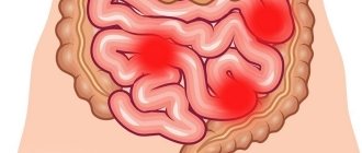
Intestinal amebiasis is the cause of ulcers.
Penetration of amoebas into the walls of the large intestine and its destruction causes ulcers to appear in these places.
There is a possibility of intense intestinal bleeding if a large vessel is localized at the site of the lesion.
This poses a great danger to human life. In the event of such a complication, there is an urgent need for surgical intervention.
All complications that are outside the intestines (abscesses of the liver, brain, lungs, etc.) are solved in a similar way through surgery. Based on this, it is important to quickly diagnose this disease. Detection of pathogen amoebas is possible in feces or in body tissues.
There is a need to analyze the detection of antibodies to this type of amoeba directly in the patient’s blood. True, in order for a patient to take a blood test, the doctor treating him must assume that the cause of the patient’s poor health may be protozoa, and not something else. It is also a fact that it is not always possible to establish a correct diagnosis in the emergency room.
This disease is quite rare and is very similar in its symptoms to other diseases. Intestinal amoebiasis may be suspected if the patient tells the doctor about his visit to the Southeast Asian region about a couple of months before and his stool resembles raspberry jelly.
However, it is strictly forbidden to confirm a diagnosis and prescribe treatment for a disease based solely on stories alone. As a rule, the next step is diagnosis: taking urine, feces and blood for analysis. If a pathogen is detected in the stool of amoebae, an amoebic infection can be confirmed.