Osgood-Schlatter disease can present as a painful lump in the area below the kneecap during childhood and adolescence as puberty begins. Osgood-Schlatter disease occurs most often in children who participate in sports, especially sports such as running, jumping, or sports that require rapid changes in movement trajectories, such as football, basketball, figure skating and gymnastics.
And although Osgood-Schlatter disease is more common in boys, the gender gap narrows as girls become more involved in sports. Osgood-Schlatter disease affects more adolescents who play sports (by a ratio of one to five). The age range of incidence has a gender factor, as girls experience puberty earlier than boys. Osgood-Schlatter disease usually occurs in boys between 13 and 14 years of age and in girls between 11 and 12 years of age. The disease usually goes away on its own as bone growth stops.
Symptoms
The main symptoms of Osgood-Schlatter disease include:
- Pain, swelling, and tenderness in the area of the tibial tuberosity, just below the kneecap
- knee pain that gets worse after physical activity—especially running, jumping, and climbing stairs—and gets better with rest
- tightness of surrounding muscles, especially the thigh muscles (quadriceps)
Pain varies depending on each individual. Some may only have mild pain when doing certain activities, especially running or jumping. For others, the pain can be constant and debilitating. Typically, Osgood-Schlatter disease occurs in only one knee, but sometimes it can occur in both knees. The discomfort may last from several weeks to several months and may recur until the child stops growing.
From the medical history
Osgood-Schlatter disease is a peculiar disease that affects young people and is localized in the tibial tuberosity and was known before all other forms of osteochondropathy. Boys get sick more often than girls; Among the patients, strong young people who play sports, primarily football, predominate. Both limbs are affected more often than with all other osteochondropathy; There is no preferred location on the right.
In most cases, the disease begins for no apparent reason, but sometimes the onset of the process is preceded by a limb injury. In the area of the tibial tubercle, swelling and slight swelling of the soft tissues appear, and a hard bone growth can be felt. When walking, especially when the patient fully bends or straightens the lower leg at the knee joint, when he climbs stairs, lifts an object from the floor or kneels, severe local pain is felt. The disease usually drags on for six months, a year or a year and a half and always ends with complete recovery.
The objective picture during clinical examination is usual - good general condition of the patient, normal temperature, strictly limited pain when pressing on the tubercle.
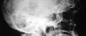
Features of interpreting radiographs for osteochondropathy
A common radiological symptom of osteochondropathy of the tibial tuberosity is a large oval or round shadow lying isolated in front of the trunk-shaped tuberosity, which has already fused with the metaphysis, in a niche or its nest-like depression. This shadow gives the impression of a freely lying body. However, this is only a bony inclusion in the cartilage and therefore, when attempting to move it under screen control, it remains motionless. Accordingly, the entire trunk-like process is deformed, expanded in places, narrowed in others. Relatively rarely, the bone layers on the anterior surface of the tubercle are outlined radiologically, sometimes very lush, with a bizarre comb-like shape.
Thus, the symptomatology of this disease consists, in essence, of the same radiological elements as the X-ray picture of osteochondropathy of the epiphyses and small spongy bones.
During the process, individual necrotic bone areas resolve, others increasingly increase in size, merge with each other, then rearrange themselves, and the tubercle acquires a normal structural pattern. In this case, it is not necessary to distinguish certain successive phases of the pathological process. Osteochondropathy of the tibial tuberosity, therefore, radiologically almost always results in complete recovery. There can be no outcome in arthrosis here.
X-ray examination in some cases takes a back seat to clinical data. The reason for this is that the pronounced clinical picture of the disease is quite compatible with the normal radiographic picture, and that some normal ossification patterns on radiographs are indistinguishable from pathological changes. Having only radiographs in front of him and not knowing the medical history, the radiologist in some cases is unable to draw the line between normality and pathology, in contrast to most osteochondropathy, where, based on the images alone, the radiologist can confidently decipher the clinical picture.
The most valuable, undoubtedly, pathological symptoms are deep structural changes with sequestration-like shadows, significant deformation of the trunk-shaped tubercle and an isolated round bone shadow in the niche of the tubercle after its fusion with the metaphysis.
Thus, the main difficulties in X-ray diagnosis of osteochondropathy of the tibial tuberosity lie in the difference between the pathological process and the normal picture.
Comparison of the “sick” side with the “healthy” side in some controversial cases has only little practical significance due to the possibility of differences in the pattern of normal ossification on both sides in the same person. It is also necessary to be cautious in recognizing osteochondropathy of the tibial tuberosity in a systemic ossification disorder. In these cases, the tibial tuberosity can be painful when pressed: true osteochondropathy of the tibial tuberosity, like any other osteochondropathy, is still a local process in the entire human body, and not a systemic one.
In such cases of systemic pathology of ossification (for example, with endocrine diseases), one should not, under the suggestion of clinical data, make a diagnosis of osteochondropathy; one should only limit oneself to a description of anatomical deviations from the norm in the area of the tubercle. Calcifications in the patellar ligament proper at the site of its attachment to the tibia should also not be confused with osteochondropathy. A true traumatic fracture or avulsion of the tibial tuberosity is rare, and at any age, and the characteristic fracture line, large displacement of the fragment and a defect in the tibia itself are easily distinguished from a pathological fracture in osteochondropathy.
One acquaintance with osteochondropathy is enough not to think about this or that osteomyelitis, tuberculosis, sarcoma, and especially about gumma - diseases that the clinician most often suspects before radiography. In controversial cases, it should be borne in mind that in all these diseases the cortical and spongy substance of the tibia and around the tubercle are affected, or rather, the tubercle is only secondarily involved in the pathological process, while osteochondropathy is strictly limited to the area of the tubercle itself.
Causes
Each tubular bone of a child (in the arm or leg) has growth zones, at the end of the bones consisting of cartilage. Cartilage tissue is not as strong as bone tissue and is therefore more susceptible to damage, and excessive stress on the growth zones can lead to swelling and pain in this area. During physical activity that involves a lot of running, jumping and bending (football, basketball, volleyball and ballet), the child's hip muscles stretch the tendon - the quadriceps muscle, which connects the kneecap to the tibia. This type of repetitive stress can cause small tears in the tendon from the tibia, resulting in the swelling and pain characteristic of Osgood-Schlatter disease. In some cases, the child's body tries to close this defect by growing bone tissue, which leads to the formation of a bone lump.
Classification
In the orthopedic environment, this pathology is usually classified according to the degree of its severity and the severity of the observed external and internal symptoms. Regarding this, there are three degrees of Schlatter’s disease, namely:
- initial – visual manifestations in the form of a lump-like growth under the knee are absent or minimal, pain in the area of the knee joint is episodic, mild and occurs mainly at the time of physical activity on the leg;
- an increase in symptoms - swelling of the soft tissues around the affected knee appears, a lump becomes visually visible directly below it, pain syndrome manifests itself during the period of loads on the leg and for a certain period of time after them;
- chronic - a lump-like formation is clearly visible under the knee, which is most often surrounded by swelling, discomfort and pain in the joint is persistent and is observed even at rest.
Risk factors
The main risk factors for developing Osgood-Schlatter disease are age, gender and participation in sports.
Age
Osgood-Schlatter disease occurs during puberty and growth of the body. The age range is different for boys and girls because maturation begins earlier in girls. Osgood-Schlatter disease usually develops in boys aged 13-14 years and in girls aged 11-12 years. Age ranges differ by sex because girls experience puberty earlier than boys.
Floor
Osgood-Schlatter disease is more common in boys, but the gender gap is closing as more girls gradually take up sports.
Sports activities
Osgood-Schlatter disease occurs in nearly 20 percent of adolescents who participate in sports, while it occurs in only 5 percent of adolescents who do not participate in sports. The disease occurs mainly when playing sports that require a lot of jumping and changing the trajectory of movement. This is for example:
- Football
- Basketball
- Volleyball
- Gymnastics
- Figure skating
- Ballet
Osgood–Schlatter disease
Basically, Osgood-Schlatter disease manifests itself in active children who are actively involved in traumatic sports such as football, volleyball, hockey, track and field athletics, where there is a sharp increased load on the joints of the lower extremities.
According to statistics, adolescent boys between 13 and 14 years of age are most susceptible to Osgood-Schlatter disease. But such results are only due to the fact that, as a rule, girls do not play sports as intensively. Although in recent years the incidence of the disease in girls has increased markedly, moreover, due to earlier puberty, the disease can manifest itself as early as 11-12 years.
The prognosis for this disease is very favorable, since with age the growth of bone tissue in the child’s body stops, and all symptoms usually go away without serious consequences.
Symptoms
The first visible manifestations of Osgood-Schlatter disease are as follows:
- acute pain in the knee, at the junction of the tendon of the patella and the tibia;
- the pain intensifies with physical activity, walking, squats, sudden movements, is felt with external palpation, and at the same time practically goes away at rest;
- swelling and swelling of nearby muscles and tissues, especially those located above the affected joint. There are usually no complaints about knee injuries. General body temperature, as a rule, does not increase.
Symptoms can be present in either one knee or both at the same time, but the latter is much less common.
The disease manifests itself individually and with varying degrees of severity - some children notice discomfort in the joint only during active movements or playing sports, while in others the pain can be acute and constant, sometimes even unbearable. But, as the child develops, as his bone and muscle tissue grows, the disease recedes on its own after a few weeks or months.
Causes
During intense sports activities, especially those accompanied by sharp flexion and extension of the knee joints, the quadriceps tendon is overstretched, and the growing and still fragile cartilages of the tubular bone of the lower limb are damaged. Too much stress on these areas leads to swelling and increased sensitivity of the joint, and if physical exercises continue to be repeated, then even tendon tears and microtraumas are possible, provoking excessive growth of bone tissue in the area of damage, which leads to the appearance of a bone growth.
Risk factors
Based on the above, the following factors should be taken into account when making a diagnosis:
- Age. Osgood-Schlatter disease begins exclusively during puberty, which is typical for boys at the age of 13-14 years, and for girls, due to their earlier development, at 11-12 years.
- Floor. Teenage boys are more susceptible to Osgood-Schlatter disease, but due to the fact that more and more girls are starting to play sports from an early age, they can already be included in the risk zone.
- Playing sports. Osgood-Schlatter disease can also occur in children who do not play sports, but are quite mobile and active. The probability of illness in such children is only 5 percent, while for children who periodically participate in competitions where the main loads are running, intense walking, squats, and jumping, the risk of contracting the disease is already about 20 percent.
Complications
The prognosis for Osgood–Schlatter disease is generally favorable. Symptoms of the disease can be treated relatively well with physical therapy, therapeutic exercises and the prescription of non-steroidal anti-inflammatory drugs. Surgical intervention in this situation is used extremely rarely. In some cases, after healing, a visible bone growth may remain in the lower leg area, which will look like a slight swelling and will not in any way affect the functionality of the knee joint in the future. There may also be increased sensitivity of the affected area due to changes in weather conditions.
Diagnostics
To make an accurate diagnosis, it is necessary to clarify some mandatory aspects of the medical history:
- presence of clinical manifestations of the disease, detailed description of symptoms;
- whether the child has had previous injuries and whether they are related to his current condition;
- presence of physical activity, playing sports, active lifestyle;
- whether there were cases of similar diseases in family members, whether heredity in general is favorable;
- whether the child took any medications.
After the doctor has examined and palpated the knee joints, and detected the typical symptoms of Osgood-Schlatter disease, an X-ray examination is usually performed to confirm or refute changes at the junction of the patella tendon.
Treatment
At the first symptoms of Osgood-Schlatter disease, it is necessary to provide the sick child with complete rest, avoid any physical activity, and it is also recommended to apply cold compresses to the joint area.
If pain and clinical manifestations are severe, drug treatment is prescribed. This is the prescription of analgesics, non-steroidal anti-inflammatory drugs, calcium supplements, physiotherapy (in particular, electrophoresis). Mud applications, paraffin baths, therapeutic massage, and exercise therapy are also shown.
In the future, it is strongly recommended to limit sports activities that involve stress on the knee joints, or replace these sports with other, less traumatic ones. During training, it is advisable to use knee braces or elastic bandages. Sanatorium-resort treatment during the rehabilitation period is also welcome.
There is nothing more important than the health of our children.
Therefore, mandatory compliance with all these recommendations usually leads to the rapid disappearance of the symptoms of Osgood-Schlatter disease and complete recovery after the end of the growth of the bone tissue of the child’s body. Author: K.M.N., Academician of the Russian Academy of Medical Sciences M.A. Bobyr
Diagnostics
For diagnosis, the history of the disease is of great importance and the doctor needs the following information:
- Detailed description of the child's symptoms
- Relationship between symptoms and physical activity
- Information about past medical problems (especially previous injuries)
- Information about medical problems in the family
- All medications and nutritional supplements that the child takes.
To diagnose Osgood-Schlatter disease, the doctor will examine the child's knee joint, which will determine the presence of swelling, tenderness, and redness. In addition, range of motion in the knee and hip will be assessed. Of the instrumental diagnostic methods, radiography of the knee joint and tibia is most often used, which allows one to visualize the area of attachment of the patellar tendon to the tibia.
Treatment with folk remedies
With the permission of the attending physician and in addition to traditional methods of treating Schlatter's disease, the use of folk remedies is allowed, which mainly boil down to the use of various compresses and rubbing that relieve pain and inflammation. The following recipes have proven themselves well in this direction.
Honey compress
To make such a product, natural fresh honey should be mixed in equal proportions with medical alcohol and heated in a water bath until the honey is completely liquefied. Immediately after this, you need to moisten a clean piece of gauze in this mixture, apply it to the problem joint and wrap it first with cellophane and then with a warm cloth (preferably wool). Such procedures can be carried out twice a day for a month, keeping the compress on the knee for approximately 2 hours.
St. John's wort and yarrow
A kind of ointment is prepared from a crushed mixture of these herbs (in equal proportions), for which they are mixed with rendered pork fat, and then heated over low heat for 15 minutes. After cooling, the ointment is considered ready for use and can be rubbed into the skin around the injured knee 2-3 times a day.
Garlic
Two medium heads of garlic are peeled, passed through a garlic press and mixed with 400 ml of regular apple cider vinegar. Before use, this drug should be infused for a week in a dark glass container, where it can then be stored for six months. The method of application is to rub a small volume of this tincture into the damaged knee area 2-3 times a day.
Burdock
Finely chop a few fresh burdock leaves, place them on clean gauze and wrap it around the painful part of the leg for 3 hours. This dry compress is placed at night and applied once every 24 hours for one month (instead of burdock, you can take cabbage or plantain leaves).
Onion
Grate two small peeled onions on a fine grater and mix them with 1 tsp. granulated sugar. The resulting mixture is used for night compresses for about a month.
Healing oils
Camphor, clove, eucalyptus, menthol oil and aloe juice should be carefully mixed in equal proportions. This mixture should be rubbed into the skin over the damaged area several times a day, and then wrapped with a warm cloth.
Treatment
Osgood-Schlatter disease usually resolves on its own, and symptoms disappear after bone growth has completed. If the symptoms are severe, then treatment includes medication, physiotherapy, and exercise therapy.
Drug treatment involves prescribing pain relievers such as acetaminophen (Tylenol, etc.) or ibuprofen. Physiotherapy can reduce inflammation and relieve swelling and pain.
Exercise therapy is necessary to select exercises that stretch the quadriceps muscle and hamstrings, which reduces the load on the area where the patellar tendon attaches to the tibia. Hip strengthening exercises also help stabilize the knee joint.
Description
The bones of children and adolescents have a special area where bone grows called the growth plate. Growth plates are areas of cartilage located near the ends of bones. When the baby is fully grown, the growth plates harden into hard bone.
Some growth plates serve as attachment sites for tendons, tendinous tissues that connect muscles to bones. A bony bump called the tibial tubercle covers the growth plate at the top of the tibia. A group of muscles at the front of the thigh (called the quadriceps muscle) attaches to the tibial tubercle (see photo).
When a child is active, the quadriceps muscles pull on the patellar tendon, which in turn pulls on the tibial tubercle. In some children, this repeated traction on the tubercle leads to inflammation of the growth plate. The protuberance or tubercle of the tibia may become very prominent.
Prevention
Prevention of the first occurrence or re-development of Schlatter's disease in general consists of controlling the intensity of physical activity performed by a child or adolescent on the lower extremities, especially if he is actively involved in sports, dancing, etc. This largely depends on the parents, since young people are rarely aware of the adequacy of their own training and can constantly overexert themselves. Also, an important role in the preservation of joints and the entire skeletal system during the period of its growth is played by nutritious nutrition, which should include the entire complex of minerals and vitamins . In addition, it is imperative to undergo full professional treatment for any injuries sustained by children, even if at first glance they seem insignificant.