Among diagnostic cardiac procedures that are informative, it is worth highlighting echocardiography or ultrasound examination of the heart. This is a hardware procedure performed using high-frequency sound waves. It is indispensable for accurately identifying problems that are directly related to the work of the myocardium and the entire cardiovascular system.
The effectiveness of echocardiography is directly related to the evaluation of its results. Competent interpretation of cardiac ultrasound is a direct path to a speedy diagnosis and drawing up an effective treatment plan. Entrust the solution to the problem to an experienced specialist, contact us for qualified medical support!
Why is cardiac echocardiography needed?
There are a myriad of reasons why a doctor might recommend an echocardiogram. A harmless and inexpensive procedure is indispensable for:
- constant pain in the chest, rapid heartbeat, disturbing in a calm state, after minor physical exertion;
- hypertension (stable high blood pressure);
- swelling of the lower extremities, when kidney disease is not diagnosed;
- breathing difficulties that occur after physical activity;
- feeling of a foreign object in the chest.
The attending doctor can prescribe a procedure, and then conduct a full interpretation of the ultrasound of the heart, in case of constant dizziness, arrhythmia, atherosclerosis, pericarditis, heart muscle defects, coronary artery disease. Indications are often multiple pregnancy, hereditary predisposition, medical examination at the enterprise.
Ultrasound cardiography is performed for both adults and children. When a baby is suspected of having abnormalities in myocardial development, shortness of breath appears without symptoms of an acute respiratory infection, loss of consciousness is observed, it is imperative to check the functioning of the main organ. The pediatrician can refer for diagnostics when, during the use of a phonendoscope, extraneous sounds were noticed against the background of normal contractile activity.
The above-mentioned cardiac examination also plays an important role for a teenager. The test is aimed at assessing the normal development of the organ during puberty, because it is at this time that a sharp increase in body size is often observed.
Interpretation of ultrasound of the heart
In order for the interpretation of cardiac echocardiography to be carried out without errors and completely, the patient’s age, general state of health, and the presence of chronic diseases (pancreatitis, tonsillitis, asthma, vasculitis, etc.) are taken into account. It is impossible to determine the pathology on your own (without knowledge and experience). Only a doctor can correctly assess the answer. There is no place for experiments and guesswork. It is inappropriate to look for answers on the Internet on the websites of companies with a dubious reputation. Trust your health to professionals who provide guarantees and value each patient.
Study parameters
Thanks to ultrasonic waves, you can accurately determine:
- myocardial parameters (sizes of all its parts);
- tissue structure, wall density;
- rhythms, abbreviations, etc.
Imaging will help detect scarring, thrombosis, benign and malignant tumors. The test informs about the condition of the mitral valve, blood volumes and the level of vascular blockage.
By interpreting cardiac echocardiography, the presence/absence of the following diseases can be determined:
- ischemia, when there is a persistent disturbance of blood supply against the background of vascular blockages;
- necrosis, during which tissue death occurs (infarction);
- blood pressure above or below normal (hypo-, hypertension);
- defect, that is, a structural defect of an acquired/congenital type;
- decompensation, when a whole syndrome of failures is noticeable;
- valvular dysfunction;
- rhythmic disruptions;
- rheumatism, when inflammation is observed;
- pericarditis, in which there is inflammation of the membrane;
- stenosis, which indicates a narrowing of the aortic lumen;
- vegetative-vascular dystonia.
Competent image decoding is the basis for success. After all, it is thanks to him that one can establish the fact whether there is a disease or not.
Among the research parameters, several main areas are distinguished. When interpreting cardiac ultrasound in children and persons over 13 years of age, the diameter of the left ventricle and left ventricle, the thickness of the posterior wall of the left ventricle, and more are determined. Each number has a meaning individually. Also, all indicators are taken into account together, because many of them are related to each other and often form a single clinical picture.
How it happens
A painless examination is carried out in a hospital setting or at home, if the situation requires it. The manipulation takes from 20 to 45 minutes. This is a safe way to assess the functioning of the heart muscle and blood vessels, which has no contraindications and is not harmful to health. Step by step it usually looks like this:
- The visitor bares his torso, undresses to the waist and takes a horizontal position, lying on his back with his head towards the diagnostician.
- A special gel is applied to the chest area to help conduct ultrasound waves better.
- A special sensor is used, thanks to which the diagnostician carries out an inspection. The detector is moved slowly across the area being examined to ensure that nothing is missed. The attentiveness of the diagnostician will help with echocardiography decoding.
- The specialist can correct the patient’s position and condition, for example, asking him to hold his breath, roll over, raise his arm, bend his knees, and more.
The response is assessed by a competent specialist, a doctor. It is important that all indicators are taken into account, as well as the individual parameters of the patient’s body. The time of the diagnostic procedure and the state of health, that is, the well-being of the person being diagnosed, are taken into account. Standards and actual figures are reviewed. A comparison is being made. Only after a thorough study can a diagnosis be made and a course of therapy formed. A person who has nothing to do with modern cardiology will not be able to understand the indicators, even if he uses a plate with standard figures for this. Only a properly qualified doctor should be involved in forming a conclusion!
Differences in men, women and children
Normally, data by age and gender is different. EchoCG interpretation helps to analyze the full cycle of myocardial activity (1 systole + 1 diastole). With a heartbeat of 70 beats in 60 seconds, 1 cycle is normally 0.85 seconds.
Adult women and men, as well as children, have different normal values. For example, the heart mass of a representative of the stronger sex aged 25-30 years is about 135 g. In a woman - up to 100 g. The EDV of the left ventricle in men normally reaches 193 ml, in women it does not exceed 136 ml.
The LV wall mass index is an indicator of the ratio of the weight of the organ to the surface area of the human body. The strong half of humanity is characterized by indicators from 71 to 95 g/m2, the fair sex is characterized by a range from 71 to 90 g/m2.
When is it prescribed?
Ultrasound examination (echocardiography or EchoCG) is an informative and easy-to-use method, without which it is difficult to imagine diagnostics in modern cardiology. With its help, a sonologist (better known as an “uzist” or “ultrasound specialist”) can assess in real time the condition and functions of the organ and its surrounding tissues. Non-invasive diagnostics determines structural abnormalities and changes that can appear in various pathologies and developmental defects.
Thanks to its informativeness and high safety, cardiac ultrasound has become widely used in the diagnosis of cardiac diseases in adults and children. With its help, diseases of the myocardium, heart valves, pericardium, large vessels, including the aorta and pulmonary trunk, as well as other structural units of the cardiovascular and related systems are identified. Other benefits of such research include:
- lack of difficult-to-implement training recommendations;
- the ability to monitor current processes over time;
- affordable cost and high speed of execution;
- availability of mobile equipment for diagnosing urgent cases.
A referral for echocardiography is given to adult patients by a general practitioner or cardiologist, and to children by a pediatrician or pediatric cardiologist. The examination can be carried out for preventive purposes, in the presence of risk factors for the development of cardiac pathologies, for direct indications in the presence of obvious symptoms or diagnosed disorders.
Indications for use
The main advantage of echocardiography is the ability to diagnose pathologies even before the appearance of their first symptoms. But in general, indications for cardiac ultrasound include:
- complaints of shortness of breath, weakness, dizziness, swelling;
- cases of loss of consciousness, rapid heartbeat;
- difficulty breathing, blue fingertips and lips during physical activity;
- severe arrhythmia, so-called chest pain;
- detection of noises during auscultation;
- diseases with a risk of damage to the cardiovascular system, for example, rheumatism;
- often high blood pressure, diagnosed hypertension;
- the presence of changes in the electrocardiogram, for example, myocardial hypertrophy;
- suspicion of congenital defects in children, etc.
In some cases, echocardiography is performed once to confirm or refute existing suspicions, in others repeatedly at certain intervals to reliably track changes.
In addition, thanks to the active development of medical equipment and the expansion of its functionality for cardiac ultrasound, the indications for ultrasound may also be much wider. Thus, many models of modern devices are capable of combining classical ultrasound with Doppler ultrasound. This allows the specialist, during diagnosis, to track the direction and speed of blood flow in the chambers, which is important for a comprehensive assessment of the functioning of the organ and determination of disorders in its valve apparatus.
Contraindications
Along with safety and ease of implementation, another advantageous difference of ultrasound examination is the absence of absolute contraindications. In practice, specialists note only the relative difficulties they encounter when conducting diagnostics. Usually this:
- violations of the integrity of the skin in the area where the sensor is located (wounds, external manifestations of dermatological disorders, etc.);
- pronounced hair growth in the chest area;
- open injuries and deformations of the chest;
- extreme obesity, significant accumulation of fatty deposits in the examined area;
- performing ultrasound of the heart in newborns, etc.
There are no side effects or complications when performing ultrasound diagnostics. The safety of the method allows it to be used several times a week. There are no contraindications for it during pregnancy, since there is no threat to the health of the expectant mother and fetus.
But it is worth considering that some patients, in extremely rare cases, report an allergic reaction to the gel used for smooth sliding and better contact of the sensor with the body. In general, gels for ultrasound diagnostics are hypoallergenic, but exceptions to the rules may still exist. Therefore, when visiting a sonologist, it is better to immediately warn about a possible reaction. You can avoid it by replacing the gel, washing the examined area with soap immediately after diagnosis, or pre-applying baby cream to the skin, which will prevent the gel composition from penetrating the pores.
Interpretation of EchoCG results in adults
The diagnostic conclusion is made only by the attending physician. The document in written (sometimes electronic) form is transferred to the patient in the diagnostic room, after which it is recorded in the hospital record. The individual indicators of the person undergoing the examination are entered into a special table, which already contains the normal figures for a specific age and gender.
Trying to decipher an ultrasound of the heart on your own is stupid and unsafe. The combination of several deviations may indicate different pathologies. You can’t do without experience and knowledge here.
| Cardiac parameter | Men's answers | Women's answers |
| LVMM—left ventricular myocardial mass | 135-180 g | 95-142 g |
| LVMI—left ventricular mass index | 71-92 g/m2 | 71-88 g/m2 |
| ESD – LV end-systolic size | 31-42 mm | 31-42 mm |
| ESD – LV end-systolic size | 31-42 mm | 31-42 mm |
| EDV - end diastolic size | 46-58 mm | 45-58 mm |
| Volume of fluid in the pericardial cavity | 10-30 ml | 10-30 ml |
| LV wall thickness in diastole | up to 11 mm | up to 10 mm |
| Blood ejection volume during LV systole | 60-100 ml | 60-100 ml |
| Wall thickness of the RV - right ventricle | 5 mm | 4.8-5 mm |
| LA size - left atrium | 18.5-33 mm | 17.5-33 mm |
| LA end-diastolic volume | 50-82 ml | 38-57 ml |
| End-diastolic volume of the RA – right atrium | 20-100 ml | 20-100 ml |
| Thickness of the IVS - interventricular septum in systole | 5-9.5 mm | 5-9.0 mm |
| IVS thickness in diastole | 7.5-11 mm | 7.5-11 mm |
| Aortic opening area | 20-35 mm2 | 20-35 mm2 |
| Thickness of the outer membrane of the pericardium | 1.2-1.7 mm | 1.2-1.7 mm |
Only a cardiologist can evaluate an ultrasound of the heart; the results are interpreted according to standard values, taking into account the general health of the patient, the medications he is taking and other factors. The size, weight, volume and location of the organ, the condition of the valves and tissues are studied. Parameters for contractions, blood vessels and blood flow, wall thickness and other important nuances are recorded. The speed of blood distribution through the chambers of the heart helps to determine Doppler measurements. It is often performed in conjunction with traditional ultrasound examination.
What does the specialist see?
During an echocardiogram, the doctor can evaluate the functioning of the heart using several criteria. Each of them has certain norms, and a deviation in one direction or another indicates the presence of various pathologies.
Ultrasound allows you to evaluate the following indicators:
- main characteristics of the heart chambers;
- characteristics of the ventricles and atria;
- functioning of valves and their condition;
- condition of the walls of blood vessels;
- direction and intensity of blood flow;
- characteristics of the heart muscles during the period of relaxation and contraction;
- is there exudate in the pericardial sac?
To make a diagnosis, doctors use certain standards of echocardiography, but sometimes minor deviations in one direction or another are allowed. It depends on the age, weight of the patient and other individual characteristics.
Important! The interpretation of the results obtained should be carried out exclusively by a cardiologist. Once you have the conclusion in your hands, you should not try to establish a diagnosis yourself.
Norms of cardiac echocardiography in children
In pediatrics, standard indicators are directly related to the patient’s body area. It can be determined using a formula using the child’s height and weight data. During diagnostics, review and note information on:
- cavities of the right/left ventricle, as well as the left atrium;
- wall density;
- aorta, especially its ascending section;
- hemodynamics;
- the septum between the heart ventricles.
Competent interpretation of cardiac echocardiography in children requires care and responsibility in all situations.
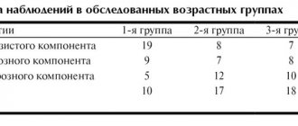
| Parameters for diagnosing myocardium in newborns | Male | Female |
| Left ventricular EDR | From 19 to 25 mm | From 18 to 24 mm |
| Left atrium diameter | From 13 to 18 mm | From 12 to 17 mm |
| Left ventricle diameter | From 6 to 14 mm | From 5 to 13 mm |
| Left ventricular ESR | From 12 to 17 mm | |
| MZhP (thickness) | From 3 to 6 mm | |
| Right ventricular wall thickness | From 2 to 3 mm | |
| Blood flow speed | About 1.3 m per second | |
| Ejection fraction varies in infants | From 65 to 75% | |
Please note that sometimes after cardiac echocardiography has been performed, interpretation is carried out directly in the diagnostician’s office. The fact is that often the hardware examination is carried out by the attending physician himself, which significantly saves time for the patient and helps the doctor quickly make the correct diagnosis.
Sometimes the list of parameters in the response is different. This happens if the research was carried out with devices of different manufacture and power, that is, devices with different technical characteristics and capabilities.
What abbreviations are used in the protocol?
Having received the EchoCG protocol completed by a specialist, the patient is faced with abbreviations that are incomprehensible to him. For example, MPAP is the average pressure in the pulmonary artery, CO and DO are the short and long axis. The most commonly used abbreviations can be seen in the figure.
Basic abbreviations that are indicated in the ultrasound protocol
In most cases, it is impossible to make a diagnosis based only on the results of the protocol. The specialist takes into account such features as ultrasound indicators, the patient’s medical history, the chronology and intensity of the development of symptoms, and other nuances. Taken together, these data help to accurately determine a particular pathology.
Deviations from the norm
There are a huge number of indicators indicating deviations from the norm. Eg:
- Stenosis is said to occur when the valve opening is reduced in diameter, making it difficult to pump blood.
- If the valve leaflets interfere with the reverse blood flow, perform their natural function inadequately, insufficiency is suspected.
- Pericarditis is detected when, with a normal fluid level of 20-30 ml, all 500 ml are traced.
The list of deviations from the norm and possible diagnoses associated with this can be endless. But you shouldn’t take deciphering an ultrasound of the heart so lightly. Here it is inappropriate to guess and assume without the help of a doctor. Mistakes lead to irreparable consequences. Therefore, experiments should be abandoned.
Preparing for the study
There are no special recommendations for preparing for an ultrasound examination. But in order for the results to be most reliable during ultrasound of the heart, it is necessary:
- exclude physical activity the day before;
- a day before, stop drinking coffee, alcohol, and strong tea;
- Avoid eating spicy and fatty foods before eating;
- do not eat for a couple of hours so that the stomach is not full and does not put pressure on the diaphragm;
- do not smoke 2-3 hours before;
- before entering the office, rest a little and calm down in the corridor;
- if there is thick hair on the chest, shave it off if possible;
- Before making an appointment, tell your doctor what medications you are taking and, if necessary, stop taking them for a while;
- lying on the couch, do not worry and remain as calm as possible;
- strictly follow all recommendations of a specialist.
In case of urgent need, echocardiography can be performed without any preparation. When planning a visit to a specialist, you do not need to take anything with you, in particular towels. Everything you need for a cardiac ultrasound is in the treatment room.