Disruption of calcium metabolism due to deviations in the functionality of the endocrine glands can lead to serious complications. Calcium is a mineral necessary for the body to form and maintain healthy bone tissue. The body contains 1.5 kg of this mineral.
Calcium enters the body with food and is also produced by the parathyroid glands. The endocrine system regulates the metabolic processes between calcium and phosphorus. With hormonal deficiency, there is an increase in phosphate in the blood with a concomitant decrease in calcium levels. Dysfunction of the gland can also lead to the reverse process, when calcium levels are increased. Both types of deviations are not considered normal and cause the development of pathological processes.
Characteristics
In the human body, a violation of calcium metabolism provokes the development of various diseases. Calcium is considered an important element in the process of regulating the nervous excitability of muscle tissue, the process of hemostasis, in the stabilization of cell tissue membranes, and in the transduction of intracellular signals.
In medicine, there are two forms of disturbance of mineral metabolism in the endocrine system:
- hypocalcemia (hypoparathyroidism);
- hypercalcemia (hyperparathyroidism).
The second type of disease is more dangerous and requires more complex treatment. In most cases, if the pathology is advanced, doctors resort to surgery.
In order to understand what could provoke a change in the production of calcium by the parathyroid glands into the blood, how to deal with this disorder and what the consequences may be, each pathology should be considered separately.
Calcium and phosphorus metabolism in the body and its disorders
Calcium metabolism
The functions of calcium in the body include:
· structural (bones, teeth);
· signaling (intracellular second messenger);
· enzymatic (coenzyme of blood clotting factors);
· neuromuscular (control of excitability, release of neurotransmitters, initiation of muscle contraction).
The main role in the metabolism of calcium in the human body belongs to bone tissue. In bones, calcium is represented by phosphates - Ca3(PO4)2 (85%), carbonates - CaCO3 (10%), salts of organic acids - citric and lactic (about 5%). Outside the skeleton, calcium is found in the extracellular fluid and is practically absent in the cells. The dense matrix of bone, along with collagen, includes calcium phosphate, a crystalline mineral compound close to hydroxyapatite Ca10(PO4)6(OH)2. Some Ca2+ ions are replaced by Mg2+ ions, a small part of OH– ions are replaced by fluorine ions, which increase bone strength. The mineral components of bone tissue are in a state of chemical equilibrium with calcium and phosphate ions in the blood serum. Bone cells can accelerate the deposition or, conversely, dissolution of mineral components with local changes in pH, concentration of Ca2+, HPO42- ions, and chelating compounds (D. Metzler, 1980). The adult human body contains 1-2 kg of calcium, 98% of which is found in the skeleton (A. White et al., 1981). It makes up about 2% of body weight (approximately 30 mol). The level of calcium in the blood is 9-11 mg/100 ml (2.2-2.8 mmol/l), in the extracellular fluid - about 20 mg/100 ml. The regulation of calcium exchange between extra- and intracellular fluid is carried out by parathyroid hormone, calcitonin, and 1,25-dioxycholecalciferol. As the concentration of calcium ions decreases, the secretion of parathyroid-stimulating hormone (PTH) increases, and osteoclasts increase the dissolution of mineral compounds contained in the bones. PTH simultaneously increases the reabsorption of Ca2+ ions in the renal tubules. As a result, the level of calcium in the blood serum increases. With an increase in the content of calcium ions, calcitonin is secreted, which reduces the concentration of Ca2+ ions due to calcium deposition as a result of the activity of osteoblasts. Vitamin D is involved in the regulation process; it is required for the synthesis of calcium-binding proteins necessary for the absorption of Ca2+ ions in the intestine and its reabsorption in the kidneys. A constant supply of vitamin D is necessary for the normal course of calcification processes. Changes in the level of calcium in the blood can be caused by thyroxine, androgens, which increase the content of Ca2+ ions, and glucocorticoids, which reduce it. Ca2+ ions bind many proteins, including some proteins of the blood coagulation system. The proteins of the coagulation system contain calcium-binding sites, the formation of which depends on vitamin K.
In food products, calcium is contained mainly in the form of calcium phosphate, which enters the body. In nature, calcium is found in the form of carbonate, oxalate, tartrate, phytic acid (in cereals).
Calcium deficiency in the body is often associated with the low solubility of most of its salts.
The authors associate calcification of arterial walls and the formation of stones in the gall bladder, renal pelvis and tubules with the poor
Calcium phosphates easily dissolve in gastric contents. Maximum calcium absorption occurs in the proximal parts of the small intestine and decreases in the distal parts.
The proportion of calcium absorption is more significant in children (compared to adults), in pregnant and lactating women. Calcium absorption decreases with age and with vitamin D deficiency.
Blood plasma contains fractions of protein-bound (non-diffusing) calcium (0.9 mmol/l) and diffusing: ionized (1.1-1.4 mmol/l) and non-ionized (0.35 mmol/l). Ionized calcium is biologically active; it penetrates cells through membranes; the non-ionized form is associated with proteins (albumin), carbohydrates and other compounds. Inside the cells, the concentration of free calcium is low. Thus, the total concentration of Ca2+ ions in the cytoplasm of erythrocytes is about 3 μM, of which free ions account for less than 1 μM. The concentration gradient of calcium ions on opposite sides of the membrane (from 102 to 105) is maintained by a calcium pump. The very slow reverse diffusion of ions into the cell opposes the pump. Ca2+ refers to secondary messengers - intracellular substances, the concentration of which is controlled by hormones, neurotransmitters, and extracellular signals. Low levels of calcium in cells are maintained by calcium pumps (calcium ATPases) and sodium-calcium exchangers. High activation of Mg2+-, Ca2+-ATPase is associated with conformational changes in the calcium pump, leading to Ca2+ transfer. A sharp increase in calcium content in the cell occurs when calcium channels or intracellular calcium stores open (the concentration increases to 500-1000 nM with 10-100 nM in an unstimulated cell). The opening of channels can be caused by depolarization of membranes, the action of signaling substances, neurotransmitters (glutamate, ATP), secondary messengers (inositol-1,4,5-triphosphate, cAMP) (Y. Kolman, K. G. Rehm, 2000). The level of calcium in cells increases (5-10 times) in the form of short-term fluctuations (high concentrations of calcium have a cytotoxic effect). In cellular organelles and the cytoplasm of cells there are a large number of proteins that can bind calcium and act as a buffer. The action of calcium is mediated by “calcium sensors” - special calcium-binding proteins - annexin, calmodulin, troponin. Calmodulin is found in all cells and, when four calcium ions bind, it transforms into an active form that can interact with proteins. C2+ affects the activity of enzymes, ion pumps, and cytoskeletal components due to the activation of calmodulin.
Hypoalbuminemia does not affect the level of ionized calcium, which varies within a narrow range and thereby ensures the normal functioning of the neuromuscular system. With increasing pH, the proportion of bound calcium increases. During alkalosis, hydrogen ions dissociate from the albumin molecule, which leads to a decrease in the concentration of calcium ions. This may cause clinical symptoms of hypocalcemia, despite the fact that the concentration of total plasma calcium is not altered. The opposite picture (increased concentration of calcium ions in plasma) is observed in acute acidosis. Globulins also bind calcium, although to a lesser extent than albumin.
Components of the regulation of calcium levels in blood plasma include:
· skeleton (calcium reservoir);
· kidneys;
· excretion of calcium through the intestines with bile;
· parathyroid hormone, calcitonin (their secretion is determined by the level of calcium in the plasma);
· 1,25-dioxycholecalciferol.
The extracellular calcium pool is renewed approximately 33 times during the day (W. J. Marshall, 2002), passing through the kidneys, intestines and bones. And even a small change in any of these flows has a significant effect on the concentration of calcium in the extracellular fluid, including blood plasma. Calcium, which is part of the secretions of the digestive tract, is partially reabsorbed along with dietary calcium.
Disorders of calcium metabolism are accompanied by disturbances of phosphate metabolism and are clinically manifested in changes in the bone skeleton and neuromuscular excitability.
There is an inverse relationship between the content of calcium and phosphorus in the blood serum (a simultaneous increase is observed in hyperparathyroidism, a decrease in rickets in children). With an increased content of phosphorus in food, non-absorbable tribasic calcium phosphate is formed in the gastrointestinal tract. The daily calcium requirement for an adult is 20-37.5 mmol (0.8-1.5 g), in pregnant and lactating women it is twice as high (M. A. Bazarnova et al., 1986). 35 mmol of calcium enters the food canal daily, but only half is absorbed, 50 times slower than sodium, but more intense than iron, zinc, and manganese. Absorption occurs in the small intestine (maximum in the duodenum). Calcium gluconate and lactate are best absorbed. Optimum absorption is observed at pH=3.0. Calcium combines with fatty and bile acids and enters the liver through the portal vein. Transport through the enterocyte membrane into the blood is facilitated by vitamin D. Absorption is reduced by a lack of phosphates (the calcium/phosphorus ratio is important). Absorption is affected by the concentration of Na+, the activity of alkaline phosphatase, Mg2+-, Ca2+-ATPase, and the content of calcium-binding protein. Calcium is normally excreted from the body through the intestines. Every day, about 25 mmol Ca2+ is secreted into the food canal by the salivary, gastric and pancreatic glands (M. A. Bazarnova et al., 1986). Calcium excretion in feces persists even with a calcium-free diet (as part of bile). About 270 mmol of Ca2+ is filtered in the kidneys per day. 90% of the calcium filtered in the kidneys is reabsorbed, so overall little is excreted in the urine (excretion increases with increasing calcium concentration in the blood and leads to the formation of kidney stones). Daily excretion ranges from 1.5 to 15 mmol and depends on the circadian rhythm (maximum in the morning), hormone levels, acid-base status, and the nature of food (carbohydrates increase calcium excretion). As the mineral framework of bones is reabsorbed, calcium reabsorption decreases. Bones are a reservoir of calcium: in hypocalcemia, calcium comes from the bones and, conversely, in hypercalcemia, it is deposited in the skeleton.
Calcium ions are important for the course of many processes:
· neuromuscular excitation;
· muscle contraction;
· blood clotting;
· permeability of cell membranes;
· activity of many enzymes and lipid peroxidation.
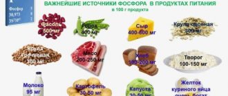
The main sources of calcium are milk, dairy products (cottage cheese, hard cheeses), fish, eggs. It is also found in green vegetables and nuts. One of the sources of calcium is drinking water (up to 350-500 mg in 1 liter). 10-30% of calcium comes from drinking water (V.I. Smolyar, 1991). The bioavailability of calcium is improved by fermented milk products and animal proteins; it is reduced by dietary fiber, alcohol, caffeine, excess fat (insoluble compounds are formed), phosphates, and oxalates. An increased content of magnesium and potassium in food inhibits the absorption of calcium: they compete with calcium for bile acids. Vitamin D supplements promote calcium absorption. When treating osteoporosis, simultaneously with the prescription of calcium supplements, it is necessary to compensate for the deficiency of proteins, calciferol, and vitamins.
Hypercalcemia is the result of increased calcium entry into the extracellular fluid from resorbed bone tissue or from food in conditions of decreased renal reabsorption. The most common cause of hypercalcemia (90% of cases) is primary hyperparathyroidism and malignant neoplasms. Often hypercalcemia is not clinically apparent. Rare causes of hypercalcemia include (W. Clutter, 1995) granulomatous diseases (including sarcoidosis), hypervitaminosis D, thyrotoxicosis, the use of thiazide diuretics, lithium drugs, milk-alkali syndrome, prolonged immobility, hereditary hypocalciuric hypercalcemia, renal failure. Clinical symptoms of hypercalcemia include:
· lack of appetite, nausea, vomiting, abdominal pain (stomach and duodenal ulcers, pancreatitis develop), constipation;
· weakness, fatigue, weight loss, muscle weakness;
· personality changes, deterioration in concentration, drowsiness, coma;
· arrhythmias, shortening of the QT interval on the ECG;
· nephrocalcinosis, renal calculi, vascular calcification, cornea;
Polyuria, dehydration, renal failure.
The most common cause of decreased total serum calcium concentration is hypoalbuminemia.
Calcium metabolism in the body is not disturbed if the content of free calcium is within normal limits. The concentration of free calcium in the serum decreases with hypoparathyroidism, resistance to parathyroid hormone (pseudohypoparathyroidism), vitamin deficiency D, renal failure, severe hypomagnesemia, hypermagnesemia, acute pancreatitis, necrosis of skeletal muscles (rhabdomyolysis), tumor disintegration, and multiple transfusions of citrated blood. Clinical manifestations of hypocalcemia include: paresthesia, a feeling of numbness, muscle cramps, laryngeal spasm, behavioral abnormalities, stupor, positive Chvostek and Trousseau symptoms, prolongation of the QT interval on the ECG, cataracts. Moderate hypocalcemia may be asymptomatic.
Hypercalciuria develops with increased dietary calcium intake, vitamin D overdose (increased intestinal resorption), tubular disorders (idiopathic hypercalciuria, renal tubular acidosis), with increased bone tissue breakdown (myeloma, bone tumors, phosphate diabetes, osteoporosis, hyperparathyroidism) .
Hypocalciuria is observed with hypoparathyroidism, hypovitaminosis D, hypocalcemia, and decreased glomerular filtration.
The role of phosphorus in the human body
The adult human body contains about 670 g of phosphorus (1% of body weight), which is necessary for bone formation and cellular energy metabolism. 90% of phosphorus, like calcium, is found in the skeleton - bones and teeth (M.A. Bazarnova et al., 1986). Together with calcium, they form the basis of the hard substance of bone. In bones, phosphorus is represented by sparingly soluble calcium phosphate (2/3) and soluble compounds (1/3). Most of the remaining amount of phosphorus is found inside cells, 1% is in the extracellular fluid. Therefore, the level of phosphorus in the blood serum does not allow us to judge its total content in the body.
Phosphates are structural elements of bone tissue and participate in energy transfer in the form of high-energy bonds (ATP, ADP, creatine phosphate, guanine phosphate and others). Phosphorus and sulfur are two elements in the human body that are part of various high-energy compounds. With the participation of phosphoric acid, glycolysis, glycogenesis, and fat metabolism are carried out. Phosphorus is part of the structure of DNA and RNA, which ensure protein synthesis. It is involved in oxidative phosphorylation, which results in the formation of ATP, and the phosphorylation of some vitamins (thiamine, pyridoxine and others). Phosphorus is also important for the functioning of muscle tissue (skeletal muscle and cardiac muscle). Inorganic phosphates are part of the buffer systems of plasma and tissue fluid. Phosphorus activates the absorption of calcium ions in the intestine. The daily requirement for phosphorus is 30 mmol (900 mg), in pregnant women it increases by 30-40%, during lactation it doubles (M. A. Bazarnova et al, 1986). According to V.I. Smolyar (1991), the need for phosphorus in adults is 1600 mg per day, in children - 1500-1800 mg per day.
the human body with plant and animal foods in the form of phospholipids, phosphoproteins and phosphates.
Plant products (in particular, legumes) contain a lot of phosphorus, but its digestibility is low. Its important sources are meat and fish. In the stomach and intestines, phosphoric acid is split off from organic compounds. Absorption of 70-90% of phosphorus occurs in the small intestine. It depends on the concentration of phosphorus in the intestinal lumen and the activity of alkaline phosphatase (its inhibition reduces phosphorus absorption). Alkaline phosphatase activity increases vitamin D, and phosphate absorption increases parathyroid hormone. Absorbed phosphorus enters the liver, participates in phosphorylation processes, and is partially deposited in the form of mineral salts, which then pass into the blood and are used by bone and muscle tissue (creatine phosphate is synthesized). The normal course of ossification processes and the maintenance of normal bone structure depend on the exchange of phosphates between blood and bone tissue.
In the blood, phosphorus is found in the form of four compounds: inorganic phosphate, organic phosphorus esters, phospholipids and free nucleotides. In blood plasma, inorganic phosphorus is present in the form of orthophosphates, but its concentration in serum is assessed directly (1 mg% phosphorus = 0.32 mmol/l phosphate). It penetrates through semi-permeable membranes and is filtered in the glomeruli. The concentration of inorganic pyrophosphate in blood plasma is 1-10 µmol/l. The content of inorganic phosphorus in the blood plasma of adults is 3.5-4 mg of phosphorus/100 ml, it is slightly higher in children (4-5 mg/100 ml) and in women after menopause. Plasma also contains hexose phosphates, triose phosphates and others. The skeleton is a reservoir of inorganic phosphorus: when its content in plasma decreases, it comes from the skeleton and, conversely, is deposited in the skeleton when its concentration in plasma increases. It is recommended to determine the concentration of phosphorus in the blood serum on an empty stomach: food rich in phosphorus increases it, and carbohydrates and glucose infusion decrease it. Phosphorus is excreted from the body through the intestines and kidneys in the form of calcium phosphate. 2/3 of soluble mono- and disubstituted sodium and potassium phosphates and 1/3 of calcium and magnesium phosphates are excreted in the urine. About 208 mmol of phosphate is filtered in the kidneys per day, and 16-26 mmol is excreted. The ratio of mono- and disubstituted phosphorus salts depends on the acid-base state. With acidosis, monosubstituted phosphates are excreted 50 times more than dibasic phosphates. During alkalosis, disubstituted phosphate salts are intensively formed and released.
Parathyroid hormone reduces the level of phosphorus in the blood serum, inhibiting its reabsorption in the proximal and distal tubules, increasing excretion in the urine. Calcitonin has a hypophosphatemic effect, reducing reabsorption and increasing excretion. 1,25(OH)2D3, enhancing the absorption of phosphate in the intestine, increases its level in the blood, promotes the fixation of phosphorus-calcium salts by bone tissue. Insulin stimulates the entry of phosphate into cells and thereby reduces its content in the blood serum. Growth hormone increases phosphate reabsorption, vasopressin increases excretion.
The exchange of phosphorus and calcium is closely interrelated. It is believed (V.I. Smolyar, 1991) that the ratio between phosphorus and calcium is 1:1-1.5 optimal for joint absorption from food. Hypercalcemia, by reducing the secretion of parathyroid hormone, stimulates the reabsorption of phosphates. Phosphate can combine with calcium and lead to calcium deposition in tissues and hypocalcemia.
If phosphorus metabolism is disrupted, an increase and decrease in phosphorus levels in the blood are detected. Hyperphosphatemia is often observed in renal failure and occurs in hypoparathyroidism, pseudohypoparathyroidism, rhabdomyolysis, tumor breakdown, metabolic and respiratory acidosis. Hyperphosphatemia inhibits the hydroxylation of 25-hydroxycalciferol in the kidneys. Moderate hypophosphatemia is not accompanied by significant consequences. Severe hypophosphatemia (less than 0.3 mmol/l (1 mg%) is accompanied by dysfunction of erythrocytes, leukocytes, muscle weakness (the formation of ATP, 2,3-diphosphoglycerate is impaired). It is observed with alcohol abuse and withdrawal, respiratory alkalosis, impaired absorption of intestines, taking phosphate binders, resuming food intake after fasting, overeating, severe burns, treatment of diabetic ketoacidosis (W. Clutter, 1995). In diabetic ketoacidosis, hypophosphatemia is not a sign of depletion of phosphate stores. Moderate hypophosphatemia (1.0-2 .5 mg%) can be observed with glucose infusion, vitamin D deficiency in food or decreased absorption in the intestine, hyperparathyroidism, acute tubular necrosis, after kidney transplantation, hereditary hypophosphatemia, Fanconi syndrome, paraneoplastic osteomalacia, increased extracellular fluid volume. Respiratory alkalosis can cause hypophosphatemia by stimulating phosphofructokinase activity and the formation of phosphorylated glycolytic intermediates. Chronic hypophosphatemia leads to rickets and osteomalacia.
Hypophosphatemia is manifested by loss of appetite, malaise, weakness, paresthesia in the extremities, and bone pain. Hypophosphaturia is observed with osteoporosis, hypophosphatemic renal rickets, infectious diseases, acute yellow liver atrophy, decreased glomerular filtration, increased phosphorus reabsorption (with PTH hyposecretion).
Hyperphosphaturia is observed with increased filtration and decreased reabsorption of phosphorus (rickets, hyperparathyroidism, tubular acidosis, phosphate diabetes), hyperthyroidism, leukemia, poisoning with salts of heavy metals, benzene, phenol.
Homeostasis of calcium and phosphate
Hypocalcemia stimulates the secretion of parathyroid hormone and thereby increases the production of calcitriol. As a result, the mobilization of calcium and phosphates from the bones and their supply from the intestines increases. Excess phosphates are excreted in the urine (PTH has a phosphaturic effect), and the reabsorption of calcium in the renal tubules increases, and its concentration in the blood normalizes. Hypophosphatemia is accompanied by increased secretion of only calcitriol. An increase in its plasma concentration under the influence of calcitriol leads to a decrease in the secretion of parathyroid hormone. Hypophosphatemia leads to stimulation of the absorption of phosphate and calcium in the intestine. Excess calcium is excreted in the urine, since calcitriol enhances calcium reabsorption to a small extent (compared to PTH). As a result of the described processes, the normal concentration of phosphate in the blood plasma is restored regardless of the calcium concentration.
Causes of hypocalcemia
Endocrine organs are sensitive to effects on them, so calcium metabolism can be disrupted for various reasons.
A decrease in plasma Ca may occur for the following reasons:
- neck trauma with bleeding;
- complication after surgery on the thyroid gland;
- inflammatory processes;
- ionizing radiation;
- metastasis in endocrine organs;
- adrenal insufficiency;
- autoimmune diseases;
- systemic diseases.
Identifying the causes is considered the basis of treatment, since if the influence of negative factors continues, therapy will not be effective.
Causes of hypercalcemia
Excess Ca in plasma can be caused by several factors. It is not uncommon for calcium metabolism disorders to occur due to the development of malignant tumors or benign tumors.
In 80% of patients, hypercalcemia develops due to disorders resulting from the formation of a solitary adenoma on the pituitary gland. Adenoma, in turn, is formed due to stress, low blood pressure, hormone intake, etc.
In 12% of cases, the cause is the primary form of hyperplasia. 2% of patients are diagnosed with parathyroid cancer. About 6% of patients have multiple adenoma.
Reasons can also include:
- deficiency in the diet of foods containing calcium;
- violation of malabsorption functions of the intestine;
- chronic renal failure.
Treatment in this case involves eliminating the cause. If after this the indicators do not change, targeted therapy or even surgery is performed.
Pathologies of calcium metabolism
Pathogenetic mechanisms of calcium metabolism disorders can be divided into two groups: endocrine and energy-dependent. The first group includes conditions caused by dysfunction of the parathyroid glands, which play a role in regulating the level of calcium in the extracellular fluid. For parathyroid hormone, the target organs are bone tissue and kidneys. The second most important regulator of calcium metabolism is the thyroid hormone calcitonin, which maintains intracellular calcium homeostasis3. But estrogen and vitamin D are most directly involved in the metabolism of calcium in skin cells. With a decrease in the production of estrogen in women after menopause, the skin quickly ages and serious problems arise with calcium metabolism in the bones. On the other hand, limiting skin exposure to UV rays with age reduces the accumulation of vitamin D in the skin, which is formed through photobiogenesis that occurs in the skin under the influence of UV rays. Vitamin D also ensures skin resistance to UV rays. The hormonal form of vitamin D - 1.25 dihydroxycholecalciferol (calcitriol) stimulates the absorption of calcium and phosphates in the intestines and their reabsorption in the kidneys, stimulates bone remodeling, calcium transport through cell membranes, cell differentiation, and the development of the immune system. In addition to the hormones that regulate calcium metabolism (group I factors), the second group of factors produced in cells as a result of ATP transformation is of great importance in its regulation. This group of factors includes inorganic poly- and bisphosphates, which are products of normal energy metabolism in cells. With age, ATP production decreases significantly and, as a result, the formation of poly- and bisphosphates decreases, which leads to disturbances in calcium metabolism4.
A little more about the symptoms
The clinical picture in both cases is pronounced. The main area of damage is the skeletal system, which contains more than 90% of the total calcium content in the body.
Symptoms of hypercalcemia, regarding bone tissue:
- pseudo- and simple form of gout;
- deformation;
- pain;
- muscle weakness and atrophy;
- fractures even with simple bruises;
- cystic bone formations.
In the severe stage, there is a sensation of “pins and needles”, numbness and burning in certain parts of the body, and temporary paralysis of the pelvic muscles may occur.
Hypocalcemia is characterized by seizures that can affect not only the limbs, but also the muscles of the chest. The danger comes from a hypocalcemic crisis, which causes serious complications.
Introduction
In the second half of our century, significant discoveries were made in the field of mineral metabolism and its endocrine regulation. The new knowledge gained about the regulation and disorders of calcium metabolism has made it possible to understand the pathogenesis of various clinical syndromes caused by these disorders. Such syndromes include rickets, osteochondrosis, osteoporosis, especially postmenopausal with a high risk of vertebral fractures, visceral and skeletal forms of hyperparathyroidism, osteodystrophy, urolithiasis, atherosclerosis, Fahr's disease with calcium deposition in the brain, calcification, dermatomyositis and others.
Calcium is a phosphatophil by nature and is found in various tissues, in particular in the mitochondria of cells in the form of phosphate salts (calcium hydroxyapatite). If apatites are the most sparingly soluble calcium salts, then compounds with inorganic bisphosphates (pyrophosphates) can become insoluble (in bones) or soluble (in soft tissues), depending on the conditions, i.e. inorganic bisphosphates are natural regulators of calcium metabolism in the body. With a gradual decrease in the activity of numerous regulatory mechanisms with age, calcium metabolism is disrupted and insoluble calcium salts are deposited in soft tissues. The question arises, is skin an exception? To answer this question, a study was conducted on changes in calcium content in the cells of various layers of the skin depending on age.
Hypercalcemic crisis
A rare but dangerous complication is hypercalcemic crisis. In an acute condition, the patient’s entire nervous system is disrupted and blood clotting accelerates. Increased blood density implies increased risks for the patient’s life, as this can lead to the formation of blood clots or complete cardiac arrest.
Hypercalcemic crisis has symptoms:
- maintaining temperature above 40°C;
- internal bleeding;
- signs of fever;
- severe itching of the skin.
Disturbances of the central nervous system lead to the occurrence of psychosis, and subsequently shock. The patient's condition leads to the fact that he does not understand what is happening. If medical assistance is not provided in a timely manner, respiratory paralysis begins, followed by cardiac arrest and death.
Physiological effects of calcium[edit | edit code]
Nerves and muscles[edit | edit code]
A moderate increase in calcium concentration in the extracellular fluid may not be accompanied by clinical signs of dysfunction of the nervous and muscular systems. However, with severe hypercalcemia, the excitability of nerves and muscles decreases, which leads to the development of muscle weakness, drowsiness, and even coma. On the contrary, a moderate decrease in calcium concentration increases excitability, leading to the appearance of Chvostek and Trousseau symptoms, tetanic convulsions and laryngospasm. It is believed that the entry of Ca into cells occurs: 1) through facilitated diffusion (using a carrier), 2) due to Na+/CaJ+-o6MeHa, 3) through channels. The permeability of the latter is regulated by hormones and mediators, and in many cells these channels are voltage-dependent. In the liver and skeletal muscle, intracellular calcium is reversibly taken up by the endoplasmic and sarcoplasmic reticulum, respectively.
Calcium ions play an important role in electromechanical coupling. The action potential causes the release of calcium from the sarcoplasmic reticulum of the muscle cell. The released calcium binds to troponin, removing the blocking effect of tropomyosin on the interaction of actin with myosin, and thereby activating the contraction process. When calcium is reabsorbed by the sarcoplasmic reticulum, the blocking action of tropomyosin is restored and the muscle relaxes.
Calcium ions are also necessary for exocytosis, so they play an important role in coupling stimulation with secretion in most exocrine and endocrine glands. The secretion of catecholamines by the adrenal medulla, the release of neurotransmitters at synapses, and the release of certain other biologically active substances (eg, histamine by mast cells) all require the presence of calcium. The cardiovascular system. Calcium ions play a critical role in electromechanical coupling in the heart muscle, as well as in conducting electrical impulses through certain areas of the heart, in particular in the AV node. Depolarization of cardiac muscle fibers opens voltage-gated calcium channels (so-called slow calcium channels), through which calcium enters the cell during the plateau of the action potential. A local increase in calcium concentration causes the opening of calcium channels in the sarcoplasmic reticulum, which further increases the calcium concentration in the cytoplasm and leads to contraction. In some cells, such as the AV node cells, the action potential is driven almost entirely by calcium influx through slow calcium channels.
In smooth muscle, including vascular smooth muscle, calcium mediates contraction and often generates a significant portion of the depolarizing current. Therefore, calcium antagonists have a strong effect on the contractility of the myocardium and vascular smooth muscle, as well as on the conduction of impulses in the heart. These drugs occupy an important place among antianginal, antiarrhythmic and antihypertensive drugs. Other effects. Calcium ions maintain the integrity of mucous membranes, participate in cell adhesion and ensure the functions of cell membranes. Calcium plays an important role in blood clotting mechanisms, although it is not used as a therapeutic agent for clotting disorders. Calcium chloride has an acidifying effect on urine (although ammonium salts are much more effective in this regard) and promotes diuresis.
Hypocalcemic crisis
An exacerbation of calcium deficiency is characterized by a decrease in plasma minerals to less than 2.25 mmol/l. A hypocalcemic crisis is also dangerous.
The attack can occur suddenly, but, as a rule, it is preceded by a number of symptoms:
- feeling of "goosebumps";
- tingling feeling throughout the body;
- limbs and part of the face go numb;
- severe muscle weakness;
- sudden muscle pain;
- depression;
- change in skin color.
Gradually, loss of sensitivity and other signs are replaced by muscle twitching. Initially, the patient feels only mild and isolated spasms, but quickly enough the condition develops into attacks of severe convulsions.
Muscle tone is expressed by characteristic manifestations:
- “the hand of the obstetrician”;
- "horse foot";
- "fish mouth";
- and others.
The danger arises when spasms affect the muscle tissue of internal organs: renal/liver colic, laryngospasm or bronchospasm. Impaired breathing can cause asphyxia.
At the moment of crisis, the patient feels unbearable pain, from which he loses consciousness and the ability to rationally perceive what is happening.
Calcium[edit | edit code]
Calcium is the main extracellular divalent cation. The body of healthy men and women contains about 1300 and 1000 g of calcium, respectively, of which more than 99% is in the bones. Small amounts of calcium are present in plasma and interstitial fluid, and even smaller amounts are present in cells, where the resting concentration of ionized calcium is approximately 0.1 µmol/L. Under the influence of chemical, electrical or mechanical stimuli, calcium enters cells, and its intracellular concentration reaches 1 µmol/l. At the same time, it interacts with specific calcium-binding proteins, which activate many intracellular processes. The main calcium-binding protein is calmodulin. It is a highly conserved protein, each mole of which binds 4 moles of calcium. Calcium ions are necessary for the excitation of neurons, the release of neurotransmitters, muscle contraction, the maintenance of membrane structure, blood clotting and many other physiological reactions. In addition, calcium acts as a second messenger for many hormones, mediators, etc.
The implementation of all these various functions is possible only at a certain concentration of ionized calcium. In human plasma, the calcium concentration is 8.5-10.4 mg% (2.1-2.6 mmol/l). Approximately 45% of this amount is associated with proteins (mainly albumin) and 10% forms complexes with anions of buffer systems (citrate and phosphates). The rest is ionized calcium, which has physiological activity. It is the decrease in the concentration of ionized calcium that causes the symptoms of hypocalcemia. In order to judge the concentration of ionized calcium from the total plasma calcium concentration, it is necessary to know the protein concentration. The following rough rule helps here: a deviation in plasma albumin concentration by 1 g% (the norm is 4 g%) should be accompanied by a change in total calcium concentration by 0.8 mg%.
The concentration of calcium in the blood is under strict hormonal control. Hormones influence its absorption in the intestines and excretion by the kidneys, and also regulate the entry into the blood of calcium stored in the bones. Calcium reserves. More than 99% of all calcium in the body is contained in the bones in a crystalline form resembling the mineral hydroxyapatite Ca|0(PO4)6(OH)2. Bone mineral contains other ions, including sodium, potassium, magnesium and fluoride. The amount of calcium in the bones depends on the relationship between resorption and osteogenesis - two coupled processes of constant bone turnover (see below). In addition, bones contain a labile fraction of calcium, from which it easily passes into the intercellular fluid of the bones, and from it into the blood. The speed of all these processes is influenced by medications, hormones, vitamins and other factors.
Absorption and excretion of calcium[edit | edit code]
In the United States, approximately 75% of dietary calcium comes from milk and dairy products. The daily calcium requirement for adolescents is 1300 mg, for persons under 24 years of age - 1000 mg, for men and women over 50 years of age - 1200 mg (see Part XIII, “Introduction”) (Institute of Medicine, 1997). At the same time, the median calcium intake for boys and girls aged 9 years and older is 865 and 625 mg/day, respectively, and for women after 50 years of age it gradually decreases to 517 mg/day (Institute of Medicine, 1997).
In Fig. 62.1
Figure 62.1. Calcium metabolism. Approximate values of daily calcium fluxes are given.
A diagram of calcium metabolism is shown. Calcium enters the body only from the intestines. It is only partially absorbed in the gastrointestinal tract. Calcium absorption is ensured by two mechanisms. Active vitamin E-dependent calcium transport occurs in the proximal duodenum. In addition, large amounts of calcium are absorbed by facilitated diffusion along the entire length of the small intestine. Obligatory calcium losses through the intestines are approximately 150 mg/day; this amount is contained in the secretion of the mucous membrane, bile and exfoliating intestinal cells.
The absorption of calcium is inversely proportional to its consumption: at low dietary levels, the proportion of absorbed calcium increases, partly due to increased activation of vitamin D. With age, this compensatory response weakens significantly. Some drugs (for example, glucocorticoids and phenytoin) inhibit calcium absorption. Certain compounds present in food (for example, phytic and oxalic acids) interfere with the absorption of calcium by forming insoluble complexes with it. Calcium absorption also decreases in diseases accompanied by steatorrhea, diarrhea or chronic malabsorption.
Urinary calcium excretion depends on the ratio of glomerular filtration and tubular reabsorption. Approximately 9 g of calcium is filtered per day, and more than 98% of this amount is reabsorbed. Calcium reabsorption is regulated by PTH, but is also influenced by the amount of sodium filtered, the presence of nonreabsorbable anions, and diuretics. Urinary calcium excretion is directly related to sodium intake (and, accordingly, excretion). Loop diuretics increase calcium excretion. In contrast, thiazide diuretics have the unique ability to disrupt the coupling between sodium and calcium excretion, resulting in decreased calcium excretion (Lemann et al., 1985). Protein intake also affects urinary calcium excretion, which is likely due to the effect of sulfur-containing amino acids on renal tubular function. In healthy people, the calcium content in urine depends only slightly on the amount of calcium in food. During lactation, significant amounts of calcium are excreted in milk. A small amount of this element is lost through sweat.
Bone tissue renewal[edit | edit code]
Immediately after the formation of bone tissue, a continuous process of renewal (resorption and osteogenesis) begins in it, which continues throughout life. After the cessation of linear growth, when the mass of bone tissue reaches its maximum, its further changes are ultimately determined precisely by the dynamics of the renewal processes. These processes independently occur in a huge number of separate areas - units of bone renewal (Fig. 62.2)
Figure 62.2. Bone turnover cycle.
. Renewal occurs on the bone surface, approximately 90% of which is normally inactive and covered with a thin cellular lining. In response to physical or biochemical signals, bone marrow progenitor cells move to the surface of the bone and fuse with each other to become the characteristic multinucleated osteoclasts; the latter resorb the bone layer located underneath them, forming depressions in it.
Osteoclast formation is regulated by cytokines (eg, M-CSF, IL-1, IL-6, osteoclast differentiation factor) that are produced by osteoblasts. Recent studies shed light on the mechanisms of this process (Suda et al., 1999). The protein, the production of which is necessary for the formation of active osteoclasts, is called RANK (Receptor for Activating NFkB). The natural ligand of this receptor is osteoclast differentiation factor (also called RANK ligand), located on the osteoblast membrane. By interacting with the RANK protein, osteoclast differentiation factor induces the maturation of osteoclasts. Indeed, antibodies to this factor prevent the resorption-enhancing effects of many regulators of bone turnover (Yasuda et al., 1998). Osteoclast differentiation factor not only triggers the differentiation of osteoclast precursors but also activates mature osteoclasts (Jimi et al., 1999). Osteoblasts also produce a soluble inhibitor called osteoprotegerin, which acts as a decoy receptor for osteoclast differentiation factor. Under conditions that promote increased bone resorption (for example, estrogen deficiency), the production of osteoprotegerin is inhibited, the osteoclast differentiation factor freely interacts with the RANK protein, and osteoclast formation is enhanced. When estrogen levels are restored, osteoprotegerin production increases and it prevents osteoclast differentiation factor from binding to the receptor.
As a result of resorption in the compact substance of the bone, tunnels are formed in the osteon canals, and in the spongy substance - uneven surface areas called resorption (gauship) lacunae. At the end of resorption, a cavity with a depth of about 60 microns remains, bordering in its deepest part the line of adhesion (an area of loosely organized collagen fibers). Upon completion of the resorption stage, preosteoblasts, formed from bone marrow stromal cells, arrive at the bottom of the cavity. Preosteoblasts acquire the characteristic features of osteoblasts and begin to restore resorbed bone, forming new organic matrix components - collagen, osteocalcin and other proteins. As soon as the layer of newly formed organic matrix reaches approximately 20 μm, its mineralization begins. The entire update cycle normally takes about 6 months.
If osteogenesis quantitatively corresponds to resorption, then renewal does not lead to a change in tissue mass. However, due to the insufficient efficiency of renewal, a small mass deficit remains after each cycle. Over the years, this deficiency increases, which explains the well-known decrease in bone mass that begins soon after the cessation of growth. Changing the turnover rate is a common final mechanism for the influence of various stimuli (malnutrition, hormones, drugs, etc.) on bone health. The overall rate of bone tissue renewal depends on the dynamics of the individual components of this process. Hormonal changes often result in activation of bone turnover or an increase in the number of bone turnover units. This occurs, for example, with thyrotoxicosis, hyperparathyroidism and hypervitaminosis D. Other factors, in particular high doses of glucocorticoids and ethanol, disrupt the function of osteoblasts. Finally, estrogen deficiency appears to enhance osteoclast activity (Marcus 1987; Dempster 1992).
At each moment of time, there is a certain deficiency of bone tissue mass, which is determined by the resorption foci that have not yet been filled. In response to any stimulus that changes the rate of new units of bone turnover, this deficit increases or decreases until a new equilibrium is established between resorption and osteogenesis.
Read the main article:
Physiology of bone tissue restoration
Diagnostics
Calcium metabolism disorders are determined by laboratory tests. To determine the cause of the pathology, more extensive research is required, so specialists prescribe hardware studies. Additionally, differential diagnosis can be carried out.
Examination methods:
- urine test (general) - with elevated Ca levels, a mineral will be present in the biomaterial and the alkaline reaction will be pronounced;
- blood test (biochemical) - a significant increase/decrease in ionized and total calcium is detected in the plasma;
- ultrasound examination - the entire endocrine system is examined for the presence of tumors, tissue enlargement and other abnormalities;
- radiography - used to examine bones to determine the possible development of osteoporosis, fractures, cystic formations and other pathological processes;
- densitometry - used to accurately determine the density of bone tissue;
- radiography (with contrast agent) - helps to identify ulcerative lesions of the gastrointestinal tract and parathyroid adenoma in the retrosternal space;
- computed tomography - examines the urinary system and kidneys for the presence of stones;
- magnetic resonance imaging - allows you to obtain the most accurate data about tumor formations of the thyroid glands;
- spintigraphy is a study using radioactive isotopes, with the help of which it is possible to visualize the area under study.
Diagnostics allows you to obtain the maximum amount of information to identify provoking factors, characteristics of the disease and possible complications. Treatment can begin only after receiving the examination results.

![Table 1. Bioavailability (absorption) of various organic forms of calcium within 2 hours after oral administration in the experiment [22]](https://laram-halal.ru/wp-content/uploads/tablica-1-biousvoyaemost-absorbciya-razlichnyh-organicheskih-form-kalciya-v-techenie-330x140.jpg)