GLAUCOMA
– a serious eye disease that often leads to blindness.
Today, science knows only one way to prevent blindness from glaucoma: timely recognition and proper treatment. It is usually accompanied by an increase in intraocular pressure, which gradually provokes the death of the optic nerve, impaired visual fields and blindness.
The patient often does not notice this process for a long period and turns to the doctor only when he discovers that his vision has lost its sharpness. With timely early treatment of glaucoma, the pathological process can be stopped, the death of the optic nerve and loss of vision can be avoided. Glaucoma classification:
- congenital (hereditary, intrauterine; trauma during childbirth) and acquired;
- open-angle (the drainage system of the eye is disrupted);
- closed angle (the camera angle is closed, fluid does not flow out of the eye quickly enough, intraocular pressure increases);
- mixed;
- pseudonormal pressure glaucoma (open-angle glaucoma, but with normal intraocular pressure values);
- primary and secondary, that is, provoked by ophthalmological diseases, eye surgery, taking certain medications, injuries, etc.
Drug treatment of primary glaucoma
Conservative treatment of glaucoma is carried out using medications in 3 areas:
- Ophthalmic hypotensive therapy to reduce intraocular pressure;
- Improving blood supply to the internal membranes and intraocular segment of the optic nerve;
- Normalization of metabolic processes of eye tissues to inhibit the degenerative processes of glaucoma.
It is immediately necessary to stipulate that the key issue in the treatment of the disease is the issue of normalizing the level of intraocular pressure (IOP). The remaining methods are only auxiliary in nature. To these can also be added the organization of the correct work schedule for a patient with glaucoma and his daily life.
When starting regular use of antiglaucoma drops, the patient should know what options exist for their effect on intraocular pressure:
- IOP decreases after a single application (instillation) of the drug. Each repeated instillation regularly repeats this effect;
- The effect of the drug is delayed. At first it is weakly expressed, but intensifies in the following days of regular instillation of the drug;
- There is resistance to the drug from the very beginning, and it has no effect on intraocular pressure;
- The drug has an unexpected effect, i.e. the pressure after instillation does not decrease, and sometimes even increases quite significantly. Therefore, a test is required for each antiglaucomatous drop.
In this regard, prescribing medications to lower IOP levels is the prerogative of the doctor. An ophthalmologist, when choosing the drug needed in a particular case, is able to take into account many factors that are completely unknown to the patient. Therefore, you cannot prescribe or stop antiglaucoma medications on your own! Even if the frequency of instillation is changed, it is necessary to consult with your doctor.
When prescribing a certain regimen of instillation of antiglaucoma drops, the patient must be observed by an ophthalmologist for 2-3 weeks dynamically. Further, treatment monitoring should be carried out every three months. Every 1-2 years, it is recommended to change medications, with appropriate monitoring.
Medicines used in the treatment of glaucoma are divided into two groups: drugs that accelerate the outflow of intraocular fluid and drops that inhibit its production.
Definition of disease
Glaucoma is a disease in which gradual degradation of the optic nerve and pathological changes in the retina occur . These changes lead to gradual loss of visual fields and in the long term can lead to complete blindness. Pathological processes affecting the optic nerve and retina are irreversible.
Currently, glaucoma ranks second among ophthalmological diseases leading to complete loss of vision.
In Russia alone, more than a million patients with this pathology are currently registered. Most patients do not know about their problem due to the asymptomatic course of the disease in the early stages. It is for this reason that it is so important to undergo regular eye examinations to identify this problem.
Antiglaucoma drops
I. Topical agents that accelerate the outflow of aqueous humor
1. Miotics
- The active ingredient pilocarpine is pilocarpine hydrochloride solutions 1%, 2%, 4% (Russia, Ukraine), Oftan Pilocarpine solution 1% (Finland), Isopto-carpine solutions 1%, 2%, 4% (USA), etc.
- The active ingredient is carbachol. Isopto-Carbachol solution 1.5 or 3% (USA).
2. Sympathomimetics
- The active substance is epinephrine. Solutions Glaucon 1% or 2% (USA), Epiphrine drops 0.5%, 1% or 2% (USA).
- Active ingredient: dipivefrin. Oftan-dipivefrin drops 0.1% (Finland).
3. Prostaglandins
- The active substance is latanoprost. Xalatan solution 0.005% (USA).
- The active ingredient is travoprost. Travatan solution 0.004% (USA).
II. Solutions that reduce the production of intraocular moisture
1. Selective sympathomimetics
- Active ingredient: clonidine. Clonidine solution 1.125%, 0.25% or 0.5% (Russia).
2. Beta blockers
- Non-selective adrenergic blockers (ß1,2). The active substance is timolol. Drops Oftan timolol (Finland), Timolol-DIA and Timolol-LENS (Russia), Timohexal (Germany), Arutimol (USA), Niolol (France), Okumol (India), Timoptic and Timoptic-depot - a form with prolongation action (Netherlands ), Cusimolol (Spain).
- Selective (ß1) adrenergic blockers. Active ingredient: betaxolol. Betoptik drops 0.5% and Betoptik C ophthalmic suspension 0.25% (Belgium).
3. Carbonic anhydrase inhibitors
- Active ingredient: dorzolamide. Trusopt solution 2% (USA).
- The active ingredient is brinzolamide. Azopt ophthalmic suspension 1% (USA).
III. Combination drugs
a. Active ingredients: proxodolol + clonidine, Proxofelin drops (Russia). b. Active ingredients timolol + pilocarpine, drops Fotil and Fotil forte (Finland). c. Active ingredients: pilocarpine + metypranolol, Normoglaucon drops (Germany). d. Active ingredients: dorzolamide + timolol, Cosopt drops (France).
When determining the tactics of drug treatment of glaucoma, the first choice drugs are: Timolol, Pilocarpine, Xalatan, Travatan.
Second choice drugs include: Betaxalol, Brinzolamide, Dorzolamide, Clonidine, Proxodolol, Dipivefrin, etc.
conclusions
- Glaucoma is an irreversible eye disease that leads to complete loss of vision as a result of destruction of the optic nerve and pathological processes in the retina.
- There are open-angle and closed-angle forms of the disease.
- There are many reasons why the disease occurs. Among them are abnormalities in the development of the eye, disruption of the drainage system, general diseases accompanied by circulatory disorders, eye injuries and long-term uveitis, iridocyclitis.
- In the first stages of development, the disease is asymptomatic. During attacks of glaucoma, blurred vision and pain are observed.
- Medicinal, surgical, and traditional methods of treatment do not allow one to get rid of the disease. They only slow down the pace of its development.
- To prevent glaucoma, patients are advised to visit doctors at least once a year and have their intraocular pressure measured.
- Eye drops for glaucoma: types of drugs and their features
Principles of conservative treatment of glaucoma
Treatment begins with one of the first-choice drip medications. If therapy does not bring the expected effect, a replacement is made with the next first-choice drug, or a combination treatment is carried out with a first-choice drug and a second-choice drug, or two first-choice drugs.
If intolerance or contraindications to first-choice drugs are detected, treatment can begin with one of the second-choice drugs.
In the case of prescribing combination therapy, it is optimal to prescribe combined antiglaucoma drugs.
Drugs with an identical mechanism of action are not prescribed in combination therapy.
Long-term treatment requires periodic replacement of prescribed medications.
Possible complications
If a patient with glaucoma does not receive the necessary treatment, the disease can progress rapidly and lead to the development of complications. The patient develops severe corneal atrophy, accompanied by increased lacrimation and acute pain. At the last (terminal) stage of development of the pathology, the patient loses the ability to open the affected eye. The most severe complication of glaucoma is complete loss of vision due to optic nerve atrophy.
If a patient who has lost his vision experiences pain that cannot be relieved with specialized medications, he is prescribed removal of the diseased eye. Some time after such an operation, a prosthesis may be installed in place of the eye.
Therapy of angle-closure glaucoma during an acute attack
An acute attack of angle-closure glaucoma is considered an emergency requiring emergency care. If intraocular pressure reaches 40-60 mm Hg during the development of an attack. and higher, it is not possible to reduce it to satisfactory values within 24 hours; the prognosis for vision can be disastrous. The eye is in danger of blindness!
Therefore, the main goal in the event of an acute attack is an emergency reduction in intraocular pressure. To do this:
1. Drug therapy:
- They immediately begin to instill a miotic - a 1% solution of pilocarpine according to the following scheme: for the first 2 hours from the onset of the attack, a drop of the solution is instilled every 15 minutes, for the next 2 hours, a drop of the solution is instilled every 30 minutes, then, for 2 hours, a drop per hour. Afterwards, the drug is applied 3-6 times during the day, depending on the level of existing IOP. A similar scheme is used only if the test for pilocarpine is positive (the pupil narrows when the drug is instilled once or twice). If there is no pupil reaction due to iris ischemia, treatment with pilocarpine is pointless and even dangerous;
- As an addition to miotic instillation, instillation of a 0.5% solution of timolol is carried out, which is prescribed drop by drop twice a day;
- For oral administration, acetazolamide (diacarb) is recommended in a dosage of 0.25-0.5 g. up to 3 times a day. Together with systemic carbonic anhydrase inhibitors, a 2% solution of dorzolamide (the drug Trusopt) is often prescribed three times a day or a 1% suspension of brinzolamide (the drug Azopt) twice a day;
- Osmotic diuretics are used intravenously or orally (usually 1.5-2 g/kg of a 50% glycerol solution). In case of unsatisfactory reduction in pressure, intramuscular or intravenous loop diuretics (20-40 mg furosemide) are used;
- If intraocular pressure does not decrease despite the therapy, a “lytic mixture” is administered intramuscularly, including: 2.5% solution of chlorpromazine (1-2 ml), 2% solution of diphenhydramine (1 ml) or promethazine (2 ml), 2% promedol solution (1 ml). When administering the mixture, the patient must remain in bed for 4 hours due to the risk of orthostatic collapse (a sharp decrease in blood pressure).
2. Distraction procedures:
- Hot foot baths, cupping, mustard plasters, saline laxatives, leeches to the temple (simultaneously with drug therapy);
3. Carrying out laser iridectomy (iridotomy) on both eyes, in order to remove the block and normalize the outflow path of intraocular fluid (to relieve an attack) and prevent recurrent attacks.
4. If it is impossible to stop the attack on the first day, a basal iridectomy is prescribed - a surgical operation.
Prevention
The main prevention of glaucoma is regular medical examinations by an ophthalmologist. They need to pay special attention to people over 40 years of age. They need to visit a specialist at least twice a year. Mandatory monitoring of eye pressure should be carried out for patients who suffer from farsightedness.
Until the age of 40, medical examination is mandatory for those people who suffer from pathologies of the vegetative-vascular system, increased cranial pressure, as well as for patients who have suffered eye injuries. Their risk of developing the disease increases.
Also, for the purpose of general disease prevention, you need to comply with the requirements for visual hygiene, do eye exercises that improve blood circulation in the tissues, and eat food containing an increased amount of vitamins and minerals necessary for the body.
Laser treatment of glaucoma
Laser glaucoma surgery is used to eliminate intraocular blocks that occur when intraocular fluid drains from the eye.
Lasers in glaucoma surgery began to be widely used in the 70s of the last century. Today, argon laser systems (with wavelengths of 488 and 514 nm), as well as neodymium YAG lasers (with a wavelength of 1060 nm) or semiconductor (diode) lasers (with a wavelength of 810 nm) are most often used.
When exposed to a laser, a local burn is applied to the trabecular area, which subsequently causes atrophy and scarring of its tissue (coagulation), or a microexplosion is performed, with tissue rupture by a shock wave (destruction).
There are many laser operations, however, only two are most widespread:
- Laser trabeculoplasty.
- Laser iridotomy (iridectomy).
Laser surgeries have their advantages and disadvantages. The benefits of laser treatment include:
- Restoring the outflow of intraocular moisture naturally;
- Carrying out the intervention under local drip anesthesia without the use of general anesthesia;
- Performing the operation on an outpatient basis;
- Short rehabilitation period;
- Absence of complications inherent in traditional glaucoma surgery;
- Relatively low cost.
The disadvantages of laser treatment for glaucoma include:
- Limited effect of the operation and its reduction in advanced cases of the disease;
- The occurrence of a reactive syndrome, with an increase in intraocular pressure immediately after laser intervention, as well as the subsequent development of the inflammatory process;
- Risk of damage to the tissue of the posterior epithelium of the cornea, iris vessels, and lens capsule;
- The occurrence of synechiae (adhesions) in the iridotomy area (angle of the anterior chamber).
When performing laser iridectomy (iridotomy), a small hole is created in the peripheral part of the iris. The operation is prescribed if there is a functional pupillary block, and its implementation allows you to open the angle of the anterior chamber and equalize the pressure in each of the chambers of the eye. The use of iridotomy is possible in the case of angle-closure glaucoma (primary, secondary), or in a mixed form of the disease. Sometimes it is required after surgery for glaucoma.
Laser iridectomy is often performed on the second eye for prophylactic purposes during an acute attack of primary angle-closure glaucoma.
The operation is performed under local anesthesia (introducing a solution of lidocaine, inocaine, etc. into the eye). A special goniolens is placed on the eye, which allows laser radiation to be focused on a selected area of the iris. Iridectomy can be performed in any quadrant and is performed in several stages in different sectors of the iris in thinned areas.
In some cases, it is not possible to obtain a through hole in the iris, or it closes very quickly due to the formation of synechiae and pigment deposition. In this regard, repeated intervention is required.
Laser trabeculoplasty is performed by applying a series of burns to the inner trabecular surface. This effect improves the permeability of the trabecular diaphragm for aqueous humor and reduces the risk of Schlemm’s canal block. The mechanism of action of the operation is characterized by tension and shortening of the trabecular diaphragm due to wrinkling of tissue at the burn site, with widening of the trabecular slits in the areas between burns.
The operation is prescribed in the surgery of primary open-angle glaucoma, when drug therapy is ineffective. The procedure is performed using local anesthesia. During this procedure, a goniolens is placed on the eye. According to the particularly popular technique of linear trabeculoplasty today, one row of burns is performed in the area of Schlemm’s canal.
Causes
Various factors can lead to the development of pathology, including:
- congenital anomalies of the structure of the eye chamber;
- blood pressure disorders, for example, hypotension;
- taking certain medications, including corticosteroids;
- eye injuries;
- intraocular tumors;
- protracted eye diseases, including uveitis, iridocyclitis;
- change in the position or volume of the lens;
- pathologies of the drainage system of the eye;
- poor circulation in the chamber of the eye.
Also, certain pathologies of the musculoskeletal system, for example, osteochondrosis, can lead to the development of glaucoma. With such diseases, the functioning of the vascular system, which ensures the functioning of the optic nerve, deteriorates, which leads to glaucoma and blindness.
Contraindications
With drug treatment:
- individual intolerance to the drug;
- patient resistance to the drug;
- a paradoxical effect - instead of reducing blood pressure, the medication causes an increase.
With laser treatment:
- late stages of glaucoma;
- terminal phase of the glaucomatous process.
During surgery:
- some concomitant ophthalmological pathologies;
- if the risk of decreased visual acuity exceeds the expected positive result of the operation.
Diagnostics
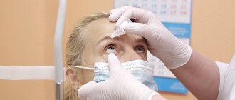
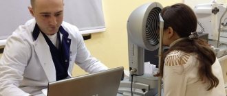
At the initial appointment, the ophthalmologist will carefully examine the eyes, ask the patient about warning symptoms, inquire about the medical history, and find out the risk factors that contribute to the manifestation of the pathology. In addition to this, he will carry out the following diagnostic procedures:
- Measure intraocular pressure using tonometry. All manipulations are simple and painless.
- Conducts testing for optic nerve damage using special instruments and equipment.
- Tests visual fields by examining features of the peripheral visual system.
- It will measure the thickness of the cornea (pachymetry), which will provide additional information in the diagnosis of glaucoma.
Dr. Trubilin’s clinic uses traditional and latest methods for diagnosing eye problems. An integrated approach is used for a comprehensive examination and making the most accurate diagnosis.
With us you can:
- undergo a visual acuity test;
- measure intraocular pressure;
- examine visual fields;
- do optical coherence tomography.
Additionally, biomicroscopy and gonioscopy of the affected area are performed. Based on the screening results, the doctor makes a diagnosis and prescribes individual treatment for glaucoma. When drawing up a scheme of restorative procedures, the patient’s age, medical history, as well as related details are taken into account.


