Colonoscopy is a diagnostic method that is used to assess the condition of the colon mucosa. The examination is carried out using a special instrument - a colonoscope. It is a thin flexible tube with a video camera at the end. The colonoscope is inserted into the intestines through the anus.
- Execution Goals
- Preparing for a colonoscopy
- Diet before colonoscopy
- Indications and contraindications
- Diagnostic colonoscopy
- Therapeutic manipulations during colonoscopy
- Colonoscopy in a state of medicated sleep
- Methodology
- Recording the procedure on video
- Recovery after colonoscopy
- Possible complications
- Alternative diagnostic methods
- Colonoscopy results
Execution Goals
Using a colonoscope, the doctor can detect ulcers, polyps, foci of inflammation, tumors, and sources of intestinal bleeding. In addition, during the study it is possible to obtain tissue samples from the intestines for histological and cytological examination, as well as to remove polyps or other formations.
Colonoscopy, diagram
It is recommended to have your first colonoscopy at age 50. Further recommendations are given individually, depending on the results of the study. Unfortunately, most colon tumors are detected at late stages. There is only one reason here - late diagnosis of the tumor. The only way to detect a tumor at an early stage is regular examination of the colon, which allows us to identify pre-tumor pathology and remove it, or carry out other necessary treatment so that it does not degenerate into cancer. The key to curing colon cancer is early diagnosis. Early diagnosis can be achieved using endoscopy. [1]
Diet before colonoscopy
An important part of preparing for a colonoscopy is proper diet in the preceding days and on the day of the examination. In general terms, the recommendations are as follows:
- A few days before the procedure, you need to switch to a low-fiber diet, reduce the consumption of fresh vegetables and fruits, dried fruits, nuts, and whole grains.
- 1–3 days before and on the day of the procedure, you need to give up solid food. Broths, clear fruit juices (for example, clarified juice from apples, white grapes), tea and coffee, and jelly are allowed. It is recommended to drink more fluid the day before.
- You should not eat or drink anything 2–4 hours before the procedure The procedure can only be carried out under medicated sleep conditions on an empty stomach.
In parallel with the diet, in the afternoon before, the intestines are prepared for the procedure using laxatives. Recommendations regarding types of drugs and dosage regimens may vary from doctor to doctor. [2,3]
Preparation with Flit
Fleet is a drug that appeared relatively recently, its popularity is rapidly growing, many patients have been convinced of the effectiveness of the drug from their own experience.
Rules for preparation using the drug Flit
- Take in two doses.
- 45 ml of the product is diluted in half a glass of water and consumed immediately after breakfast;
- the second dose (45 ml) - in the evening after a diet dinner.
- If the patient has time, you can take another dose in the morning on the day of the examination, no later than 8-00.
- Breakfast and dinner should include only 1 glass of water.
- Lunch should be dietary and include no more than 750 ml of water.
- Each dose of Flit should be washed down with 1-3 glasses of cold water; after taking the drug, the liquid can be consumed in unlimited quantities.
- Fleet begins to act approximately 30 minutes after administration.
Indications and contraindications
Indications for colonoscopy are:
- blood and mucus in the stool;
- presence of relatives with colon cancer;
- previous colon surgery;
- chronic abdominal pain of unknown etiology;
- suspicions of cancer, ulcerative colitis, Crohn's disease;
- elevated temperature for a long period of time, accompanied by anemia and weight loss.
Regular colonoscopy is also recommended for all people over 50 years of age for the early detection of tumors, polyps and other diseases of the colon.
Contraindications to the study are the active stage of Crohn's disease or ulcerative colitis.
Diseases diagnosed using colonoscopy
The technique allows you to diagnose pathologies of the gastrointestinal tract:
- colitis of different etiology (origin);
- polyps and pseudopolyps;
- diverticulosis;
- benign tumors;
- oncological diseases.
Examination using a minimally invasive method allows you to obtain the most informative results. And this, in turn, is the key to a correct diagnosis and the prescription of correct and effective treatment.
Diagnostic colonoscopy
A diagnostic colonoscopy is performed to identify certain pathological formations in the intestines. There are two special types of this study:
- A screening colonoscopy is recommended for all people over 50, even if they have no symptoms. For some bowel diseases, screening may need to start at a younger age. This type of screening is used for early diagnosis of polyps that can develop into a malignant tumor and intestinal cancer.
- Control colonoscopies are periodically performed in people with a history of polyps, colon cancer, and inflammatory bowel diseases. [1.4-6]
Intestinal polyps discovered during colonoscopy
Having discovered a pathological neoplasm in the intestine, the doctor removes it using a special instrument inserted through the colonoscope and sends it to the laboratory for histological and cytological examination. This procedure is called a biopsy .
The Euroonko clinics in Moscow and St. Petersburg are running a promotion for “Gastro- and colonoscopy under intravenous sedation.” The cost of the complex service includes endoscopic examinations of the stomach and colon, light anesthesia (sedation, “medicated sleep”), a preliminary appointment with a specialist (candidate of medical sciences or doctor of medical sciences), a three-hour stay in a comfortable room. More details about the program.
How will you feel during the procedure?
In order to reduce discomfort, local anesthetics (ointments and gels) are used. There may be some discomfort during the test, but there should be no pain. The examination can be painful in case of inflammatory diseases of the rectum. If it is impossible to perform a colonoscopy due to severe pain or severe destructive processes in the colon, massive adhesions in the abdominal cavity and children under 10 years of age, this study is performed under general anesthesia (anesthesia).
So, during a colonoscopy, you will have a feeling of fullness of the intestines with gases, which causes the urge to defecate. At the end of the study, the air introduced into the intestine is removed through the endoscope channel. Moderate pain that occurs when the intestine is stretched with air will gradually disappear.
Therapeutic manipulations during colonoscopy
During an endoscopic examination of the colon, the doctor may perform some therapeutic procedures:
- Remove polyp.
- Stop intestinal bleeding, for example, using electrocoagulation or clipping.
- Eliminate stenosis (narrowing of the intestinal lumen) caused by a malignant tumor or other causes. A special balloon is inserted into the narrowed area and inflated to expand the lumen, and then a stent is installed - a tube with a mesh wall made of metal or plastic (this procedure is called stenting ). [7,8]
Removal of an intestinal polyp during colonoscopy
During the work of the endoscopy department at Euroonko, more than a hundred endoscopic stenting of the colon was performed under the guidance of an endoscopist, Doctor of Medical Sciences Mikhail Sergeevich Burdyukov. This minimally invasive procedure helps restore intestinal patency in inoperable cancer.
Often, a colonoscopy is initially performed as a diagnostic procedure and, if pathological changes are detected, it becomes a therapeutic procedure.
Colonoscopy in a state of medicated sleep
Endoscopic examination of the intestine is usually painless, but the patient may experience discomfort from a feeling of bloating (this goes away immediately after the procedure) and the advancement of the probe through the loops of the intestine. In Euroonco clinics it is possible to carry out the procedure in a state of medicated sleep. In this case, the patient is given a special sedative, under the influence of which he is plunged into deep sleep. After approximately 40 minutes, the effect of the drug ends, and within 5-10 minutes after waking up the patient can walk and talk, and after an hour he can go home.
Types of anesthesia
In the standard version of the procedure, the probe itself is lubricated with an anesthetic. When it comes into contact with the intestinal walls, the medicine reaches them, is absorbed and reduces pain. However, if indicated or simply desired by the patient, anesthesia is used. It comes in two varieties:
- Medication sleep.
The safest way. The patient falls into a half-asleep, hears the doctor perfectly, retains the ability to follow his instructions, but experiences discomfort to a much lesser extent or does not experience it at all. The drugs are as modern as possible, the load on the body is small. - General anesthesia.
It puts a strain on the cardiovascular system, so it is not recommended for people with cardiovascular diseases and those who have recently suffered a stroke or heart attack. It has a negative effect on the lungs, so it is also not recommended for asthmatics and other patients. During general anesthesia, the patient is unconscious, does not experience any discomfort, and does not respond to any requests. After waking up, needs observation for at least 24 hours.
Before you determine which type is right for you, you will need to talk to an anesthesiologist and get tested.
Methodology
A diagnostic colonoscopy lasts on average 30 minutes. If therapeutic manipulations are required, the procedure time increases.
In order for the patient to undergo the procedure without pain and discomfort, sedation is used - the patient is immersed in a state of “medicated sleep”.
The patient must be completely undressed from the waist down. He is placed on his left side, with his legs bent and knees brought to his stomach. The doctor lubricates the colonoscope with Vaseline, carefully inserts it through the anus and slowly advances it, examining the intestinal mucosa. In this case, the intestines are inflated with gas to provide a better view. At Euroonko, carbon dioxide is used for this, because it acts as an antispasmodic, is absorbed through the intestinal walls faster than oxygen and nitrogen and is excreted from the body.
The image from the colonoscope camera is transmitted to the device screen. Videos of the study are recorded and saved on a computer.
After the intestines are examined, the doctor carefully removes the colonoscope. The study is completed.
Who is it suitable for?
Colonoscopy under anesthesia is definitely performed if:
- Inflammatory processes occur in the intestines. Ulcers, colitis, enteritis are accompanied by pain; if the probe inaccurately touches the intestinal walls, this will cause too much discomfort to be ignored.
- The patient is less than twelve years old. It is difficult for the child to relax the muscles enough for the examination to be effective and produce results, in addition, pain during the process can be delayed and create unpleasant associations with hospitals in general.
- There are adhesions in the intestines formed by connective tissue. When air enters the intestine, this also provokes severe pain, which cannot be written off.
- The patient has a psychiatric diagnosis that will not allow him to sufficiently prepare, relax, and in some cases even understand what is happening.
- The patient has a low pain threshold and knows that the examination will be too unpleasant for him.
In a private clinic, a colonoscopy under anesthesia can be done simply at will, without indications.
Recovery after colonoscopy
Hospitalization is not necessary, and colon endoscopy can be performed on an outpatient basis. The patient can leave the clinic as soon as he recovers from anesthesia. But you are not allowed to drive on your own on this day. You need to take one of your relatives to the clinic so that you can be escorted home.
Notes and recommendations regarding the recovery period after the procedure:
- When the effect of sedation wears off, a feeling of cramping and fullness of the intestines may appear. It will soon pass. To help the gas leave the intestines faster, it is recommended to walk.
- For 24 hours after the procedure, you should avoid drinking alcohol, driving vehicles, and doing work that requires concentration.
- Unless otherwise advised by your doctor, you can begin eating as usual immediately after the sedation ends. A special diet is prescribed after removal of polyps and other surgical procedures.
- It is recommended to rest for one day after the study, then you can do all your usual activities without restrictions, and go to work.
- If a biopsy was performed during the test, you may notice small amounts of blood in your stool for 1–2 days afterward.
- If you need to take blood clot preventative or other medications continuously, ask your doctor how long after your colonoscopy you can start taking them again.
- Since your bowels have been cleansed, another bowel movement may occur within a few days. It depends on your diet. If you do not have stool for a very long time after the procedure, consult your doctor.
You should immediately seek medical help if symptoms such as high fever, bleeding from the anus, or abdominal pain appear.
Possible complications
Colonoscopy is a safe test. The risk of complications during a diagnostic examination of the colon is minimal. The risks of surgical endoscopy of the colon are less than one percent, which allows us to speak with confidence about its safety. Complications during endoscopic examination of the colon are very rare, including:
- Allergic reactions to sedative medications.
- Bleeding after biopsy, polyp removal.
- Perforation (rupture of the wall) of the intestine.
Alternative diagnostic methods
Other tests can also be used for the early diagnosis of malignant tumors of the colon:
- Fecal occult blood test (annually). The accuracy of the study is 62–79%. Out of 10 people with colon cancer, only 6-8 will test positive. False-positive results are possible if the patient has recently eaten red meat, foods rich in vitamin C, or is taking drugs from the group of non-steroidal anti-inflammatory drugs. If the test shows a positive result, endoscopy is required to clarify the diagnosis.
- Flexible sigmoidoscopy (every 5 years) is an endoscopic examination of the rectum and lower third of the colon. It is performed in the same way as a colonoscopy, but during it, a smaller part of the intestine is examined. Helps identify 70–80% of polyps and malignant tumors in the rectum and lower colon.
- Fecal DNA testing combined with stool occult blood testing (every 3 years). Can detect up to 92% of malignant tumors and up to 42% of precancerous conditions.
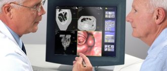
- Virtual colonoscopy (CT colonography) (every 5 years) - multislice computed tomography (MSCT), during which the colon is filled with air. The detection rate of tumors larger than 1 cm using this study is 94%, polyps measuring 6–9 mm is 65%. Polyps smaller than 6 mm are not detected. If a polyp is found during CT colonography, endoscopy will still have to be performed to remove it. [9,10]
An image taken during CT colonography
Thus, colonoscopy is the most accurate and informative method of screening for colon cancer, while it makes it possible to immediately carry out a biopsy and some therapeutic manipulations.
Colonoscopy results
Further tactics depend on whether certain pathological neoplasms have been detected.
No pathological changes were found
In such cases, no additional diagnostic procedures are required. The doctor will recommend repeating the procedure in 5–10 years. If the intestines were poorly prepared and feces remain in it, which prevented a full examination of the mucous membrane, repeat endoscopy will be recommended after a year.
If 1-2 polyps measuring less than 1 cm are found with a low risk of malignancy
The doctor will recommend repeating the test in 5 years.
If “dangerous” polyps are detected
Your doctor may recommend a repeat colonoscopy sooner than five years later in the following cases:
- more than two polyps were found in the colon;
- polyp size more than 1 cm;
- polyps were found, while there are fecal masses in the intestine, which complicate its full examination;
- according to the results of the biopsy, the removed polyps belong to histological types that have a high tendency to malignant degeneration;
- Cancer cells were found in the polyp.
If polyps are identified that could not be removed endoscopically
Such patients should consult a doctor. Perhaps the question of abdominal surgery will arise.
If cancer is detected
An examination is prescribed that helps assess the stage and other characteristics of the malignant tumor. The patient is referred to the oncology clinic, treatment begins in accordance with the protocols. If the doctor was unable to guide the colonoscope to the end of the colon
X-ray with barium enema or CT colonography may be prescribed.
Book a consultation 24 hours a day
+7+7+78
Bibliography:
- S. V. Kashin, E. L. Nikonov, N. V. Nekhaikova, D. V. Lileev. Standards for quality colonoscopy (a manual for doctors). Russian Research Medical University named after. N.I. Pirogova. Moscow, 2022.
- Subbotin A.M. Is ideal preparation for a colonoscopy a reality or a goal on the horizon? Stages of improving its quality. Regular issues of "RMZh" No. 28 dated December 10, 2010.
- Shulpekova Yu. O. High-quality preparation for colonoscopy is the key to accurate diagnosis // MS. 2022. No. 15.
- Ivashkin V.T., Shelygin Yu.A., Khalif I.L., Belousova E.A. and others. Clinical recommendations of the Russian Gastroenterological Association and the Association of Coloproctologists of Russia for the diagnosis and treatment of ulcerative colitis. Coloproctology, 2022, No. 1 (59).
- All-Russian Union of Public Associations, Association of Oncologists of Russia. Clinical guidelines for the diagnosis and treatment of patients with colon cancer. Moscow, 2014.
- Nikiforov P.A. , Blokhin A.F. , Vakhlakov A.N. , Vinogradova N.N. , Gribunov Yu.P. , Danko A.I. , Nikitina S.A. 20 years of experience in the use of colonoscopy in the diagnosis of colon tumors. Regular issues of "RMZh" No. 28 dated December 28, 2003.
- K. A. Forde. Therapeutic colonoscopy. World J Surg. Nov-Dec 1992;16(6):1048-53. doi: 10.1007/BF02067060.
- Krushelnitsky V. S., Korochanskaya N. V., Durleshter V. M., Gabriel S. A., Guchetl A. Ya. Colonoscopy in the diagnosis and treatment of polypoid formations of the colon // EiKG. 2013. No. 6.
- V. N. Volkov, V. A. Ovchinnikov Pros and cons of virtual colonoscopy // Bulletin of the Smolensk State Medical Academy. 2012. No. 1.
- Zholdybay Zh.Zh., Amankulov Zh.M., Abdrasilova Zh.S., Karimbaeva A.M., Sadibekova A.K. Virtual colonoscopy // Bulletin of KazNMU. 2018. No. 3.
Colonoscopy, CS, hose, intestine and other names have this study among patients. The research method allows you to examine the lower intestines. The endoscope is passed through the anus, examining from the rectum to the ileocecal angle (the transition of the small intestine to the large intestine) with examination of the final section of the ileum (small) intestine.
CS (Colonoscopy) is a method of examining the rectum, large intestine (from the sigmoid to the dome of the cecum) and the distal jejunum (the final section of the small intestine) using an endoscope (a medical device that allows you to look inside the lower parts of the gastrointestinal tract through the anus ). During CS, you can examine: the rectum, sigmoid colon, descending colon, transverse colon, ascending colon, cecum, ileocecal junction (the natural valve between the small and large intestines - the Bauhinian valve) and the distal small intestine throughout 10-15 cm. As with gastroscopy, the condition of the mucous membrane covering these hollow organs is assessed.
Colonoscopy is used to diagnose the following diseases
- Inflammatory diseases of the large intestine: Crohn's disease;
- Nonspecific ulcerative colitis;
- Other infectious and non-infectious colitis;
Colonoscopy can detect
- Inflammation;
- Atrophy;
- Polyps;
- Neoplasms (tumors, cancer);
- Erosion;
- Ulcers;
- Foreign bodies;
- Diverticula;
- Vascular tumors - hemangiomas;
- And other rarer finds.
Indications for performing a Colonoscopy are:
- Determination of the nature of the pathology revealed by other examination methods (X-ray, Irigoscopy, MRI, MSCT);
- Confirmation and/or clarification of the diagnosis;
- Clarification of the localization of the pathological process;
- Clarification of the prevalence of the pathological process;
- Biopsy;
- Evaluation of treatment effect;
- Therapeutic manipulations;
- Determining the source of bleeding;
- Stop bleeding;
- Foreign body removal;
- Diagnosis and treatment of anastomositis in the early postoperative period;
- Intraoperative assistance.
- Screening for colorectal cancer.
There are the following contraindications for CS. As they say: “Consult a specialist!”
Contraindications for Colonoscopy
1) Absolute contraindications (the risks of performing a CS exceed the diagnostic value of the study):
- Myocardial infarction in the acute stage (2 weeks after the heart attack);
- Stroke in the acute period;
- Cardiovascular and/or respiratory failure of the 3rd degree;
- Uncontrollable attack of bronchial asthma;
- Severe and extremely severe somatic condition;
- Hemorrhagic shock and other types of shock;
- Peritonitis;
- Intestinal perforation;
- Severe form of developing colitis with toxic magacolon.
In all these cases, CS is possible only for health reasons. When the results of the study exceed the risks from possible complications. The decision to perform a CS is made by a council of doctors together with an endoscopist, weighing all the pros and cons.
2) Relative contraindications:
- Arrhythmias (paroxysmal);
- Angina attack;
- Crohn's disease and UC in severe cases;
- Abdominal abscesses and infiltrates with abscess formation;
- Abdominal aortic aneurysm;
- Hydrothorax;
- Tense ascites;
- Paraproctitis;
- Hemorrhoids and anal fissures;
- Diverticulitis is inflammation of the diverticula of the large intestine;
- Inguinal and umbilical hernias with the possibility of loops of the large and/or small intestine coming out and their subsequent strangulation;
- Within 14 days after abdominal surgery on the abdominal organs.
Relative contraindications increase the risks during CS. But at the same time, the diagnostic value of the study is higher than these risks. The issue of risk assessment and CS is decided by the endoscopist.
3) Diseases that significantly limit Colonoscopy, impair tolerability and reduce the scope of the study:
- Adhesive disease;
- Severe diverticulosis;
- Hirschsprung's disease;
- Umbilical and inguinal hernias.
Risks arising during colonoscopy
- Related to preparing for the study:
- Violation of water and electrolyte balance;
- Individual intolerance to the drug;
- Weakening the effect of oral medications.
- Related to premedication and local anesthesia:
- Allergic reactions to lubricating gel or anesthetic gel (Lidocaine). Angioedema-type reaction and/or other allergic reactions accompanied by swelling of the mucous membrane of the respiratory tract with asphyxia and respiratory failure;
- Cardiac arrest or arrhythmia (Lidocaine).
- Related to the administration of anesthesia:
- Breathing disorders;
- Cardiac dysfunction;
- Exacerbation of chronic diseases;
- And others (better ask your anesthesiologist).
- Related to the endoscope:
- Perforation of the colon. More often, ruptures occur in places of pathological narrowing and in the region of the splenic angle and rectosigmoid.
- Bleeding both into the lumen of the colon and into the abdominal cavity. Into the intestinal lumen, more often when the mucosa is changed as a result of the presence of ulcers or tumors. Into the abdominal cavity in case of rupture of adhesions or injury by an endoscope to the liver or, more often, the spleen;
- Pneumatosis intestinalis, due to excessive inflation with air.
- Related to manipulations during the study:
- Bleeding during biopsy, usually in the area of the bottom of ulcers and fissures;
- Perforations.
- Related to the quality of processing of endoscopic equipment:
- Infection;
- Allergic reactions to detergent residues.
Pros and cons of Colonoscopy as a diagnostic method
«+»
- “Gold standard” for diagnosing many diseases of the large intestine and rectum;
- Possibility of collecting material for morphology (biopsy);
- Possibility of carrying out therapeutic manipulations;
- High information content and accuracy;
- Screening for early cancer and precancerous conditions.
«-»
- Although small, the invasiveness of the technique (compensated by the high quality of modern endoscopic technology and staff skills);
- Unpleasant sensations (compensated by conducting research under anesthesia);
- There are risks (compensated by compliance with standards and protocols for conducting research and processing equipment).
A few words about “unpleasant sensations”
When performing a CS, the patient experiences the following “indescribable” sensations:
- Pulling sensations in the abdomen, discomfort from the sensation of “stretching the intestines” can sometimes reach the pain threshold (associated with the passage of an endoscope and the tension of the intestines when straightening and/or collecting loops). Pain usually occurs when the loops are excessively stretched by the endoscope or when the intestinal loops are fixed by adhesions to each other;
- The feeling of intestinal distension, “swelling” is the same as with flatulence (gas) in the intestines (associated with the use of inflation with air to straighten the intestinal lumen and folds). Pain can occur when the intestines become overly inflated with air due to overstretching of the intestinal wall;
- There may be sensations of movement and/or movement inside the abdomen (occur when advancing and manipulating the endoscope);
- Sometimes there is a feeling of urge to defecate (associated with residual contents in the intestine after preparation and/or distention with air).
There are no other sensations during a CS. All sensations are strictly individual and depend on many factors. Painful sensations usually occur due to: poor preparation, the presence of diseases limiting the performance of colonoscopy, or violation of the technique of performing the examination itself. But if you listen to the recommendations of medical personnel and prepare well for the study, then the procedure takes place with the minimum required time and a minimum of discomfort.
How should you behave during a Colonoscopy?
- Prepare well for your research. The procedure is carried out after cleansing the intestines. For cleaning, special “lavash” solutions (saline or PEG-based polyethylene glycol), laxatives or cleansing enemas (more often used in a hospital) are used.
- Try to breathe deeply and evenly, relax your stomach, and follow the commands of the medical staff. During the examination, you may need to change your body position on the couch (on your left side, back, right side).
- If you experience pain and/or severe discomfort, notify the medical staff. Do not tolerate it under any circumstances!
- To facilitate the passage of the endoscope and reduce discomfort/pain, manual assistance from a nurse may be required; follow her instructions.
Only point 1 concerns everyone who performs a Colonoscopy under anesthesia. The rest of the sensations and interactions with the medical staff will go unnoticed by you.
Finally, a few words about the quality of Colonoscopy
- Quality bowel preparation!
- Follow a diet free of ballast substances 3 days before the start of preparation for the CS.
- Strict adherence to the regimen of taking bowel cleansing medications recommended by the doctor for preparation.
- Use of defoamers (simethicone) when taking the last portion of lavage solution.
- All this improves the quality of preparation and simplifies the examination of the mucous membrane of the large intestine.
- “Fast” does not equal “Quality”. A complete examination of the large intestine is performed upon reaching the dome of the cecum (ideally, the colonoscope is inserted into the ileum through the bauhinium valve). The examination is carried out both when the endoscope is inserted and when it is removed. A qualitative indicator is a detailed examination of the large intestine while withdrawing the endoscope for at least 6 minutes. Only during this time is it possible to examine and find the greatest number of changes in the mucous membrane of the large intestine.
- Qualitative indicators are the achievement of the level of the dome of the cecum during examination and photographic recording of the achievement of the dome and altered areas of the mucosa.
- The use of auxiliary techniques: chromoendoscopy, zoom endoscopy, narrow-spectrum examination (NBI, i-scan, etc.) and other techniques. These techniques increase the detail and contrast of the endoscopic image.
- Use of endoscopes with high image resolution, HD and higher. Availability of photo and video recording of the resulting images of the mucous membrane.
- Carrying out a biopsy. The final endoscopic conclusion is a conclusion confirmed morphologically based on the results of the pathologist’s assessment of biopsy specimens.
- Application of modern endoscopic classifications when interpreting results and writing a study protocol.
The author of the article will undergo a screening CS at the age of 45 years (recommendations of the Russian Endoscopic Society and the American Society of Gastroenterology).