Intestinal obstruction is a life-threatening pathology characterized by impaired passage of chyme (partially digested food) through the intestine. The reason for this may be various conditions that cause obstruction of the intestinal lumen, spasms, circulatory or innervation disorders. Timely access to a surgeon and establishment of an accurate diagnosis is crucial.
At CELT you can consult a surgeon.
- Initial consultation – 3,000
- Repeated consultation – 2,000
Make an appointment
Classification
The classification of intestinal obstruction is based on various factors. Accordingly, one or another treatment tactic is selected.
Depending on the cause of the pathological condition, the following types of disease are distinguished:
Dynamic intestinal obstruction (due to spasm or, conversely, decreased tone of the intestinal wall).
Mechanical, which is divided into forms:
- strangulation;
- obstructive;
- ischemic (vascular);
- mixed.
According to the level of localization of the process, the following types of obstruction are distinguished:
- high small intestinal;
- low small intestinal;
- colonic
By severity:
- partial;
- complete.
According to clinical signs:
- spicy;
- subacute;
- chronic course.
Stages of intestinal obstruction:
- It’s not for nothing that the stage of “ileus cry” bears this name. It is characterized by severe abdominal pain and lasts 2-12 hours.
- Intoxication is a period of “light” interval, when the pain syndrome subsides and intestinal peristalsis weakens. The feeling of better health is deceptive. The clinic is characterized by bloating, asymmetry of the abdomen, lack of stool and gas.
- The terminal stage is characterized by poor circulation and the development of peritonitis (inflammation of the peritoneum). It usually occurs 36 hours after the onset of the disease.
Causes of intestinal obstruction
There are many reasons that contribute to the occurrence of intestinal obstruction. Thus, its spastic form develops as a result of painful contraction of the wall caused by helminths, foreign bodies, abdominal injuries, acute inflammation of the pancreas, renal and biliary colic, pleuropneumonia, pneumothorax and other pathologies. The development of the spastic form can also be caused by various lesions of the nervous system (trauma, pathologies of the spinal cord, strokes, etc.) and circulatory disorders.
Paralysis of the intestinal wall, characteristic of the paralytic form of the disease, develops as a result of peritonitis, various poisonings, including food poisoning, after operations under anesthesia, etc.
Obstructive intestinal obstruction is caused by various kinds of obstacles to the movement of chyme. These can be fecal stones, gall bladder stones, helminths, foreign bodies, tumors growing both inside the intestine and compressing the wall from the outside.
The etiology of strangulation intestinal obstruction lies not only in mechanical compression of the intestine, but also in compression of the mesenteric vessels. This occurs as a result of the development of volvulus, strangulation hernia, intussusception, the formation of intestinal loops, which can be facilitated by a long mesentery of the intestine, scars and adhesions, rapid weight loss, severe overeating after fasting and other pathological conditions. Vascular intestinal obstruction develops due to acute disruption of mesenteric circulation as a result of thrombosis or embolism.
Causes
The causes of volvulus can be different. Often, intussusception occurs due to indigestion, the formation of a tumor in the intestinal tract or fecal stones. In addition, the disease can be triggered by poor nutrition and excessive consumption of solid foods. This includes nuts and persimmons. Injuries to the abdominal area can cause volvulus. The cause of such injuries can be an accident, a severe bruise due to a fall from a height, or simply a blow to the stomach. Intussusception can also occur due to a variety of parasites living in the human body. These include: pinworms, opisthorchiasis, roundworms and worms. A large number of polyps can lead to volvulus. Tumors that obscure the intestinal lumen do not allow stool to pass normally and, thus, provoke intussusception. Poisons that can cause damage to the nervous system of the intestine will certainly cause volvulus.
Clinical manifestations
Manifestations of the disease develop quickly, and it is extremely important to diagnose it in a timely manner.
Characteristic clinical symptoms of intestinal obstruction can be identified:
- severe pain;
- delayed bowel movement (lack of stool and gas);
- vomit;
- flatulence.
Delayed bowel movements and lack of natural gas release are another sign characteristic of colonic obstruction and are accompanied by bloating.
Abdominal pain is a mandatory symptom of the disease. The pain syndrome is always pronounced and marks the beginning of the development of the disease; it can be constant or cramping in nature with a gradual increase in intensity, up to the development of painful shock. The patient takes a characteristic body position that brings relief - he presses his legs to the anterior abdominal wall.
Vomiting, which does not bring relief, is also characteristic of this pathology, however, you should pay attention to some features of this symptom. Repeated vomiting at the very beginning of the disease may indicate that the problem is localized in the small intestine. Pathology located in the lower sections is accompanied by infrequent vomiting, which appears at the stage of intoxication.
If the patient is not helped at the onset of the disease, intoxication occurs, the clinical signs of which are:
- increased body temperature;
- development of peritonitis;
- increased frequency of breathing (shortness of breath);
- increased heart rate (tachycardia);
- sepsis;
- urinary disturbance;
- dehydration.
In the terminal stage of the disease, death can occur.
3.Diagnosis of the disease
Diagnosis of intestinal necessity is carried out by studying general symptoms and analyzing the medical history. The doctor may ask you about any digestive problems or previous surgeries or procedures in this area. A general examination is needed to check for tenderness and distension of the abdomen.
In addition, X-rays
which may show a blockage in the small or large intestine.
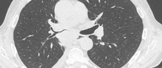
A computed tomography scan
of the abdominal area will help determine whether the blockage is complete or partial.
About our clinic Chistye Prudy metro station Medintercom page!
Diagnostics
Diagnosis of the disease begins first of all with an objective medical examination of the patient. Tympanitis is noted by percussion along with a dull sound; characteristic noises are heard on auscultation at the onset of the disease (with increased peristalsis) and with the progression of intestinal obstruction (when there is no peristalsis). By palpation, you can determine a distended intestinal loop; in the last stage of the disease, tension in the muscles of the anterior abdominal wall. Rectal and vaginal examination during examination provides information about the presence of a tumor formation blocking the intestinal lumen.
An objective examination only presupposes a diagnosis; it is finally confirmed by laboratory and instrumental examination methods:
- General blood test (an increase in the level of leukocytes is detected, indicating an inflammatory process).
- Blood chemistry.
- Plain X-ray of the abdominal cavity (a characteristic feature is the detection of Kloiber's cups) and X-ray with contrast to diagnose the level of the lesion.
- Colonoscopy (if indicated!) and/or irrigoscopy (x-ray of the colon) examines the large intestine, and sometimes allows one to identify the cause of intestinal obstruction.
- Ultrasound of the abdominal organs and kidneys is prescribed when the presence of tumors or inflammatory foci is suspected, however, visualization is often difficult due to severe pneumatization.
In difficult situations, laparoscopic surgery is performed, which makes it possible to clarify the diagnosis and eliminate the cause of the disease.
Intestinal obstruction is differentiated from acute appendicitis, perforated ulcer, renal colic and other acute diseases of the abdominal organs.
Internal rectal intussusception and rectocele
What is internal rectal intussusception and rectocele?
Internal rectal intussusception is a type of rectal prolapse (rectal prolapse). Rectal prolapse can be external, when all layers of the wall of the overlying sections of the rectum emerge from the anus, and internal. Internal intussusception of the rectum is the introduction of the overlying parts of the rectum or sigmoid colon into the lumen of the underlying parts of the rectum without going beyond the anus. In 15–30% of cases, internal invagination of the rectum is combined with rectocele - protrusion of the anterior wall of the rectum towards the vagina and prolapse of the posterior wall of the vagina.
What are the causes of internal rectal intussusception and rectocele?
Risk factors for internal rectal intussusception and rectocele are:
- Pregnancy and pathological course of childbirth (large fetus, perineal ruptures, prolonged standing of the fetal head in the pelvic cavity)
- Constipation
- Chronic lung diseases accompanied by cough (and, as a result, increased intra-abdominal pressure)
- Connective tissue dysplasia (congenital systemic “weakness” of connective tissue)
What is the mechanism of internal rectal intussusception and rectocele?
Internal intussusception of the rectum is based on the penetration of the overlying sections of the rectum or sigmoid colon into the lumen of the underlying sections of the rectum. Normally, all pelvic organs, including the rectum and vagina, are fixed by ligaments and fascia, which allows them to function adequately. When the uterosacral ligaments are weakened, stretched or ruptured (for example, after childbirth) and due to the weakness of the tendon center of the perineum, the stretching of the rectum and rectovaginal fascia, which is so necessary for the evacuation of the contents of the rectum, may be impaired. The walls of the rectum lose their fixation and begin to curl into the underlying sections, forming intussusception. If there is a defect in the rectovaginal fascia, this may be accompanied by the synchronous development of a rectocele.
What complaints do patients report?
Patients with internal rectal intussusception and rectocele complain of constipation. Internal intussusception of the rectum and rectocele, along with dyssynergia of the anal sphincter, are the causes of the so-called proctogenic constipation. Proctogenic constipation is characterized by a persistent urge to defecate and difficulty in emptying the rectum. Patients often complain of the need for prolonged straining, the sensation of a foreign body in the rectum and vagina, the feeling of an obstruction in the rectum, the feeling of incomplete emptying of the rectum, the need for manual assistance during defecation (pressure with the hand on the perineum or back). Some patients note multi-stage defecation and the need to change body position to have a bowel movement.
What examination is needed?
The diagnosis of internal intussusception of the rectum and rectocele can be suspected when questioning the patient’s complaints. The patient is examined on a gynecological chair. When examining the vagina in a speculum, a rectocele is defined as a protrusion of the posterior vaginal wall that increases with straining. During a digital examination of the anal canal and rectum, the doctor can assess the condition of the rectal mucosa, the tone of the anal sphincter, the condition of the pelvic floor muscles, and the presence of a defect in the rectovaginal fascia (cause of rectocele). In some cases, with a digital examination of the rectum, it is possible to determine the intussusception when the patient strains. Further, the examination is supplemented with instrumental research methods. Anoscopy allows you to visually assess the condition of the mucous membrane of the anal canal and distal rectum. Sigmoidoscopy allows you to visualize excessive folding of the rectal mucosa and identify internal intussusception of the rectum. Proctodefecography is the main method for diagnosing internal rectal intussusception. In addition, it allows you to determine pelvic floor prolapse and accompanying rectocele. To exclude concomitant pathology of the large intestine, all patients must undergo colonoscopy or irrigoscopy with double contrast. Functional methods of instrumental diagnostics are of great importance in the diagnosis of obstructive defecation syndrome (proctogenic constipation). Sphincterometry, anal profilometry and electromyography provide the most complete picture of the functional state of the internal and external anal sphincters and allow us to exclude anal dyssynergia as a cause of proctogenic constipation.
What treatment methods are there?
In all patients with internal intussusception of the rectum and the clinical picture of obstructive defecation syndrome (proctogenic constipation), treatment begins with conservative measures aimed at treating constipation and normalizing stool. Typically, recommendations for the treatment of constipation include drinking an adequate amount of fluid, plant foods containing large amounts of plant fiber, an adequate physical activity regimen, and the use of laxatives. If signs of anal sphincter dyssynergia are detected, it is possible to use biofeedback therapy (biofeedback method). The treatment method is based on providing the patient with visual information about the condition and functioning of the pelvic floor and anal sphincter muscles. This is realized by installing various sensors in the rectum, on the skin of the perineum and displaying data on the functioning of the pelvic floor muscles on the monitor screen located in front of the patient’s eyes. Thus, the patient herself can visually assess the effectiveness of relaxation or contraction of certain muscle groups when simulating the act of defecation. The biofeedback therapy method is effective in 70% of patients; long-term treatment effects are noted by about 50% of patients.
When is surgery necessary?
Surgical treatment is indicated for patients in whom conservative treatment methods have been ineffective.
What surgical treatment methods exist?
Depending on the access, all surgical interventions for internal intussusception of the rectum and rectocele are divided into abdominal (performed from the abdominal cavity) and perineal (performed from the perineum, vagina and rectum). The choice of surgery depends on the severity of rectal intussusception, the presence or absence of rectocele, the patient's age, and the preference and experience of the surgeon.
What methods of abdominal surgery exist?
Suture rectopexy (fixation of the rectum) according to Kümmel-Zerenin involves fixing the rectum with several sutures to the sacrum. Despite the low frequency of relapses, this technique can provoke constipation or intensify existing disorders of the evacuation function of the rectum in 50% of operated patients. There are also so-called anterior loop and posterior loop methods of rectopexy (fixation of the rectum) with synthetic mesh prostheses to the sacrum. The main disadvantage of these methods is the fairly frequent occurrence of disorders of the motor-evacuation function of the colon. When internal invagination of the rectum is combined with rectocele, it is possible to perform sacrovaginopexy (sacrocolpopexy) - fixation of the anterior wall of the rectum and posterior walls of the vagina to the promontory of the sacrum with a synthetic prosthesis located in the rectovaginal and vesicovaginal space. Despite the low relapse rate, the rate of postoperative complications of this technique is 20–25%. In case of dolichosigma (elongation of the sigmoid colon), internal intussusception of the rectum caused by sigmocele and slow transit of intestinal contents through the left parts of the large intestine, it is advisable to combine rectopexy (fixation of the rectum) with resection of the left parts of the large intestine. All of the above operations can be performed either with an open surgical approach or laparoscopically.
What methods of perineal surgical interventions exist?
In order to correct rectocele (prolapse of the posterior vaginal wall) and internal intussusception of the rectum, the installation of a posterior intravaginal sling is used - a synthetic mesh prosthesis of a ribbon shape, the central part of which is fixed to the cervix, the vaginal vault (if the uterus has been removed) and the intestinal-vaginal fascia, and the ends are passed through the walls of the pelvis in the area of the sacrospinous ligaments. Thus, the prosthesis fixes the uterus, the posterior wall of the vagina and the rectum to the walls of the pelvis, restoring the natural tension of the patient’s own fascial-ligamentous structures of the pelvis. Also, with internal invagination of the rectum, it is possible to perform transanal proctoplasty using the Longo method - circular muco-submucosal resection of the rectum using a special circular stapler. The effectiveness of the Longo operation for internal intussusception of the rectum lies in the removal of excess mobile mucous membrane of the rectum, which forms the intussusception. Operations performed through the perineal approach are less traumatic and easier to tolerate for patients.
Our doctors
Gordeev Sergey Alexandrovich
Surgeon, Candidate of Medical Sciences, doctor of the highest category
42 years of experience
Make an appointment
Lutsevich Oleg Emmanuilovich
Chief Surgeon of CELT, Honored Doctor of the Russian Federation, Chief Specialist of the Moscow Department of Health in Endosurgery and Endoscopy, Corresponding Member of the Russian Academy of Sciences, Head of the Department of Faculty Surgery No. 1 of the State Budgetary Educational Institution BPO MGMSU, Doctor of Medical Sciences, Doctor of the Highest Category, Professor
43 years of experience
Make an appointment
Prokhorov Yuri Anatolievich
Surgeon, head of the surgical service of CELT, candidate of medical sciences, doctor of the highest category
33 years of experience
Make an appointment
Treatment tactics
At the first symptoms of the disease, the patient must be immediately hospitalized in a hospital. Before being examined by a surgeon, it is prohibited to take painkillers or any other medications, or to cleanse the stomach and intestines, so as not to blur the clinical picture and cause harm.
If intestinal obstruction is diagnosed in its initial stage, conservative treatment methods can be used:
- Emptying the stomach and intestines from food and feces using probing and siphon enema.
- Taking antispasmodics in the presence of spasms or drugs that stimulate peristalsis in case of intestinal paresis.
- Intravenous drip infusions of saline solutions to achieve water-electrolyte balance.
- Colonoscopy for diagnostic and therapeutic purposes.
If a conservative approach to treatment does not eliminate the disease, it is necessary to think about mechanical obstruction or the development of complications and resort to surgical intervention.
The following surgical treatment options are used:
- unraveling intestinal loops;
- cutting adhesions;
- hernia repair in the presence of a hernia;
- excision of a necrotic area of the intestine;
- the application of a colostomy to remove feces outside (in case of malignant neoplasms and the impossibility of anastomosis).
After the operation, drug correction is carried out, which, as a rule, includes antibiotic therapy, detoxification measures, restoration of peristalsis, etc. The main goal of treatment is to save the patient’s life and prevent possible complications that may develop if the disease is not diagnosed and treated in a timely manner. The most life-threatening consequences are pain shock, sepsis and peritonitis.
Treatment
Intussusception in children and adults is eliminated surgically.
When diagnosed with intussusception, treatment can occur in several ways:
- laparotomy (for this type of operation the abdominal wall is cut);
- disenvagination (manually straightening the invaginated parts of the intestine);
- removal of a section of intestine if necrosis is present;
- laparoscopy (includes endoscopic examination and surgical intervention, performed on the organs of the abdominal region and pelvis. The operation is performed using video equipment and surgical instruments. Today, operations of this kind are more in demand, since they are less traumatic, and after them there are not many scars and huge scars);
- treatment of pathologies due to which intussusception occurred - tumors, polyps, fecal stones.
Complications
Complications that may occur due to volvulus:
- fecal obstruction in the intestines;
- poisoning of the body due to the absorption of toxins into the blood associated with stagnation of intestinal contents;
- inflammation of the abdominal cavity;
- intestinal rupture;
- reappearance of intussusception symptoms.
After surgery and treatment of volvulus, relapse may occur. If this happens, you have to remove the part of the intestine that is susceptible to intussusception.
Preventive actions
The effectiveness of treatment depends on the etiology of the disease and the qualifications of the surgeon, because accurate diagnosis and timely and correct treatment measures minimize the likelihood of complications. It is also necessary to take into account the age of the patient: older patients have an increased risk of complications.
Prevention is aimed at treating diseases that cause intestinal obstruction:
- control of helminthiasis;
- timely diagnosis and removal of tumors;
- prevention of adhesions;
- preventing traumatic injuries;
- healthy eating;
- the right way of life.
The multidisciplinary clinic CELT has a powerful diagnostic and treatment base and experienced specialists of the highest level.
How to treat rectal prolapse in adults
To eliminate rectal prolapse, conservative and surgical treatment is performed. Patients are recommended to follow a diet and perform a set of exercises to strengthen the muscles of the pelvic floor, anal sphincter and perineum in order to normalize stool. To prevent the disease from progressing, the patient completely eliminates physical activity.
DRUG THERAPY
In the first stages of the disease, the use of conservative treatment is recommended, when rectal retraction occurs independently. The goals of therapy are to reduce unpleasant symptoms; eliminate constipation and diarrhea; restore the tone of the anal sphincter and rectum.
The list of medications is small. In most cases, medications are prescribed that can regulate bowel movements. We are talking about laxative suppositories or oral medications (tablets, powders for preparing drinks. If the patient experiences severe pain, it is necessary to start taking painkillers.
SURGICAL INTERVENTION
Surgical treatment is used in stages 3 and 4 of rectal prolapse, as well as in cases where conservative therapy is ineffective. In medicine, it is customary to talk about several methods that secure the rectum in a physiologically correct position. All methods are differentiated into several groups, the difference of which is associated with the principle of influence on organs.
Let's consider the main methods of surgical treatment of rectal prolapse, represented by methods of narrowing the anus; operations of rectopexy or attachment of the distal rectum to fixed parts of the pelvis; colopexy - transperitoneal fixation of the distal sigmoid colon to fixed structures of the pelvis or abdominal wall; operations aimed at strengthening the pelvic floor and perineum; partial or complete resection of prolapsed intestine.
COMPLICATIONS
The most common form of complication of rectal prolapse is strangulation of the prolapsed section of the rectum. If treatment is not started in a timely manner, there is a risk of developing necrosis of the strangulated tissues.
Our services
The administration of CELT JSC regularly updates the price list posted on the clinic’s website. However, in order to avoid possible misunderstandings, we ask you to clarify the cost of services by phone: +7
| Service name | Price in rubles |
| Appointment with a surgical doctor (primary, for complex programs) | 3 000 |
| X-ray of the abdominal cavity (survey) | 2 400 |
| Diagnostic laparoscopy | 40 000 — 60 000 |
All services
Make an appointment through the application or by calling +7 +7 We work every day:
- Monday—Friday: 8.00—20.00
- Saturday: 8.00–18.00
- Sunday is a day off
The nearest metro and MCC stations to the clinic:
- Highway of Enthusiasts or Perovo
- Partisan
- Enthusiast Highway
Driving directions