Glaucomatous optic neuropathy (GON) is usually considered from the perspective of mechanical, vascular and metabolic theories, including numerous risk factors that increase the likelihood of progression of glaucomatous lesions. Determining the priority of one factor or another is always controversial. Only one thing is obvious: in the event that an individual level of intraocular pressure (IOP) is reached and at the same time progression of GON is noted, it is necessary to identify other, most likely influencing factors.
Ophthalmologists are well aware that even when stable IOP compensation is achieved through medication or surgery, every 5th patient continues to experience a decline in visual function [1–3]. And risk factors such as arterial hypotension, low perfusion pressure, vasospastic syndrome, diabetes mellitus, and myopia play a significant role in this [4–9]. All this makes us take seriously the need for neuroprotective therapy in order to stabilize the glaucomatous process and create conditions for preserving visual functions. We consider the need for stable normalization of ophthalmotonus to be an important and undoubted condition for carrying out this therapy.
Meanwhile, the difficulties that ophthalmologists face when deciding on neuroprotective therapy are also well known [10]. Firstly, it is necessary to act on structures that are affected but still retain their morphology and partly their function. Targeted delivery is very important, as is its timeliness. But, as practice shows, taking advantage of the “therapeutic window” is a big problem. Secondly, drugs borrowed from neurological practice and used in ophthalmology often have a large number of undesirable effects. And taking into account the elderly age of patients with glaucoma, who are usually burdened with concomitant pathology, this takes on special significance. Thirdly, glutamate NMDA receptors are widely represented in the central nervous system and ill-considered use of appropriate drugs increases the likelihood of suppression of the physiological functions of the brain.
Considering the multifactorial nature of the progression of GON, ophthalmologists usually recommend complex therapy, prescribing drugs of various pharmacological groups [10-16]. This is often done when they plan to study the effectiveness and safety of one of the drugs included in complex therapy.
Despite the existing difficulties in assessing the effectiveness of neuroprotective therapy due to the lack of absolutely reliable criteria for a number of structural and functional indicators, such an assessment is still possible.
The purpose of this work is to evaluate the therapeutic effectiveness of gliatilin in the complex treatment of progressive GON. The study was retrospective in nature.
Cholinomimetics[edit | edit code]
Acetylcholine (ACh) has no therapeutic use because it is rapidly degraded by acetylcholinesterase (AChE). However, its action can be imitated by other substances - direct cholinomimetics or indirectly by indirect cholinomimetics.
Direct cholinomimetics[edit | edit code]
The choline ester carbachol excites muscarinic receptors (a direct cholinomimetic), however, it is not hydrolyzed by cholinesterase. Carbachol is used topically in eye drops (for glaucoma) or systemically (for intestinal and bladder atony).
The alkaloids pilocarpine (from Pilokarpus jaborandus) and arecoline (from the areca nut Areca catechu) are quaternary amines with central cholinomimetic effects. The central effect is manifested in a slight stimulating effect. It is likely that the population of South Africa, where areca nut chewing is widespread, uses this cholinomimetic effect. Only pilocarpine is used therapeutically in the form of eye drops for glaucoma.
Indirect cholinomimetics[edit | edit code]
Selective inhibition of the enzyme acetylcholinesterase leads to an increase in the concentration of acetylcholine in the synaptic cleft and to a prolongation of its action. Substances that inhibit AChE are called indirect cholinomimetics. Blockade of the enzyme occurs in all cholinergic synapses where ACh functions. Inhibiting substances are carbamic acid esters (carbamates, such as physostigmine, neostigmine) or phosphoric acid esters (organic phosphates, such as paraoxon E600, a derivative of parathion E605).
Representatives of both groups interact with AChE in the same way as ACh and can be inhibitory substrates for AChE. The ester-enzyme complex is further hydrolyzed. The rate of acetylcholine hydrolysis is determined by deacetylation of the enzyme, which occurs quickly (in milliseconds), and therefore high enzyme activity is maintained. The hydrolysis reaction of carbamate occurs within a few hours. The enzyme is inhibited. Dephosphorylation of the enzyme is generally impossible, so it is inhibited irreversibly.
Application
. The quaternary carbamate neostigmine is used as an indirect cholinomimetic for postoperative atony of the bladder or intestines. The drug is also used for myasthenia gravis, for insufficiency of ACh at the end plate of the motor neuron, as well as for stopping the action of non-depolarizing muscle relaxants (cessation of curarization before recovery from anesthesia). Pyridostigmine has similar indications for use. The tertiary carbamate physostigmine, which crosses the BBB, is used as an antidote for poisoning with atropine-like compounds, since it can bind to AChE in the central nervous system. Neostigmine is used topically for glaucoma. Carbamates and organic phosphates are used as insecticides. They are very toxic to humans, but decompose faster than DDT.
In the early stages of Alzheimer's disease, some patients may benefit from improved cognition and slowed decline by taking acetylcholinesterase inhibitors such as rivastigmine, donepecil and galantamine, which should be given in slowly increasing doses. Peripheral side effects (inhibition of acetylcholine destruction) limit therapy. Donepicil and galantamine are not esters of carbamic acid and have a different mechanism of inhibitory action. It is possible that galantamine enhances the effect of acetylcholine on nicotinic receptors.
Material and methods
240 patients were randomly selected, 120 people in each group. Patients in both groups were comparable in age, concomitant somatic pathology, and severity of the glaucomatous process; their average age was 71.3 ± 1.6 years. In all patients, the therapists diagnosed systemic atherosclerosis with predominant damage to cerebral vessels and cerebrovascular insufficiency.
As can be seen from table. 1, in both groups the majority were patients with advanced stage glaucoma: 70.0 and 76.6%, respectively. In group 2, patients in whom normalization of IOP was achieved surgically predominated: 83.3% versus 53.3% in group 1. Therefore, in the latter there were more patients who required local antihypertensive therapy to maintain ophthalmotonus within the target pressure. However, despite the stable level of IOP in the range of 15-17 mm Hg, in all cases negative dynamics of visual functions were noted, which served as the basis for a course of stabilizing therapy.
Table 1. General characteristics of patients
The course of neuroprotective therapy included drugs of various pharmacological groups acting on different pathogenetic links. Each time, the general somatic condition of the patient and possible contraindications for taking a particular drug were taken into account. All patients received Mexidol 100 mg intramuscularly once a day for 14 days, and patients in group 1 were also given gliatilin 1000 mg/4 ml intravenously in 12-15 injections, then continued the course of taking this drug orally 1 capsule 2 times a day for 4 months.
Mexidol
has antihypoxic, membrane-protective, nootropic effects. The drug increases resistance to conditions such as hypoxia, ischemia, cerebrovascular accident, improves metabolism and blood supply to the brain, microcirculation and rheological properties of blood, and reduces platelet aggregation. The mechanism of pharmacological action is associated with antioxidant and membrane protective properties, inhibition of lipid peroxidation processes. There is positive experience of its use in ophthalmic practice [16].
Gliatilin
- a nootropic drug, a centrally acting cholinomimetic with a predominant effect on the central nervous system. The release of choline from the active substance occurs in the brain; choline is involved in the biosynthesis of acetylcholine (one of the main mediators of nervous excitation). Alphoscerate is biotransformed to glycerophosphate, which is a precursor to phospholipids.
Acetylcholine improves the transmission of nerve impulses, and glycerophosphate is involved in the synthesis of phosphatidylcholine (membrane phospholipid), resulting in improved membrane elasticity and receptor function. Gliatilin increases cerebral blood flow, enhances metabolic processes and activates the structures of the reticular formation of the brain, restores consciousness in case of traumatic brain injury. It has a preventive and corrective effect on the factors of involutional psychoorganic syndrome, such as changes in the phospholipid composition of neuronal membranes and a decrease in cholinergic activity. Thus, pharmacodynamic studies have shown that gliatilin acts on synaptic, including cholinergic, transmission of nerve impulses (neurotransmission), plasticity of the neuronal membrane, and receptor function.
All patients underwent routine ophthalmological examinations.
The state of the visual fields was assessed in several ways. Static perimetry was performed on a Humphrey Visual Field Analyzer II (HFA II) 750i (Germany). Depending on the initial visual acuity and the degree of visual impairment, a screening or threshold research program was used. When assessing the central visual field (CVF), all patients underwent correction of near visual acuity. Screening was carried out according to the FF-120 Screening program using a three-zone strategy. The threshold program for studying the visual field included the use of the Central 30−2 tests when studying the CPZ (within 30° from the point of fixation of the gaze) and Peripheral 60−2 when assessing the peripheral visual field - PPV (from 30° to 60°). At the same time, we analyzed the threshold foveal photosensitivity, the sum of decibels of threshold values in each quadrant over the entire field of view, and indicators of mean deviation (MD) and pattern standard deviation (PSD), calculated automatically by the device taking into account its own database.
The criteria for assessing the effectiveness of neuroprotective therapy are not sufficiently informative, and from the point of view of practical ophthalmology, the most accessible study of visual functions is perimetry.
All studies were carried out before the start of treatment, after 3 and 6 months.
Read also[edit | edit code]
- Anatomy and physiology of the nervous system
- Parasympathetic nervous system
- Sympathetic nervous system
- Synaptic transmission
- Acetylcholine
- Cholinergic receptors and synapses
- Anticholinergics
- Nicotine
- M-cholinergic receptor stimulants
- M-cholinergic receptor blockers
- Acetylcholinesterase inhibitors (AChE)
- Poisoning with acetylcholinesterase blockers
- Nerve transmission at neuromuscular synapses and autonomic ganglia N-cholinergic receptors
- Muscle relaxants
- Agents acting on the autonomic ganglia
- Ganglion stimulators
- Ganglioblockers
Results and discussion
In the first two weeks, during which patients received the drugs in a hospital setting, no adverse events were reported in any case.
The IOP level (Table 2) was normalized throughout the observation period and was at a level not exceeding 15 mm Hg.
Table 2. Dynamics of ophthalmotonus indicators (mm Hg) Note. Here and in the table. 3: p>0.05 compared to the original data.
This level of IOP corresponded to the target pressure recommended by the European Glaucoma Society, taking into account the stage of the glaucomatous process.
In cases where doubts arose about the actual target IOP, flowmetry was performed, which made it possible to clarify the indicators of the individual level of ophthalmotonus.
One of the criteria for assessing functional capabilities is central visual acuity (Table 3). Neuroprotective therapy, as a rule, does not affect this indicator, however, mentioning it is important as indirect evidence of the dynamics of the process.
Table 3. Dynamics of visual acuity indicators
In our case, central visual acuity remained stable. Some improvement in vision was noted in some patients in both groups.
One of the objective criteria for assessing the effectiveness of neuroprotective therapy, to a certain extent, can be considered visual field testing. We considered the indicators taken into account when assessing changes in visual functions as CPZ, foveal and total photosensitivity, PPV, MD and PSD indicators. The data obtained before and after the course of stabilizing therapy are presented in table. 4.
Table 4. Dynamics of static perimetry indicators by group
The study was conducted before the start of the course of drug therapy and 3.6 months after it. As can be seen from table. 4, all average indicators tended to improve, especially for the central and peripheral visual fields. Moreover, in the group of patients receiving gliatilin, this trend was more significant. This is all the more important because the baseline data of both groups were comparable.
We are inclined to believe that the more pronounced therapeutic effect in patients of group 1 is due to the action of gliatilin. This assumption is confirmed by observation data of a large number of patients periodically receiving such therapy at the institute.
Drug interactions of antiglaucoma drugs against the background of common chronic diseases
Glaucoma is a chronic disease that requires long-term conservative treatment using eye drops to reduce elevated intraocular pressure (IOP). The goal of such drug therapy should be to achieve the required hypotensive effect with the least amount of drugs. The expected result from prescribing the drug should be significant, and any possible side effects should be minimal. In Russia, there are over 1 million patients with glaucoma [1]. A significant number of patients with this diagnosis are elderly people suffering from systemic chronic diseases (cardiovascular, endocrine, gastroenterological, etc.), also requiring long-term treatment. In this regard, knowledge about the interactions of drugs used to treat glaucoma and other diseases is relevant in order to eliminate or at least reduce their side effects, as well as to monitor their effectiveness. The effect of eye drops is not limited to the eye. It would be wrong to think that eye drops are safer than medications taken orally. There are reports that the systemic effect of using eye drops may exceed the effect of taking the same drug orally, probably due to direct absorption into the bloodstream through the mucous membrane of the conjunctival sac and nose. Thus, the drug bypasses passage through the liver and enters directly into the systemic circulation, which provides an increase in the effect of a specific absorbed dose of the drug [2, 3]. The main groups of drugs currently used to treat glaucoma are beta-blockers (BABs), carbonic anhydrase inhibitors (CAIs), M-cholinomimetics, alpha-adrenergic agonists, and prostaglandin analogues (PA). All these drugs inevitably cause side effects, both local and general. Often, patients with glaucoma receive the same group of drugs both locally (in the form of eye drops) and orally (in the form of tablets or injections), which leads to increased side effects. In addition, interactions between different groups of drugs can lead to the leveling of the therapeutic effect of their use. Interaction of beta-blockers for the treatment of glaucoma with other drugs
BBs (timolol, betaxalol, levobunolol, carteolol, proxodolol, etc.) are the most frequently prescribed group of drugs for the treatment of patients with glaucoma. They allow you to reduce the secretion of intraocular fluid (IOH) and reduce IOP by 20–25%. However, they have quite pronounced general side effects with less pronounced local ones (keratopathy, dry eye syndrome, allergic blepharoconjunctivitis). The pharmacodynamic effects of beta-adrenergic blockade are diverse, since cells containing beta-adrenergic receptors are widely represented throughout the body and are part of the heart, lungs, kidneys, blood vessels, endocrine glands, nervous system, and blood cells [4]. Numerous systemic side effects of beta blockers in the form of eye drops are associated with the fact that they are actively absorbed into the blood, to a greater extent than their oral analogues, since the latter are metabolized in the liver. From the cardiovascular system, these are bradycardia, cardiac conduction disturbances, heart failure; respiratory system – dyspnea, bronchospasm, bronchial asthma, respiratory failure; central nervous system - dizziness, nausea, drowsiness, headache, insomnia, depression, increased symptoms of myasthenia gravis. Prescribing beta blockers to patients receiving systemic therapy with calcium channel blockers (CCBs) (nifedipine, amlodipine, verapamil, diltiazem, etc.) increases the risk of decreased blood pressure (BP), bradycardia, atrioventricular block, even asystole. Such interactions are especially pronounced when combining the use of instilled forms of beta blockers and verapamil. Therefore, when prescribing a combination of these drugs to a patient, it is necessary to choose the one that has less pronounced effects on heart rhythm and conduction [4]. When applied topically, beta blockers can lead to a deterioration in the blood lipid profile of patients, while the level of high-density lipoproteins decreases and the level of triglycerides increases. When combining beta blockers and diuretics, hypolipoproteinemia may increase, as well as an increase in the risk of developing systemic hypotension. In addition, thiazide diuretics (chlorthalidone, indapamide, metolazone) in combination with beta blockers can provoke impaired glucose tolerance in patients with diabetes mellitus (DM). The hyperglycemic effect of thiazides develops against the background of hypokalemia they cause. BBs often increase bradycardia and atrioventricular block in patients receiving cardiac glycosides. Xanthopsia (visual impairment in which everything appears yellow), usually resulting from the toxic effects of cardiac glycosides, may be exacerbated by the use of beta blockers and lead to decreased visual acuity. The combination of beta blockers with angiotensin-converting enzyme inhibitors (ACE inhibitors) (captopril, lisinopril, enalapril, etc.) is quite safe in the absence of systemic hypotension. Administration of beta blockers to patients with diabetes may enhance the hypoglycemic effect of insulin, which is associated with their ability to suppress gluconeogenesis and glycogenolysis. In addition, beta blockers may mask symptoms of acute hypoglycemia, such as agitation and palpitations. The use of beta blockers in women receiving hormone replacement therapy (HRT) may provoke the development of headaches. Against the background of the use of thyroid-stimulating hormones, acetylsalicylic acid (ASA) drugs and non-steroidal anti-inflammatory drugs (NSAIDs), the effectiveness of local use of beta blockers in patients with glaucoma is reduced. When prescribing beta blocker drops to patients with glaucoma, it is necessary to remember that with the simultaneous use of oral drugs of this group, their side effects increase. Pressing the lacrimal canaliculus with a finger and closing the eyelids after instillation will reduce the adverse systemic effects from the use of instilled forms of beta blockers [5].
Interaction of carbonic anhydrase inhibitors for the treatment of glaucoma with other drugs
The most effective ICAs for use in patients with glaucoma are some sulfonamides (dorzolamide, brinzolamide). The mechanism of action of this group of drugs is associated with a decrease in the secretion of intraocular fluid, while the level of intraocular pressure decreases by 15–20%. In the treatment of glaucoma, both local and general use of these drugs is possible. The main side effects of local ICA are blurred vision, blepharitis, dry eye, allergic blepharoconjunctivitis, keratitis, asthenopia, diplopia. Common side effects include an unusual taste in the mouth, nausea, headache, dizziness, paresthesia, arterial hypertension, pharyngitis, alopecia, nephrourolithiasis, allergic reactions. Since ICAs and their metabolites are excreted in the urine, the drugs are not recommended for use in patients with severe renal impairment (creatinine clearance <30 ml/min) [6]. When prescribing ICA eye drops to patients with glaucoma who receive oral CCBs, side effects such as nausea/vomiting, paresthesia, and general weakness may increase. The combination of ICAs with diuretics can increase hypokalemia, the development of acidosis, and increases the likelihood of developing agranulocytosis. As a result of the interaction of ICA with ACE inhibitors, it is possible to increase the suppression of the hematopoietic function of the bone marrow with the development of agranulocytosis and aplastic anemia. The combination of ICA drops with oral beta-blockers can cause unpleasant side effects such as insomnia, dizziness, nausea, vomiting, and depression. The combined use of ICA in the form of drops and ASA tablets can increase metabolic acidosis, hypokalemia, and depression. As a result of interaction with NSAIDs, the effectiveness of ICA is reduced, and suppression of the hematopoietic function of the bone marrow may also be observed. If a patient has diabetes, ICA drugs can increase blood glucose levels. When combining ICA and HRT with estrogens, women may develop headaches. The effect of thyroid replacement therapy may be reduced as a result of interaction with ICA. Prescribing ICA drops to patients with glaucoma simultaneously with oral acetazolamide increases the risk of developing systemic side effects from their combined use [5].
Interaction of M-cholinomimetics for the treatment of glaucoma with other drugs
Drugs in this group reproduce the effects of the parasympathetic nervous system mediator, acetylcholine, due to its interaction with M-cholinergic receptors. M-cholinergic receptors are localized in all organs that receive parasympathetic innervation, at the end of postganglionic parasympathetic fibers. M-cholinomimetics (pilocarpine hydrochloride, carbachol, etc.) for glaucoma improve the outflow of intraocular fluid through the trabecular apparatus, thereby reducing IOP by 20–25% of the initial value. Drugs in this group have quite pronounced side effects, both general and local. Constriction of the pupil often causes a decrease in visual acuity, especially if the patient has cataracts; Accommodative myopia often occurs in young patients. In addition, side effects such as narrowing of the anterior chamber angle, retinal detachment, development of iris cysts, cataracts, etc. are possible. Common side effects include headache, bronchospasm, slow heart rate, increased salivation, and allergic reactions [7]. When cholinomimetics are used together with CCBs and ACEIs, increased headache, vasodilation, and arterial hypotension may occur. As a result of the interaction of M-cholinomimetics and beta blockers, bronchospasm may increase in patients with bronchial asthma, and the likelihood of nausea/vomiting and arterial hypotension may also increase. In patients with thyroid dysfunction, the administration of M-cholinomimetics leads to increased tremor, muscle weakness, and the development of diarrhea. When combined with HRT estrogens, women may develop headaches. As a result of the interaction of M-cholinomimetics and ASA or NSAIDs, headaches may worsen and nausea/vomiting is possible [5].
Interaction of alpha-agonists for the treatment of glaucoma with other drugs
Selective alpha-2 adrenergic agonists (brimonidine, apraclonidine) have a dual mechanism of action on IOP in patients with glaucoma: reducing secretion and improving uveoscleral outflow. At the same time, the IOP level decreases by 20–25%. The main local side effects when using brimonidine are associated with the development of allergic blepharoconjunctivitis, keratitis and dermatitis, and possible pupil constriction. Systemic side effects often include dry mouth and nose, fatigue, headache, dizziness, drowsiness, often hypertension, less often hypotension, bradycardia/tachycardia, bronchitis, dyspepsia, nausea, hypercholesterolemia, allergic reactions [8]. When NSAIDs and ASA are combined with alpha-agonists, the effect of the latter is reduced, and blood pressure may also increase. Alpha adrenergic agonists can reduce the effectiveness of ACE inhibitors when used together. As a result of the interaction of alpha-agonists with CCBs and diuretics, increased nausea/vomiting, diarrhea, and abdominal pain may occur. When used with diuretics, palpitations and headache may occur. In patients with diabetes, alpha-agonists can stimulate hyperglycemia and reduce the effect of insulin. Thyroxine may enhance the effect of alpha-agonists with the appearance of undesirable side effects [9].
Interaction of prostaglandin analogues for the treatment of glaucoma with other drugs
When using APs (latanoprost, travoprost, etc.), a decrease in IOP is associated with an improvement in the uveoscleral outflow pathway of the IOP. At the same time, the IOP level decreases by 25–33%. Among the local side effects when using this group of drugs, changes in the color of the iris, conjunctival hyperemia, cystic edema of the macula, hyperpigmentation and lengthening of eyelashes, etc. predominate. Systemic side effects are less pronounced, because both the dosage of the active substance and, accordingly, its concentration in the blood is low. Influenza-like syndrome, increased respiratory symptoms, chronic inflammatory diseases, herpes infection, headache, arrhythmia, arterial hypertension/hypotension, and allergic reactions are rare [10]. The interaction of drugs for systemic use with AP in the form of eye drops has not been described in the literature, which may be due to the rather low concentration of the main drug substance in them. Conservative treatment of glaucoma involves prescribing instilled forms of antihypertensive drugs for a long time to maintain normal IOP levels. The effectiveness and safety of the drugs used are the most important aspects, since glaucoma is a chronic disease that requires constant treatment [11]. A significant number of common complications when using eye drops to lower IOP in the treatment of glaucoma is due not only to their widespread use, but also to an overdose of drugs, since most patients with glaucoma are elderly people with reduced visual acuity, for whom it is difficult to instill only one drop of medication. Thus, correct dosing of medications using long-acting forms of medications and tips on the safe use of eye drops will help solve many problems associated with side effects of drugs. Closing the eyes for 3 minutes after instilling eye drops can reduce systemic absorption of the drug by 50% [12, 13].
How do drugs work?
The main principle of action of antiglaucomatous drugs is to reduce IOP. Pharmaceutical concerns produce different groups of these drugs, but they are all based on eliminating hypertension, only with the help of different substances.
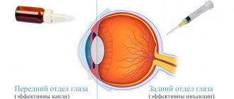
Drops for the treatment of glaucoma mainly have a local effect. They almost completely enter the choroidal vessels. But sometimes a small amount of them ends up in the blood. There, their active substance can affect the cardiovascular and respiratory systems. Before prescribing such drugs, the doctor must conduct an examination to find out whether the patient has chronic diseases of these organs. If you are intolerant to the active ingredients contained in the drops, a different treatment regimen will be prescribed.


