Lymph nodes are an organ that performs a drainage function and are a peripheral organ of the lymphatic system. She fights infections and other pathologies. The human body has about 600 lymph nodes located throughout the body. Most of them are not palpable, painless and not fused. Lymph nodes accumulate lymph cells and lymph to help overcome disease. Thus, this leads to their increase and the development of a process such as lymphadenopathy.
https://www.aafp.org/afp/1998/1015/p1313.html
Lymphadenopathy is an increase in the size of the lymph nodes, which is palpable.
(> 1 cm).
Depending on the pathology, one or more nodes may enlarge.
This condition can occur as an independent disease or as a separate symptom. In any case, it is necessary to consult a doctor and search for the reason that caused the increase. (https://cutt.ly/DbM40so)
Classification:
For localization:
- cervical lymphadenopathy;
- axillary (axillary);
- chest;
- inguinal;
- abdominal lymphadenopathy;
- pelvic lymphadenopathy
For prevalence:
- localized form;
- generalized form;
Behind the etiology
- infectious;
- non-infectious;
Features of the pathology
Considering that there are more than 600 lymph nodes in the human body, we can assume the development of pathology in any organ and system of the body, both in an adult and a child. Most often, in the practice of a hematologist at a medical center, it is necessary to diagnose lymphatic lesions in places such as:
- abdomen;
- mediastinum;
- groin;
- submandibular zone;
- neck;
- armpits.
The presence of pathology of the lymph nodes in any of these areas of the body indicates an existing underlying disease. And, unfortunately, often it can be oncology. According to statistics, in 1% of cases in patients diagnosed with lymphadenopathy, additional examination reveals malignant formations.
Classification
When determining the forms of lymphadenopathy, the location of enlarged lymph nodes is primarily taken into account. Lymphoid tissue is the main protective barrier against the spread of infectious pathogens and tumor cells. Therefore, the location of altered lymphatic formations facilitates the diagnosis of the disease that caused the lymphadenopathic reaction. Depending on the localization of the process, the following are distinguished:
- Enlargement of the submandibular lymph nodes
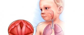
. Characteristic of pathological processes in the head and neck area - diseases of the eyes, ENT organs and paranasal sinuses, skin damage. Submandibular lymphadenopathy often signals dental problems and chronic tonsillitis. - Enlarged cervical lymph nodes
. Usually observed with respiratory infections, oral pathology, infectious mononucleosis, late stages of tuberculosis. Cervical nodes can be affected by lymphomas, lymphogranulomatosis, metastasis of thyroid cancer, lung cancer. - Enlarged supraclavicular lymph nodes
. Most often due to tumor causes. Detection of an enlarged node on the right is pathognomonic for cancer of the esophagus and lungs. The left lymph node is affected by malignant processes in the abdominal cavity, pelvis, and retroperitoneal space. - Enlarged axillary lymph nodes
. Inflammatory lesions are possible in the presence of wound infections, cat scratch disease, and brucellosis. Damage to the nodes of the axillary group is typical for breast cancer, melanoma of the upper extremities, and the installation of silicone breast implants. - Enlarged inguinal lymph nodes
. As a rule, nodes in the groin react to the development of syphilis, gonorrhea, chancroid, and other genital infections. Inguinal lymphadenopathy is also a sign of malignant lesions of the pelvic organs, lymphoma, and bubonic plague.
Somewhat less frequently, lymph nodes of other groups are involved in the process - submental, cubital (in the area of the elbow), parotid, occipital, jugular. During a routine instrumental examination, an increase in internal lymph nodes can be determined - intrathoracic (mediastinal), bronchopulmonary, para-aortic, splenic, mesenteric, retroperitoneal.
In the diagnostic plan, it is important to take into account other criteria for the classification of lymphadenopathy - the characteristics of altered lymphoid formations, the extent of the lesion. This approach allows us to assume the type of pathological process occurring in the involved nodes and the body as a whole. Important criteria for the classification of enlarged lymph nodes are:
- Dimensions
. With I degree lymphadenopathy, the diameter of the affected formations is 0.5-1.4 cm, with II degree - 1.5-2.4 cm, and with III degree - 2.5 cm or more. A significant and prolonged increase in the size of lymph nodes is more typical for malignant processes. - Soreness
. Intense pain is often caused by inflammation of the lymph nodes, especially acute purulent lymphadenitis. Formations that have undergone malignant degeneration are often painless, except in cases of hemorrhage into the necrotic center. - Density
_ Enlarged, inflamed lymph nodes are usually soft; when they suppurate, fluctuation (fluctuation of fluid) is felt during palpation. The stony consistency of the formations is typical for the metastatic process, and tight elasticity is typical for lymphomas. - Communication with each other
. A pathological formation of lymph nodes that can be felt as a single unit and move together is called a conglomerate. Lymph nodes fused together are detected in tuberculosis, sarcoidosis, lymphogranuloma venereum, lymphomas and metastasis. - Quantity
. It can affect either one or two or several nodes in one zone. In the first case, they talk about single enlarged nodes, in the second - about local lymphadenopathy. The more active the process, the more formations are affected, however, with metastasis, one large node is often detected. - Prevalence
. With local lymphadenopathy, single nodes are identified in one area, with regional lymphadenopathy - several formations in 1-2 adjacent zones. A generalized (widespread) process is characterized by damage to lymphatic structures in three or more areas.
Taking into account the pathogenesis, enlargement of lymph nodes can be primary (systemic), secondary (reactive) and inflammatory. Primary polyadenopathies develop with systemic malignancy of lymphoid tissue (leukemia, lymphogranulomatosis, non-Hodgkin's lymphoma) and benign processes (sinus histiocytosis). Reactive lesions are a response to another pathology (infection, immune disease, proliferation of tumor cells, metabolic disorders). Inflammation (lymphadenitis) occurs when infectious agents multiply in the tissue of the node.
Causes
Lymphadenopathy in children occurs as a consequence of various infectious diseases. If we distribute them into groups, then these are:
- bacterial;
- fungal;
- mycobacterial;
- chlamydial;
- viral;
- parasitic.
In addition, long-term use of certain medications can lead to the onset of the disease. Depending on how many lymph nodes are affected by the pathological process, experts classify the following types of lymphadenopathy:
- Localized when we are talking about one lymph node. They occur most often (up to 75%).
- Regional – several lymph nodes are enlarged.
- Generalized – lymph nodes in several non-contiguous areas of the body are affected.
Causes
There are many reasons for increasing size, since any pathological process in the human body leads to a reaction of the lymphatic system.
- Infections. One of the most common causes of lymphadenopathy. This includes all infections that can be in the human body: bacterial, viral, parasitic and fungal.
- oncological diseases. The second most common reason. It may be a reaction to the primary cancer or metastases. Lymphadenopathy can develop both in a malignant and benign process.
- immune diseases. This includes all systemic diseases: rheumatoid arthritis, dermatomyositis, systemic lupus erythematosus and others.
Regardless of the cause of the development of lymphadenopathy, consultation and examination by a doctor with a thorough diagnosis is necessary.
Symptoms of lymphadenopathy
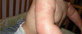
Enlarged lymph nodes can be of various sizes, from barely visible to noticeable to the naked eye. Palpation may cause pain. Sometimes there is redness of the skin over enlarged lymph nodes.
A pediatrician in Makhachkala can identify the development of lymphadenopathy in a child by a number of external signs, including:
- causeless weight loss;
- increase in temperature;
- increased sweating at night;
- weakness;
- attacks of fever;
- enlarged liver and spleen (on palpation);
- heart rhythm disturbances;
- frequent infectious diseases of the upper respiratory tract.
Inflammation of the lymph nodes
Reactive lymphadenitis is the most common cause of palpable swelling of the neck or lymph nodes in children, adolescents, and adults of any age.
Lymph nodes are located throughout the body as part of the lymphatic system, which also includes lymph capillaries, vessels, and ducts. In healthy people, in the area of the lower jaw, neck, groin, etc., it is possible to palpate under the skin several small, less than 1 cm, seals that are visually invisible. Lymphadenopathy is a noticeable enlargement of a lymph node greater than 1 cm.
Patients of different ages come to see specialists at the Miracle Doctor clinic with symptoms of lymphadenitis, who either themselves discovered enlarged lymph nodes, or were referred by another specialist who discovered characteristic signs:
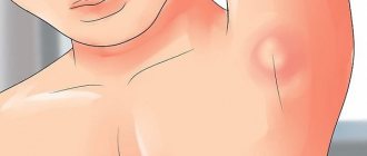
- painful subcutaneous thickening over 1 cm, located in the anatomical areas of the lymph nodes: under the jaw, on the neck, in the groin, under the armpit, etc.
- limited mobility and tight fit of the nodes to the surrounding tissues,
- the appearance of a lump is accompanied by headache, malaise, weakness, and changes in body temperature.
Causes of enlarged lymph nodes
Lymph nodes are “filtration stations” in the lymphatic system and are involved in activating the immune system when infectious pathogens enter the lymph flow.
Some components of blood plasma and foreign cells (for example, cancer cells, microorganisms) penetrate into the lymphatic vessels along with cellular material, antigens, etc.
Thanks to the filtration process, antigens also interact with lymphocytes contained in the lymph nodes. The immune response of these lymphocytes involves cell proliferation, which can cause enlarged nodes—reactive lymphadenopathy. Pathogenic microorganisms that enter the lymphatic fluid can directly infect nodes, causing lymphadenitis.
Thus, lymph node inflammation is primarily found in the context of inflammation and malignant diseases. If a patient has swollen lymph nodes, there are primarily two types of causes: benign causes, which are much more common, and malignant causes, which must be taken into account due to their serious consequences.
Depending on the duration of the course and manifestations, acute and chronic lymphadenitis are distinguished. Acute lymphadenitis can end in a purulent stage, with the formation of an abscess and the possible entry of purulent infected contents into adjacent tissues. Chronic develops after acute inflammation subsides and can be wavy in nature.
Diagnostics
The main goal is to establish the primary cause of the development of lymphadenopathy and differential diagnosis of other diseases with similar symptoms - phlegmon, purulent atheroma, etc.
Clinical examination and history taking are essential. Palpation is carried out to determine the number and degree of pain of the superficial inflamed lymph nodes, especially in the neck (including the occipital and supraclavicular regions), armpits and groin. The size of the nodes, sensitivity and degree of elasticity, mobility of the lymph nodes in the adjacent tissues are determined.
The duration of inflammation is established, the following are noted:
- possible skin injuries, especially cat scratches, rat bites;
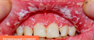
- the presence of infections in the areas drained by the affected nodes: infectious and inflammatory diseases of the upper respiratory tract, lesions or discharge from the genitals, joint pain, swelling, local soft tissue infections, dental infection;
- information on recent travel to countries with endemic infections (eg, Middle East, Africa);
- history of taking medications that contain trigger components.
Doppler ultrasound allows you to determine the length, width, shape, echogenicity of blood vessels, and therefore the degree of damage to the lymphatic system and determine the causes of the pathology.
Histological examination after diagnostic biopsy of the lymph node is usually prescribed for the differential diagnosis of malignant tumors and destructive changes in tissues.
A complete blood count may reveal an abnormal number of white blood cells, which is a reason to prescribe a tuberculin skin test and serological tests for mononucleosis, toxoplasmosis, sexually transmitted diseases, and a test for antibodies to SLE.
As a rule, the treatment of lymphadenopathy is carried out by the attending physician, who can refer the patient for consultation with a specialist.
Depending on the true cause of lymphadenopathy, antiviral and antibacterial drugs and physical therapy sessions may be prescribed. For purulent formations, surgical sanitation, installation of drainage and drug treatment are performed.
Treatment
Since adenopathy itself is not curable, treatment is aimed at eliminating the cause that provokes the development of lymphadenopathy. Patients with generalized lymphadenopathy—numerous swollen lymph nodes—usually have a systemic disease causing the inflammation, while patients with localized adenopathy usually have a local disease.
Diagnostics
To establish the true causes of enlarged lymph nodes, you need to undergo a comprehensive diagnostic examination in a clinical setting. First of all, it is necessary to conduct a thorough history taking to establish the cause of the enlarged lymph nodes in the child. Next, a study is carried out to determine the presence of tumor formations, skin lesions, and the presence of inflammatory processes.
In our medical network clinics in Makhachkala, various types of laboratory and instrumental studies are carried out. To identify the causes of lymphadenopathy in children, specialists usually conduct a set of diagnostic examinations, which includes:
- general and biochemical blood tests;
- general urine analysis;
- X-ray of the chest area;
- ultrasound examination of internal organs;
- magnetic resonance imaging (MRI);
- computed tomography (CT);
- lymph node biopsy with histological and cytological analysis.
Prevention and treatment of lymphadenopathy in Makhachkala
The essence of treatment for lymphadenopathy is to identify and treat the underlying disease. For example, if the pathology of the lymph nodes is caused by a bacterial disease, then a course of antibacterial therapy is prescribed. If the cause of lymphadenopathy is a viral infection, then antiviral treatment is carried out accordingly; in case of oncology, a complex of antitumor measures, etc. When choosing a treatment regimen, the doctor is guided by the individual characteristics of the patient’s body and the results of the examination. Self-medication is strictly prohibited. And the use of folk remedies is possible only in agreement with the attending physician. special measures to prevent lymphadenopathy in children . But the following will help minimize the likelihood of developing lymph node pathology:
- active lifestyle;
- good nutrition;
- increasing immunity;
- outdoor games;
- preventing frequent colds.
Parents are also required to take care of the child’s health, including timely visits to the doctor.